. 2010 Apr;38(1):17-26.
doi: 10.1016/j.nbd.2009.12.021. Epub 2010 Jan 4.

miR-497 regulates neuronal death in mouse brain after transient focal cerebral ischemia

Affiliations

miR-497 regulates neuronal death in mouse brain after transient focal cerebral ischemia

Ke-Jie Yin et al. Neurobiol Dis. 2010 Apr.

Abstract

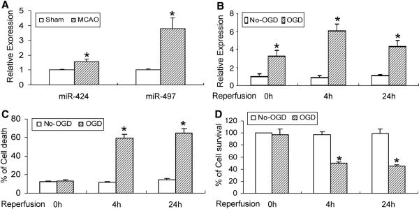
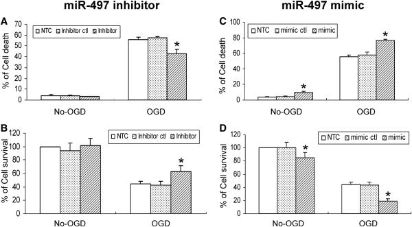
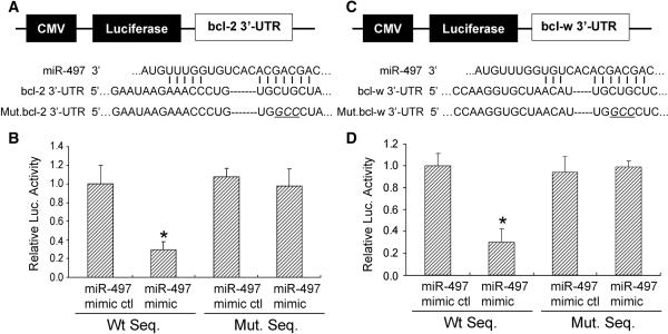
Dysfunction of the microRNA (miR) network has been emerging as a major regulator in neurological diseases. However, little is known about the functional significance of unique miRs in ischemic brain damage. Here, we found that miR-497 is induced in mouse brain after transient middle cerebral artery occlusion (MCAO) and mouse N2A neuroblastoma (N2A) cells after oxygen-glucose deprivation (OGD). Loss-of-miR-497 function significantly suppresses OGD-induced N2A cell death, whereas gain-of-miR-497 function aggravates OGD-induced neuronal loss. Moreover, miR-497 directly binds to the predicted 3'-UTR target sites of bcl-2/-w genes. Furthermore, knockdown of cerebral miR-497 effectively enhances bcl-2/-w protein levels in the ischemic region, attenuates ischemic brain infarction, and improves neurological outcomes in mice after focal cerebral ischemia. Taken together, our data suggest that miR-497 promotes ischemic neuronal death by negatively regulating antiapoptotic proteins, bcl-2 and bcl-w. We raise the possibility that this pathway may contribute to the pathogenesis of the ischemic brain injury in stroke.

Keywords: apoptosis; bcl-2; bcl-w; cerebral cortex; cerebral ischemia; microRNAs; oxygen-glucose deprivation.


Figures

Figure 1
Figure 1
Altered expression of miR-497 in mouse brain after focal cerebral ischemia and in OGD-induced neuronal death. (A) The expression of cerebral miR-497 and miR-424 is increased in mice subjected to transient MCAO and 24h of reperfusion. (B) OGD causes a time-dependent elevation in miR-497 levels, increasing at the starting point of recovery following 3h of OGD exposure and persisting at least to the 24h recovery time point after OGD. (C, D) Exposure to OGD also results in obvious mouse N2A cell death detected by LDH (C) and MTT (D) assays. OGD-induced neuronal loss is present at the 4h recovery time point, and persisted to at least the 24h recovery time point after 3h OGD. *P<0.05 vs non-OGD controls. Representative data from 3 separate experiments with similar results are shown.
Figure 2
Figure 2
miR-497 regulation of mouse N2A cell death after OGD. A miR-497 inhibitor, but not miR-497 inhibitor control attenuates OGD-induced neuronal death detected by LDH (A) and MTT (B) assays. In contrast, a miR-497 mimic, but not mimic control aggravates neuronal loss in both non-OGD and OGD conditions (C, D). * P<0.05 vs mimic or inhibitor control. Results shown are representative of 3 separate experiments with similar results.
Figure 3
Figure 3
Bcl-2 and bcl-w are target genes of miR-497 in mouse N2A cells. The protein levels of bcl-2 and bcl-w are significantly decreased after OGD. This decrease in expression is evident at the 4h recovery time point after 3h of OGD and persisted up to the 24h recovery time point after OGD (A). Treatment of mouse N2A cells with a miR-497 inhibitor effectively increases both bcl-2 and bcl-w levels after OGD (B). In contrast, a miR-497 mimic reduces the protein expression of bcl-2 and bcl-w after OGD (C). The expression of bcl-xl, one bcl-2 family protein, is not affected in mouse N2A cells after treatment with either a miR-497 mimic or inhibitor. The miR-497 mimic and inhibitor controls have no effect on the expression of bcl-2 and bcl-w. Data shown in top panels of figure 3A, B, C are representative of 3 separate experiments with similar results. Quantitative analysis of data in bottom panels of Fig 3A, B, C is expressed as mean ± SD. *p < 0.05 versus non-OGD controls or mimic and inhibitor controls.
Figure 4
Figure 4
A miR-497 binding site within the 3'-UTRs of bcl-2 and bcl-w mediates miR-497 translational repression. (A, C Top) Design of a miR-497 reporter vector containing a CMV-driven expression of a luciferase cDNA fused to a bcl-2 3'-UTR (pMIR-bcl2-3'-UTR), bcl-w 3'-UTR (pMIR-bclw-3'-UTR), mutated bcl-2 3'-UTR (pMIR-bcl2-m3'-UTR) or mutated bcl-w 3'-UTR (pMIR-bclw-m3'-UTR). MiR-497 and the miR-497-binding site in the 3'UTRs of bcl-2 (A bottom) and bcl-w (C bottom) are shown. (B, D) The 3'-UTR of bcl-2 and bclw mediate miR-497 control of bcl-2 and bcl-w expression. Mouse N2A cells were transfected with a pMIR-bcl-2-3'-UTR, pMIR-bcl-w-3'-UTR, pMIR-bcl-2-m3'-UTR, or pMIR-bcl-w-m3'-UTR respectively. The cells were also cotransfected with a miR-497 mimic or mimic control. Compared to the mimic control, the miR-497 mimic decreases expression of luciferase containing a wild type miR-497 binding site (Left 2 columns) but not a mutant binding site (Right 2 columns). Data are expressed as mean ± SEM. * P<0.05 vs mimic control. Results shown are representative of 3 separate experiments with similar results.
Figure 5
Figure 5
Bcl-2 and bcl-w are miR-497 target genes in mouse cerebral cortex after transient focal cerebral ischemia. Bcl-2 and bcl-w are downregulated following 24 h-reperfusion after 1h MCAo (A). Mini-pump assisted-cerebroventricular infusion of a miR-497 antagomir effectively inhibits cerebral miR-497 expression (B) and significantly enhances bcl-2 and bcl-w protein levels (C) in mouse cerebral cortex at the 24 h reperfusion time point after 1h MCA occlusion. In contrast, administration of an eGFP mRNA antagomir or artificial CSF (aCSF) has no effect on the expression of miR-497 and its two target proteins. Of note, the expression levels of bcl-xl, another member of the bcl-2 family is not affected in the ischemic cerebral cortex after treatment with the miR-497 antagomir. Quantitative analysis of data with representative blots shown in Fig 5A, C are in the respective bottom panels. Data are expressed as mean ± SD from 3 separate experiments. * denotes p < 0.05 versus sham or eGFP antagomir group.
Figure 6
Figure 6
The effect of miR-497 inhibition on ischemic infarction and neurological outcome. Mice were pretreated with a miR-497 antagomir, eGFP mRNA antagomir or artificial CSF (aCSF) for 48h via mini-pump assisted-cerebroventricular infusion, and then subjected to 1h MCA occlusion and 24h-reperfusion. 2% TTC-stained coronal sections were shown at different brain levels posterior to the frontal pole (A). Quantitative analysis was made on brain infarct volume in total (B), cortex (C), subcortex (D) areas (n=8) and neurological deficits (E) (n=11) in mice after stroke. In comparison to the controls, inhibition of the miR-497antagomir significantly attenuates ischemic infarct volume and improves behavioral outcome. Data are expressed as mean ± SD. * p < 0.05 vs the eGFP mRNA antagomir or artificial CSF group. (F) Schematic representation of miR-497 regulation of anti-apoptotic bcl-2 and bcl-w during ischemia-induced cell death signaling. In mouse brain, ischemic stimuli induce microRNA dysfunction, featuring activated miR-497 expression. miR-497 binds to the 3'-UTR region of bcl-2 and bcl-w, and further inhibits their activities by post-transcriptional gene silencing, thereby resulting in ischemic neuronal loss via mitochondrial apoptotic signaling.

References

    1. Bartel DP. MicroRNAs: genomics, biogenesis, mechanism, and function. Cell. 2004;116:281–97. - PubMed
    1. Bartel DP. MicroRNAs: target recognition and regulatory functions. Cell. 2009;136:215–33. - PMC - PubMed
    1. Bartel DP, Chen CZ. Micromanagers of gene expression: the potentially widespread influence of metazoan microRNAs. Nat Rev Genet. 2004;5:396–400. - PubMed
    1. Care A, et al. MicroRNA-133 controls cardiac hypertrophy. Nat Med. 2007;13:613–8. - PubMed
    1. Chen J, et al. Suppression of endogenous bcl-2 expression by antisense treatment exacerbates ischemic neuronal death. J Cereb Blood Flow Metab. 2000;20:1033–9. - PubMed

Publication types

MeSH terms