Review
. 2010 Feb;47(2):175-82.
doi: 10.1016/j.ceca.2009.12.003. Epub 2010 Jan 6.

Calcium, cellular aging, and selective neuronal vulnerability in Parkinson's disease

Affiliations
Review

Calcium, cellular aging, and selective neuronal vulnerability in Parkinson's disease

D James Surmeier et al. Cell Calcium. 2010 Feb.

Abstract

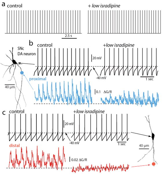
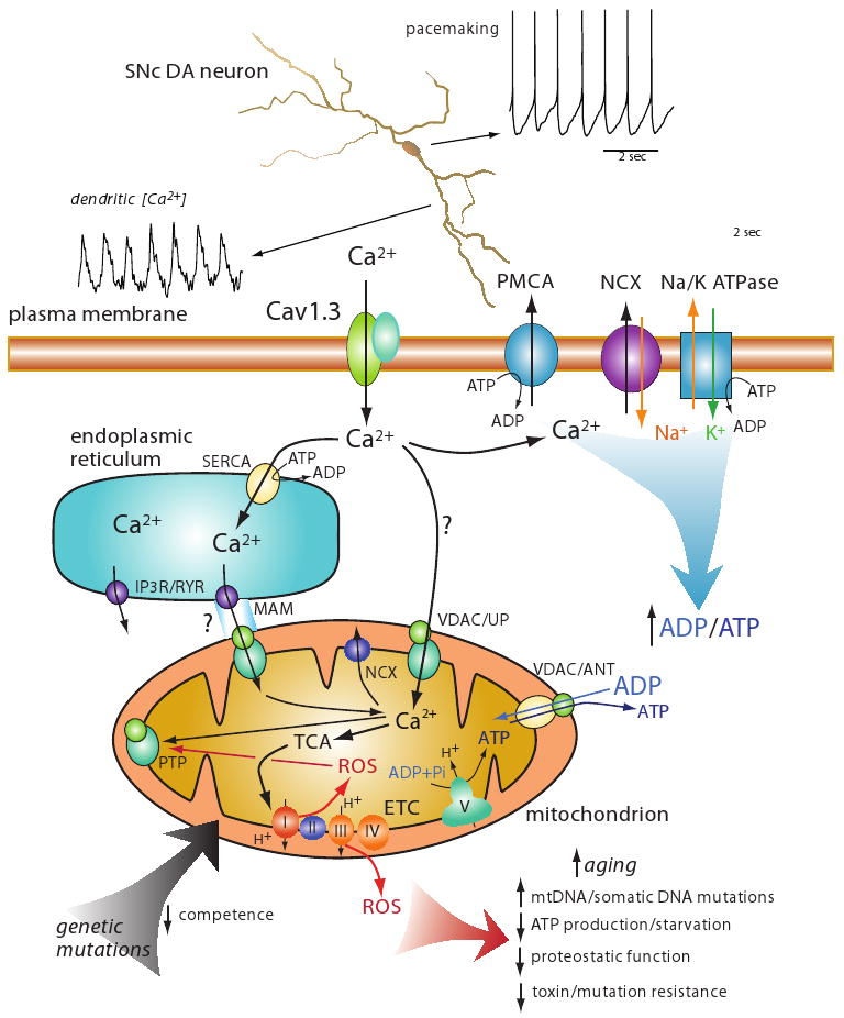
Parkinson's disease (PD) is the second most common neurodegenerative disease in developed countries. The core motor symptoms are attributable to the degeneration of dopamine (DA) neurons in the substantia nigra pars compacta (SNc). Why these neurons, and other restricted sets of non-dopamine neuron, succumb in PD is not clear. One potential clue has come from the observation that the engagement of L-type Ca2+ channels during autonomous pacemaking elevates the sensitivity of SNc DA neurons to mitochondrial toxins used to create animal models of PD, suggesting that Ca2+ entry is a factor in their selective vulnerability. Epidemiological data also supports a linkage between L-type Ca2+ channels and the risk of developing PD. This review examines the hypothesis that the primary factor driving neurodegenerative changes in PD is the metabolic stress created by sustained Ca2+ entry, particularly in the face of genetic or environmental factors that compromise oxidative defenses or proteostatic competence.


Figures

Figure 1
Figure 1. Low concentrations of DHPs suppress dendritic Ca2+ oscillations but do not slow pacemaking
a. Digitized cell-attached patch recordings from an SNc DA neuron before and after application of isradipine (5 μM). The median discharge rate before isradipine application was 2.2 Hz and 2.4 Hz after (p>0.05, n=4). b. Whole cell recording from the cell shown to the left (projection image) before and after isradipine (5 μM) application; there was no significant change in discharge rate in this cell or in 10 others. At the bottom, 2PLSM measurements of Fluo-4 fluorescence (G) at a proximal dendritic location (∼40 μm from the soma) normalized by the fluorescence of the red Alexa dye used to image the cell. c. Somatic recording during imaging at a more distal dendritic location (∼120-200 μm from the soma). Note the complete elimination of the spike associated dendritic Ca2+ transient at the distal imaging site. Similar results were obtained in 6 other neurons. From [46]
Figure 2
Figure 2. A schematic summarizing a model of how Ca2+ entry during pacemaking in SNc DA neurons might lead to mitochondrial oxidative stress, accelerated aging and eventual cell death
At the top is an image of a reconstructed SNc DA neuron showing the pacemaker driven somatic spiking and dendritic Ca2+ oscillations associated with pacemaking. This dendritic Ca2+ influx is attributable almost entirely to flux through Cav1.3 Ca2+ channels. At the bottom is a cartoon depicting the hypothetical mechanisms involved in elevating mitochondrial oxidative stress. Ca2+ influx through Cav1.3 channels is either sequestered in the endoplasmic reticulum by uptake through smooth endoplasmic reticulum Ca2+ (SERCA) pumps or taken up by mitochondria through channels created by voltage dependent anion channels (VDAC) and Ca2+ uniporter (UP); the extent to which this pathway is important is in question (hence the question mark on the arrow). Ca2+ could also enter mitochondria through this VDAC/UP channel at points of apposition between the ER and mitochondra where inositol trisphosphate receptors (IP3) and/or ryanodine receptors (IP3R/RYR) are positioned at specializations – mitochondrial associated membrane (MAM). This entry route has not been established in SNc DA neurons, hence the question mark. Ca2+ entering through Cav1.3 channels is moved back across the plasma membrane through either the Ca2+-ATPase (PMCA) or through a Na+/Ca2+ exchanger (NCX) that relies upon the Na+ gradient maintained by the Na/K ATPase at energetic cost; leading to conversion of ATP to ADP. ADP and ATP are exchanged by mitochondria with the VDAC and adenine nucleotide transporter (ANT). ADP stimulates oxidative phosphorylation. Ca2+ entering the mitochondrial matrix can stimulate enzymes of the tricarboxylic acid (TCA) cycle that produces reducing equivalents for the electron transport chain (ETC); complex I through V are shown at the inner mitochondrial membrane; complex V (ATP synthase) uses the electrochemical gradient to convert adenosine diphosphate (ADP) and inorganic phosphate to adenosine triphosphate (ATP). Electron movement along the ETC generates superoxide, leading to the production of reactive oxygen species (ROS) that can produce a variety of deleterious effects that can be viewed as accelerated aging (red arrow at the bottom). One other action of ROS is to promote opening of the mitochondrial permeability transition pore (mPTP); irreversible opening of the mPTP leads to release of pro-apoptetic factors into the cytosol. A basic question is whether reversible opening of the mPTP is possible under conditions of mild oxidative stress; mPTP opening and depolarization of the inner mitochondrial membrane could serve to diminish ROS production. Ca2+ is removed from mitochondrial through mitochrondrial Na+/Ca2+ exchangers (NCXs). Lastly, genetic mutations associated with Parkinson's disease, like those to DJ-1 or PINK1, might directly compromise the competence of mitochondria, leading to accelerated aging or increased oxidative stress.

References

    1. de Lau LM, Giesbergen PC, de Rijk MC, Hofman A, Koudstaal PJ, Breteler MM. Incidence of parkinsonism and Parkinson disease in a general population: the Rotterdam Study. Neurology. 2004;63:1240–4. - PubMed
    1. de Rijk MC, Tzourio C, Breteler MM, Dartigues JF, Amaducci L, Lopez-Pousa S, Manubens-Bertran JM, Alperovitch A, Rocca WA. Prevalence of parkinsonism and Parkinson's disease in Europe: the EUROPARKINSON Collaborative Study. European Community Concerted Action on the Epidemiology of Parkinson's disease. J Neurol Neurosurg Psychiatry. 1997;62:10–5. - PMC - PubMed
    1. Dorsey ER, Constantinescu R, Thompson JP, Biglan KM, Holloway RG, Kieburtz K, Marshall FJ, Ravina BM, Schifitto G, Siderowf A, Tanner CM. Projected number of people with Parkinson disease in the most populous nations, 2005 through 2030. Neurology. 2007;68:384–6. - PubMed
    1. Braak H, Ghebremedhin E, Rub U, Bratzke H, Del Tredici K. Stages in the development of Parkinson's disease-related pathology. Cell Tissue Res. 2004;318:121–34. - PubMed
    1. Hornykiewicz O. Dopamine (3-hydroxytyramine) and brain function. Pharmacol Rev. 1966;18:925–64. - PubMed

Publication types