. 2010 Apr 1;97(1):42-50.
doi: 10.1016/j.aquatox.2009.11.013. Epub 2009 Dec 11.

Flow cytometric analysis of BDE 47 mediated injury to rainbow trout gill epithelial cells

Affiliations

Flow cytometric analysis of BDE 47 mediated injury to rainbow trout gill epithelial cells

Jing Shao et al. Aquat Toxicol. .

Abstract

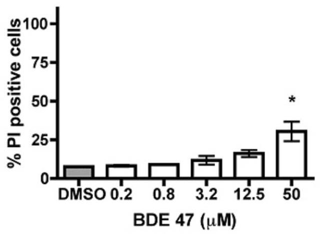
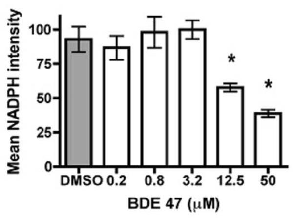
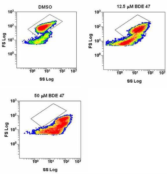
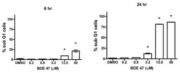
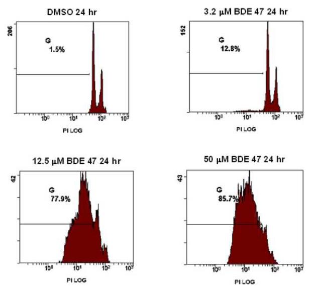
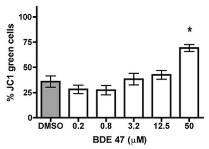
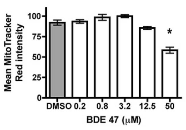
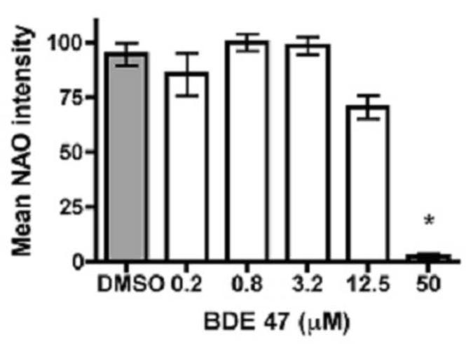
The polybrominated diphenyl ethers (PBDEs) are ubiquitous environmental contaminants whose residues are increasing in fish, wildlife and human tissues. However, relatively little is known regarding the mechanisms of cell injury caused by PBDE congeners in fish. In the present study, we employed flow cytometry-based analyses to understand the onset and mechanisms of cell injury in rainbow trout gill cells (RTgill-W1 cells) exposed to 2,2',4,4'-tetrabromodiphenyl ether (BDE 47). Substantial optimization and validation for flow cytometry protocols were required during assay development for the trout gill cell line. Exposure to micromolar concentrations of BDE 47 elicited a significant loss in RTgill-W1 cell viability that was accompanied by a decrease in NAD(P)H autofluorescence, a marker associated with disruption of cellular redox status. This loss in NAD(P)H content was accompanied by a decrease in nonyl acridine orange fluorescence, indicating mitochondrial membrane lipid peroxidation. Furthermore, low doses of BDE 47 altered cellular forward angle light scatter (FS, a measure of cell diameter or size) and side light scatter properties (SS, a measure of cellular internal complexity), consistent with the early stages of apoptosis. These changes were more pronounced at higher BDE 47 concentrations, which led to an increase in the percentage of cells undergoing frank apoptosis as evidenced by sub-G1 DNA content. Apoptosis was also observed at a relatively low dose (3.2muM) of BDE 47 if cells were exposed for an extended period of time (24h). Collectively, the results of these studies indicate that exposure of rainbow trout gill cells to BDE47 is associated with the induction of apoptosis likely originating from disruption of cellular redox status and mitochondrial oxidative injury. The current report extends observations in other species demonstrating that oxidative stress is an important mechanism of BDE 47 mediated cellular toxicity, and supports the use of oxidative stress-associated biomarkers in assessing the sublethal effects of PBDEs and their replacements in fish. The application of flow cytometry endpoints using fish cell lines should facilitate study of the mechanisms of chemical injury in aquatic species.


Figures

Figure 1
Figure 1. Effect of BDE 47 on cell viability as measured by propidium iodide (PI) fluorescence
Gill cells were incubated at 19 °C for 24 hours with 0.0 (0.2% DMSO control), 0.2, 0.8, 3.2, 12.5 and 50 μM BDE 47. Cells showing an increase in nuclear PI staining following treatment with BDE 47 indicate an increase in nuclear membrane permeability associated with cell death. Data represent mean ± SEM of 3 or more experiments. Asterisks denote significant treatment-related effects (p≤0.05).
Figure 2
Figure 2
Change in redox status associated with increasing concentration of BDE 47. Gill cells were exposed to increasing concentrations of BDE 47 in 0.2% DMSO for 24 hours and the effect on cellular redox status was determined by measuring NAD(P)H autofluorescence via flow cytometry. Data represent mean ± SEM of 3 or more experiments with asterisks denoting significant differences between controls and BDE 47-exposed cells (p≤0.05).
Figure 3
Figure 3. Alteration of forward angle light scatter (FS) and side light scatter (SS) properties following treatment with BDE 47
A reduction in the FS signal and an increase in the SS signal were observed at 12.5 μM and 50 μM following a 24 hour exposure to BDE 47 reflecting the early stages of apoptosis.
Figure 4
Figure 4. BDE 47-induced apoptosis in RTgill-W1 by sub-G1 analysis
Panel A. Exposure to BDE 47 caused significant increases in sub-G1 DNA content of gill cells at 3.2, 12.5 and 50 μM concentrations (*p < 0.01) at 24 hours post exposure. Data represent mean ± SEM of 3 experiments. In panel B are representative cytograms for cells from treatments involving DMSO (controls), 3.2 μM, 12.5 μM, and 50 μM BDE 47 exposure groups from a single experiment 24 hr following exposure.
Figure 5
Figure 5. Analysis of mitochondrial membrane potential following exposure to BDE 47
Gill cells were incubated at 19 °C for 24 hours with 0.0 (0.2% DMSO control), 0.2, 0.8, 3.2, 12.5 and 50 μM BDE 47. Panel A: Measurement of mitochondrial membrane potential (Δψm) using JC-1. Cells show an increase in membrane depolarization with treatment as seen by an increase in green fluorescence (JC-1 monomers). Panel B: MitoTracker red, which accumulates in polarized mitochondria, is reduced at the highest treatment level indicating a reduction in mitochondrial membrane potential. Data represent mean ± SEM of 3 or more experiments. Asterisks denote significant treatment-related effects (p≤0.05).
Figure 6
Figure 6. Loss of mitochondrial membrane integrity following exposure to BDE 47
Gill cells were incubated at 19 °C for 24 hours with 0.0 (0.2% DMSO control), 0.2, 0.8, 3.2, 12.5 and 50 μM BDE 47. The marked decrease of NAO fluorescence in cells at 50 μM BDE 47 indicates a decrease in mitochondrial membrane integrity. Data represent mean ± SEM of 3 or more experiments. Asterisks denote significant treatment-related effects (p≤0.05).

Similar articles

Cited by

References

    1. Ahmed SA, Gogal RM, Jr., Walsh JE. A new rapid and simple non-radioactive assay to monitor and determine the proliferation of lymphocytes: an alternative to [3H]thymidine incorporation assay. J. Immunol. Methods. 1994;170:211–24. - PubMed
    1. Antonsson B. Mitochondria and the Bcl-2 family proteins in apoptosis signaling pathways. Mol. Cell Biochem. 2004;256-257:141–55. - PubMed
    1. Antonsson B, Montessuit S, Lauper S, Eskes R, Martinou JC. Bax oligomerization is required for channel-forming activity in liposomes and to trigger cytochrome c release from mitochondria. Biochem. J. 2000;345(Pt 2):271–8. - PMC - PubMed
    1. Basova LV, Kurnikov IV, Wang L, Ritov VB, Belikova NA, Vlasova II, Pacheco AA, Winnica DE, Peterson J, Bayir H, Waldeck DH, Kagan VE. Cardiolipin switch in mitochondria: shutting off the reduction of cytochrome c and turning on the peroxidase activity. Biochemistry. 2007;46:3423–34. - PMC - PubMed
    1. Betts KS. Rapidly rising PBDE levels in North America. Environ. Sci. Technol. 2002;36:50A–52A. - PubMed

Publication types

Substances

LinkOut - more resources