. 2010 May;20(5):507-20.
doi: 10.1093/glycob/cwp203. Epub 2010 Jan 5.

Differential expression of immunomodulatory galectin-1 in peripheral leukocytes and adult tissues and its cytosolic organization in striated muscle

Affiliations

Differential expression of immunomodulatory galectin-1 in peripheral leukocytes and adult tissues and its cytosolic organization in striated muscle

Marcelo Dias-Baruffi et al. Glycobiology. 2010 May.

Abstract

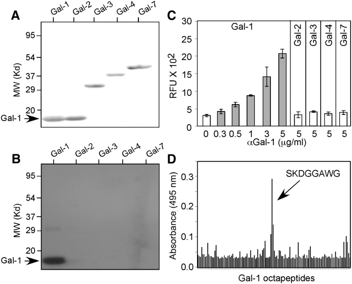
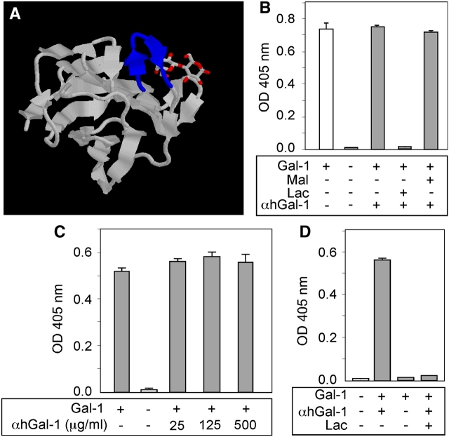
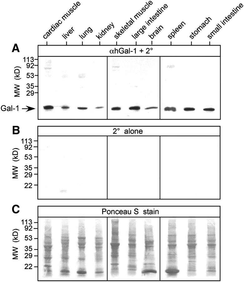
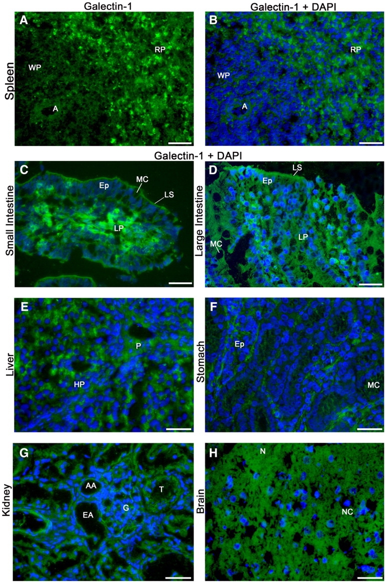
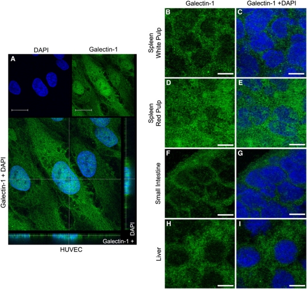
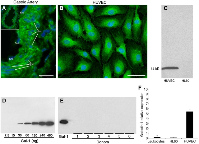
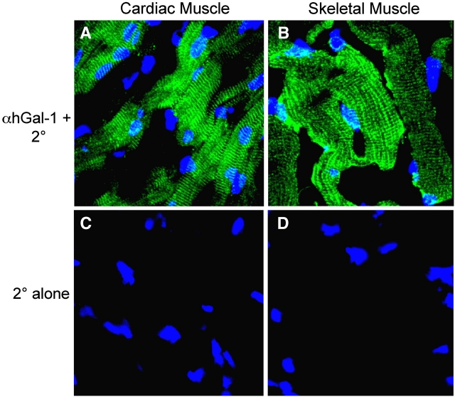
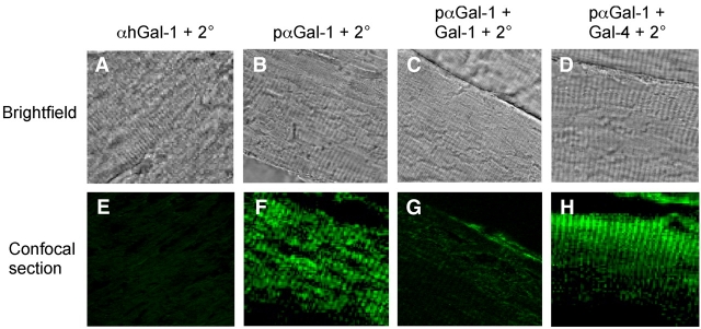
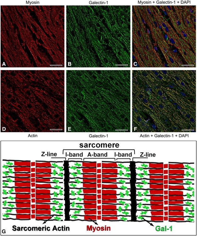
Galectin-1 (Gal-1) is important in immune function and muscle regeneration, but its expression and localization in adult tissues and primary leukocytes remain unclear. To address this, we generated a specific monoclonal antibody against Gal-1, termed alphahGal-1, and defined a sequential peptide epitope that it recognizes, which is preserved in human and porcine Gal-1, but not in murine Gal-1. Using alphahGal-1, we found that Gal-1 is expressed in a wide range of porcine tissues, including striated muscle, liver, lung, brain, kidney, spleen, and intestine. In most types of cells, Gal-1 exhibits diffuse cytosolic expression, but in cells within the splenic red pulp, Gal-1 showed both cytosolic and nuclear localization. Gal-1 was also expressed in arterial walls and exhibited prominent cytosolic and nuclear staining in cultured human endothelial cells. However, human peripheral leukocytes and promyelocytic HL60 cells lack detectable Gal-1 and also showed very low levels of Gal-1 mRNA. In striking contrast, Gal-1 exhibited an organized cytosolic staining pattern within striated muscle tissue of cardiac and skeletal muscle and colocalized with sarcomeric actin on I bands. These results provide insights into previously defined roles for Gal-1 in inflammation, immune regulation and muscle biology.


Figures

Fig. 1
Fig. 1
αhGal-1 specifically recognizes human Gal-1. (A) SDS-PAGE followed by transfer to nitrocellulose of recombinant Gal-1, Gal-2, Gal-3, Gal-4 and Gal-7 followed by Ponceau S stain. (B) SDS-PAGE followed by transfer to nitrocellulose of recombinant Gal-1, Gal-2, Gal-3, Gal-4 and Gal-7 and Western blot analysis using αhGal-1. (C) Recombinant Gal-1, Gal-2, Gal-3, Gal-4 and Gal-7 were captured on a polystyrene microtiter plate followed by detection with αhGal-1 using the concentrations indicated. (D) Octapeptides spanning human Gal-1 were generated as outlined in the Materials and methods. αhGal-1 was incubated with each pin octapeptide, and positive binding was detected with an alkaline phosphatase-labeled secondary antibody.
Fig. 2
Fig. 2
αhGal-1 recognizes Gal-1 bound to ligand. (A) Co-crystal of Gal-1 complexed with lactose as ligand (Lopez-Lucendo et al. 2004). The epitope sequence bound by αhGal-1 is highlighted in blue. (B) αhGal-1 was incubated with biotinylated Gal-1 with or without lactose or maltose as indicated for 30 min followed by incubation with laminin for 30 min. Bound Gal-1 was detected using horseradish peroxidase (HRP)-labeled streptavidin. (C) αhGal-1 was incubated with Gal-1 as in (B) (at the indicated concentrations) followed by incubation with laminin and detection of bound Gal-1 with HRP-streptavidin. (D) Gal-1 was incubated with laminin for 30 min followed by detection using αhGal-1.
Fig. 3
Fig. 3
Gal-1 exhibits expression in multiple tissues. Equal wet weights of each tissue were solubilized in SDS and subjected to SDS-PAGE. (A) Immunoblot of each respective tissue as indicated using αhGal-1 and secondary antibody (2°). (B) Incubation of membranes with secondary antibody alone (2° alone). (C) Ponceau S stains of each membrane prior to incubation with αhGal-1 in (A).
Fig. 4
Fig. 4
Gal-1 exhibits diffuse cytosolic localization in various adult tissues. Frozen sections of porcine tissue were subjected to immunofluorescence analysis using αhGal-1 and secondary antibody (2°) followed by analysis on a Leica DM 6000 M microscope. Deconvolution was performed using the Leica LAS AF software (version: 1.8.0 build 1346). (A) αhGal-1 stain of spleen (A, arteriole; WP, white pulp; RP, red pulp); (BH) αhGal-1 and 4′,6′-diamidino-2-phenylindole (DAPI) stain of (B) spleen (A, arteriole; WP, white pulp; RP, red pulp); (C) small intestine (MC, mucous cell; Ep, epithelium; LS, lumenal surface; LP, lamina propria); (D) large intestine (MC, mucous cell; Ep, epithelium; LS, lumenal surface; LP, lamina propria); (E) liver (P, portal space; HP, hepatica plate); (F) stomach (MC, mucous cell; Ep, epithelium cells forming gastric glands); (G) kidney (T, tubule; AA, afferent arteriole; EA, efferent arteriole; G, glomerulus); (H) brain (NC, nerve cells; N, neuropil and glia). Bar = 25 µm
Fig. 5
Fig. 5
Gal-1 exhibits diffuse cytosolic as well as nuclear localization in cells from different tissues. (A) Confocal analysis of HUVEC cells immunostained with αhGal-1 and DAPI as indicated. Gal-1 is localized in the nucleus and cytoplasm as seen in the confocal transversal section (Z axis image) in the bottom and side of panel (A). Bar = 20 μm. (BI) Confocal analysis through the middle sections, as the plane of focus cuts through the nucleus sections of the spleen white pulp, spleen red pulp, small intestine and liver. The sections were stained with αhGal-1 (B, D, F and H) or double-stained with DAPI (C, E, G and I) as indicated. Bar = 5 µm.
Fig. 6
Fig. 6
Gal-1 expression occurs in vascular tissue but not leukocytes. (A) Deconvolution image of frozen sections of a porcine gastric artery stained with αhGal-1 and DAPI using low (insert, ×20) and high magnification (×60) as outlined in Materials and methods. The endothelial cells (End) and smooth muscle cells (Smc) are indicated in the panel. Bar = 20 µm. (B) Human umbilical vein endothelial cells (HUVECs) were subjected to immunofluorescence analysis using αhGal-1 and DAPI and Leica DM 6000 M microscope, as in Figure 4. Bar = 25 µm. (C) Western blot analysis using αhGal-1 of extracts from HL60 cells or HUVECs as indicated. (D) Western blot analysis using ahGal-1 of various concentrations of recombinant Gal-1 as indicated. (E) Western blot analysis using ahGal-1 of recombinant Gal-1 or peripheral leukocytes from several donors. Western blot results are representative of three independent experiments. (F) Determination of the relative expression of cDNA for Gal-1 from total mRNA from pooled peripheral leukocytes, HL60, or HUVEC cells. Results were obtained using real-time PCR and represent accumulative data from three independent experiments performed in triplicate
Fig. 7
Fig. 7
Gal-1 displays organized cytosolic localization in striated tissues. Frozen sections of cardiac or skeletal muscle tissue for porcine tissue were subjected to confocal analysis using αhGal-1 and secondary antibodies or secondary alone as indicated. (AB) αhGal-1 stain of (A) cardiac muscle and (B) skeletal muscle. (CD) Staining with the secondary antibody alone is shown for (C) cardiac muscle and (D) skeletal muscle
Fig. 8
Fig. 8
Gal-1 exhibits discrete localization within skeletal muscle tissue as detected using a polyclonal anti-Gal-1. (AD) Brightfield images of paraformaldehyde (PFA)-fixed skeletal muscle subjected to PFA, fixed skeletal muscle subjected to immunohistochemical analysis of muscle tissue using (A) αhGal-1, (B) pαGal-1, (C) pαGal-1 pre-incubated with recombinant Gal-1 and (D) pαGal-1 pre-incubated with recombinant Gal-4. (EH) Confocal analysis of PFA-fixed skeletal muscle subjected to immunofluorescence analysis of muscle tissue using (E) αhGal-1, (F) pαGal-1, (G) pαGal-1pre-incubated with recombinant Gal-1 and (H) pαGal-1 pre-incubated with recombinant Gal-4
Fig. 9
Fig. 9
Gal-1 colocalizes with sarcomeric actin but not myosin within skeletal muscle tissue. (AC) Representative images of the immunofluorescence analysis of paraformaldehyde (PFA) fixed skeletal muscle using a Leica DM 6000 M microscope, as in Figure 4. (A) anti-myosin, (B) pαGal-1 and (C) merge of panels (A) and (B), or (D) anti- sarcomeric actin, (E) pαGal-1 and (F) merge of panels (D) and (E). Bar = 25 µm. (G) This is a schematic of striated muscle showing the predicted location of Gal-1 in the I-band region, as depicted by the green symbols

Similar articles

Cited by

References

    1. Ahmed H, Du SJ, Vasta GR. Knockdown of a galectin-1-like protein in zebrafish (Danio rerio) causes defects in skeletal muscle development. Glycoconj J. 2009;26:277–283. - PMC - PubMed
    1. Ahmed H, Fink NE, Pohl J., Vasta GR. Galectin-1 from bovine spleen: biochemical characterization, carbohydrate specificity and tissue-specific isoform profiles. J Biochem. (Tokyo) 1996;120:1007–1019. - PubMed
    1. Antia R, Ganusov VV, Ahmed R. The role of models in understanding CD8 T-cell memory. Nat Rev Immunol. 2005;5:101–111. - PubMed
    1. Baum LG, Seilhamer JJ, Pang M, Levine WB, Beynon D, Berliner JA. Synthesis of an endogeneous lectin, galectin-1, by human endothelial cells is up-regulated by endothelial cell activation. Glycoconj J. 1995;12:63–68. - PubMed
    1. Blois SM, Ilarregui JM, Tometten M, Garcia M, Orsal AS, Cordo-Russo R, Toscano MA, Bianco GA, Kobelt P, Handjiski B, Tirado I, Markert UR, Klapp BF, Poirier F, Szekeres-Bartho J, Rabinovich GA, Arck PC. A pivotal role for galectin-1 in fetomaternal tolerance. Nat Med. 2007;13:1450–1457. - PubMed

Publication types