. 2010 Feb 1;123(Pt 3):321-30.
doi: 10.1242/jcs.062075. Epub 2010 Jan 5.

Cdc20 is required for the post-anaphase, KEN-dependent degradation of centromere protein F

Affiliations

Cdc20 is required for the post-anaphase, KEN-dependent degradation of centromere protein F

Mark D J Gurden et al. J Cell Sci. .

Abstract

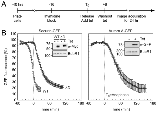
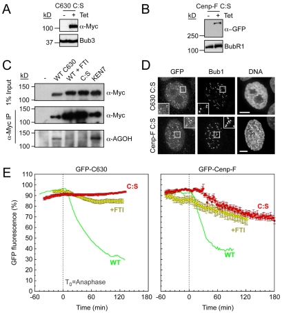
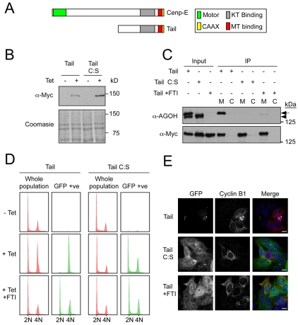
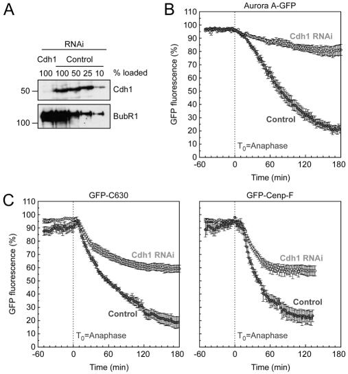
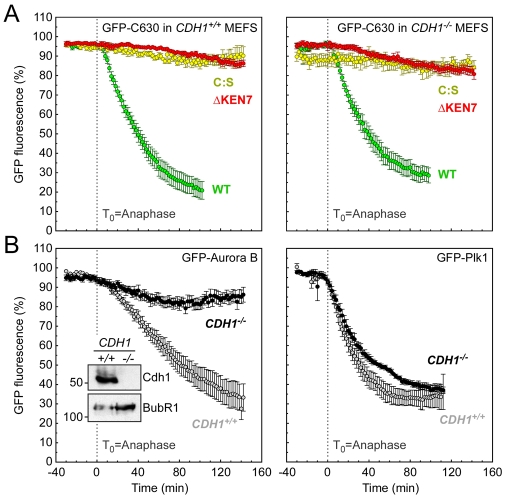
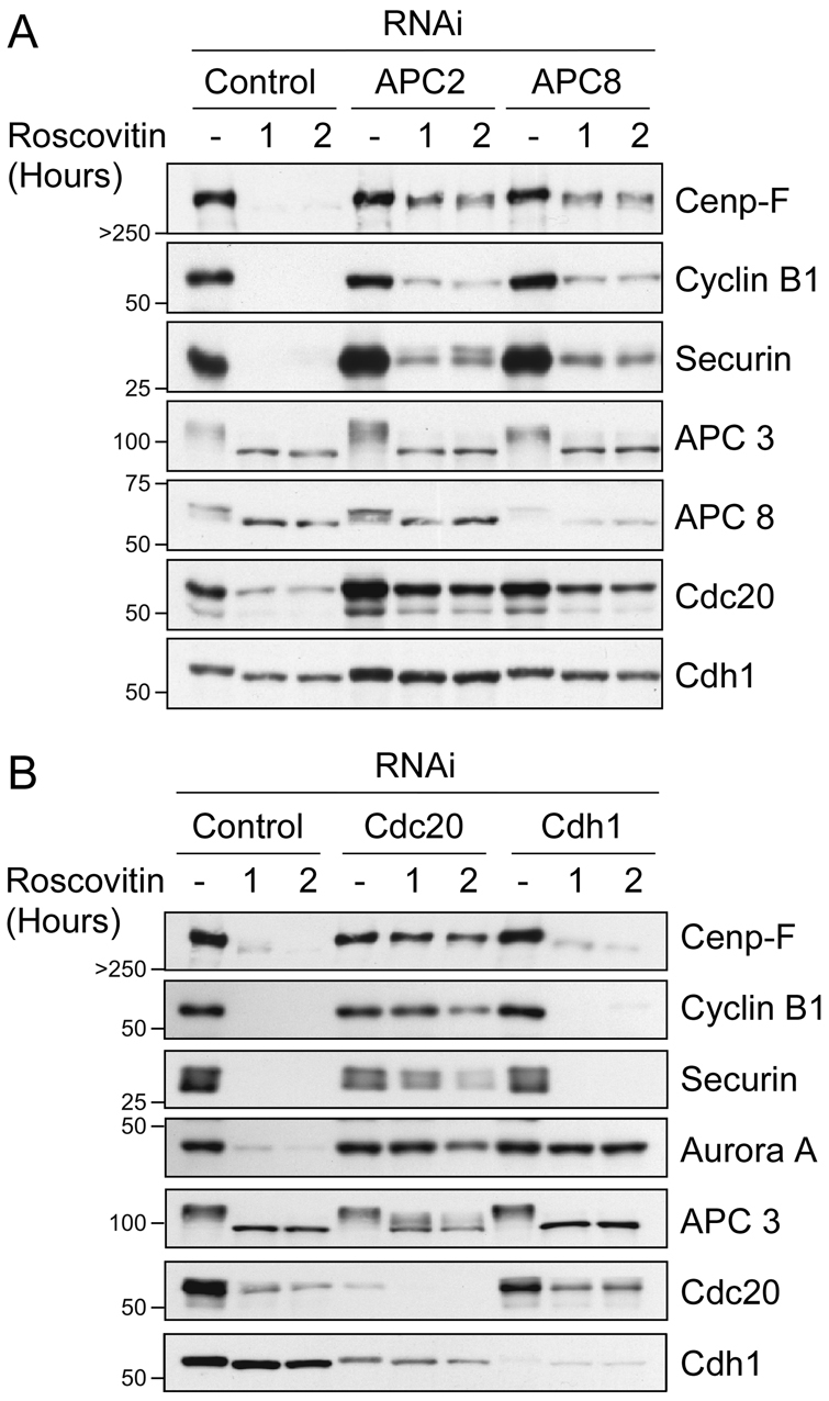
Progression through mitosis and cytokinesis requires the sequential proteolysis of several cell-cycle regulators. This proteolysis is mediated by the ubiquitin-proteasome system, with the E3 ligase being the anaphase-promoting complex, also known as the cyclosome (APC/C). The APC/C is regulated by two activators, namely Cdc20 and Cdh1. The current view is that prior to anaphase, the APC/C is activated by Cdc20, but that following anaphase, APC/C switches to Cdh1-dependent activation. However, here we present an analysis of the kinetochore protein Cenp-F that is inconsistent with this notion. Although it has long been appreciated that Cenp-F is degraded sometime during or after mitosis, exactly when and how has not been clear. Here we show that degradation of Cenp-F initiates about six minutes after anaphase, and that this is dependent on a C-terminal KEN-box. Although these two observations are consistent with Cenp-F being a substrate of Cdh1-activated APC/C, Cenp-F is degraded normally in Cdh1-null cells. By contrast, RNAi-mediated repression of APC/C subunits or Cdc20 does inhibit Cenp-F degradation. These findings therefore suggest that the APC/C does not simply 'switch' upon anaphase onset; rather, our observations indicate that Cdc20 also contributes to post-anaphase activation of the APC/C. We also show that the post-anaphase, KEN-box-dependent degradation of Cenp-F requires it to be farnesylated, a post-translational modification usually linked to membrane association. Because so many of the behaviours of Cenp-F are farnesylation-dependent, we suggest that this modification plays a more global role in Cenp-F function.


Figures

Fig. 1.
Fig. 1.
GFP-tagged SecurinΔD and Aurora A are degraded after anaphase. (A) Timeline showing experimental strategy. (B) Line graphs showing the mean degradation over time of securin-GFP (WT, light grey circles, n=15 cells from four experiments), securinΔD-GFP (ΔD, dark grey circles, n=14 from four experiments) and Aurora-A—GFP (dark grey circles, n=7 from one experiment). Immunoblots show the expression of the GFP-fusion proteins. BubR1 was used as a loading control. Note that the lower mobility forms are more apparent in the securinΔD-expressing line. This is because securin is also degraded in interphase by a phosphorylation-mediated SCF-dependent mechanism (Gil-Bernabe, 2006). The ΔD mutant seems refractory to SCF-dependent degradation, resulting in the accumulation of the hyper-phosphorylated forms.
Fig. 2.
Fig. 2.
GFP-tagged Cenp-F is degraded after anaphase in a KEN-dependent manner. (A) Schematic of Cenp-F showing the domain encoding C630. (B) Immunoblots showing the tetracycline-induced expression of GFP-tagged C630 (WT), C630 ΔKEN6 and C630 ΔKEN7 mutants. Bub3 was used as a loading control. (C) Immunoblot showing the tetracycline-induced expression of GFP-tagged Cenp-F (WT) and Cenp-F ΔKEN7. BubR1 was used as a loading control. (D) Immunofluorescence of mitotic cells expressing GFP-tagged C630 and C630 ΔKEN7, stained as indicated. Enlargements of highlighted (boxed) areas show kinetochore localisation as judged by Bub1. Scale bars: 5 μm. (E) Immunofluorescence of mitotic cells expressing GFP-tagged Cenp-F and Cenp-F ΔKEN7, stained as indicated. Enlargements of highlighted (boxed) areas show kinetochore localisation as judged by Bub1. Scale bars: 5 μm. (F) Line graphs showing the mean degradation over time of GFP-C630 (WT, green circles, n=27 from five experiments), GFP-C630 ΔKEN6 (yellow circles, n=12 from four experiments), GFP-C630 ΔKEN7 (red circles, n=14 from four experiments), GFP—Cenp-F (WT, green circles, n=8 from two experiments) and GFP—Cenp-F ΔKEN7 (red circles, n=14 from four experiments).
Fig. 3.
Fig. 3.
Degradation of Cenp-F is farnesylation-dependent. (A,B) Immunoblots showing the tetracycline-induced expression of GFP-tagged C630 C:S and GFP-tagged Cenp-F C:S. Bub3 and BubR1 were used as loading controls. (C) Anti-Myc immunoprecipitates of GFP-C630 (WT), GFP-C630 treated with FTI, GFP-C630 C:S and GFP-C630 ΔKEN7, from cells treated with AGOH. The precipitates were immunoblotted with anti-Myc and anti-AGOH, as indicated. (D) Immunofluorescence of mitotic cells expressing GFP-C630 C:S and GFP—Cenp-F C:S, stained as indicated. Enlargements of highlighted (boxed) areas show kinetochore localisation as judged by Bub1. Scale bars: 5 μm. (E) Line graphs showing the mean degradation over time of GFP-C630 treated with FTI (yellow circles, n=16 from four experiments), GFP-C630 C:S (red circles, n=30 from three experiments), GFP—Cenp-F treated with FTI (yellow circles, n=9 from five experiments), and GFP—Cenp-F C:S (red circles, n=13 from four experiments). Note that WT C630 and WT Cenp-F from Fig. 2 are shown as green lines merely for comparison.
Fig. 4.
Fig. 4.
Turnover of the Cenp-E tail is farnesylation-dependent. (A) Schematic of Cenp-E showing the tail domain. (B) Immunoblot showing the tetracycline-induced expression of GFP-tagged Cenp-E tail and tail C:S in DLD1 cells. Coomassie blue staining is shown as a loading control. (C) Anti-Myc immunoprecipitates of GFP—Cenp-E tail, GFP—Cenp-E tail treated with FTI and GFP—Cenp-E tail C:S from cells treated with AGOH. The precipitates were then immunoblotted with anti-Myc and anti-AGOH, as indicated. Arrow indicates Myc-tail protein. Arrowhead indicates an unknown farnesylated endogenous protein. M, Myc IP; C, control IP. (D) DNA content of asynchronous DLD1 cultures showing either whole cell populations (red) or only the GFP-positive cells. (E) Immunofluorescence of asynchronous cells expressing GFP—Cenp-E tail, GFP—Cenp-E tail C:S and GFP—Cenp-E tail treated with FTI, then imaged to detect GFP and cyclin B1 (red). Scale bars: 10 μm.
Fig. 5.
Fig. 5.
Cdh1 RNAi partially suppresses Cenp-F degradation. (A) Immunoblot showing the repression of Cdh1 24 hours after transfection with siRNA duplexes. BubR1 was used as a loading control. (B) Line graphs showing the mean degradation over time of Aurora-A—GFP treated with control (light grey, n=8 from two experiments) and Cdh1 RNAi (dark grey, n=10 from two experiments). (C) Line graphs showing the mean degradation over time of GFP-C630 treated with control (light grey, n=6 from four experiments) and Cdh1 RNAi (dark grey, n=10 from four experiments), and GFP—Cenp-F treated with control (light grey, n=5 from four experiments) and Cdh1 RNAi (dark grey, n=12 from four experiments).
Fig. 6.
Fig. 6.
Cenp-F is degraded normally in Cdh1-null iMEFS. (A) Line graphs showing the mean degradation over time of GFP-C630 (WT) in wild-type (green circles, n=19 from three experiments) and CDH1−/− iMEFs (green circles, n=13 from four experiments); GFP-C630 C:S in wild-type (yellow circles, n=13 from two experiments) and CDH1−/− iMEFs (yellow circles, n=8 from one experiment); and GFP-C630 ΔKEN7 in wild-type (red circles, n=12 from two experiments) and CDH1−/− iMEFs (red circles, n=10 from three experiments). (B) Line graphs showing the mean degradation over time of: GFP—Aurora-B in wild-type (grey circles, n=11 from two experiments) and CDH1−/− iMEFs (black circles, n=14 from two experiments); GFP-Plk1 in wild-type (grey circles, n=9 from two experiments) and CDH1−/− iMEFs (black circles, n=3 from two experiments).
Fig. 7.
Fig. 7.
Inhibition of APCCdc20 blocks Cenp-F degradation. Immunoblot showing degradation of securin, cyclin B1, Aurora A and Cenp-F following RNAi repression of (A) APC 2 and APC 8, or (B) Cdc20 and Cdh1. The blots are derived from one experiment, representative of three independent experiments revealing highly comparable results.

References

    1. Ameri K., Harris A. L. (2008). Activating transcription factor 4. Int. J. Biochem. Cell Biol. 40, 14-21 - PubMed
    1. Ashar H. R., James L., Gray K., Carr D., Black S., Armstrong L., Bishop W. R., Kirschmeier P. (2000). Farnesyl transferase inhibitors block the farnesylation of CENP-E and CENP-F and alter the association of CENP-E with the microtubules. J. Biol. Chem. 275, 30451-30457 - PubMed
    1. Bomont P., Maddox P., Shah J. V., Desai A. B., Cleveland D. W. (2005). Unstable microtubule capture at kinetochores depleted of the centromere-associated protein CENP-F. EMBO J. 24, 3927-3939 - PMC - PubMed
    1. Brown K. D., Coulson R. M., Yen T. J., Cleveland D. W. (1994). Cyclin-like accumulation and loss of the putative kinetochore motor CENP-E results from coupling continuous synthesis with specific degradation at the end of mitosis. J. Cell Biol. 125, 1303-1312 - PMC - PubMed
    1. Cheeseman I. M., Niessen S., Anderson S., Hyndman F., Yates J. R., 3rd, Oegema K., Desai A. (2004). A conserved protein network controls assembly of the outer kinetochore and its ability to sustain tension. Genes Dev. 18, 2255-2268 - PMC - PubMed

Publication types

MeSH terms