. 2010 Apr 1;19(7):1263-75.
doi: 10.1093/hmg/ddq003. Epub 2010 Jan 6.

Mechanisms of mosaicism, chimerism and uniparental disomy identified by single nucleotide polymorphism array analysis

Affiliations

Mechanisms of mosaicism, chimerism and uniparental disomy identified by single nucleotide polymorphism array analysis

Laura K Conlin et al. Hum Mol Genet. .

Abstract

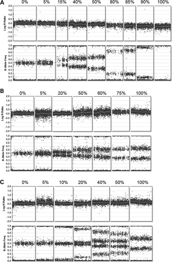
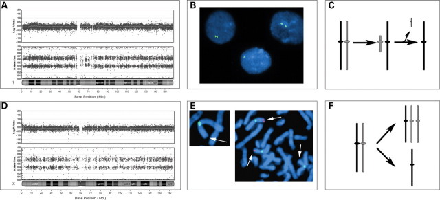
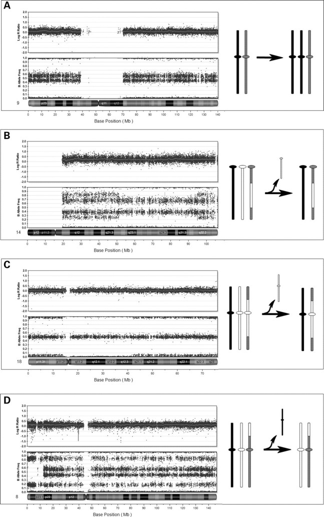
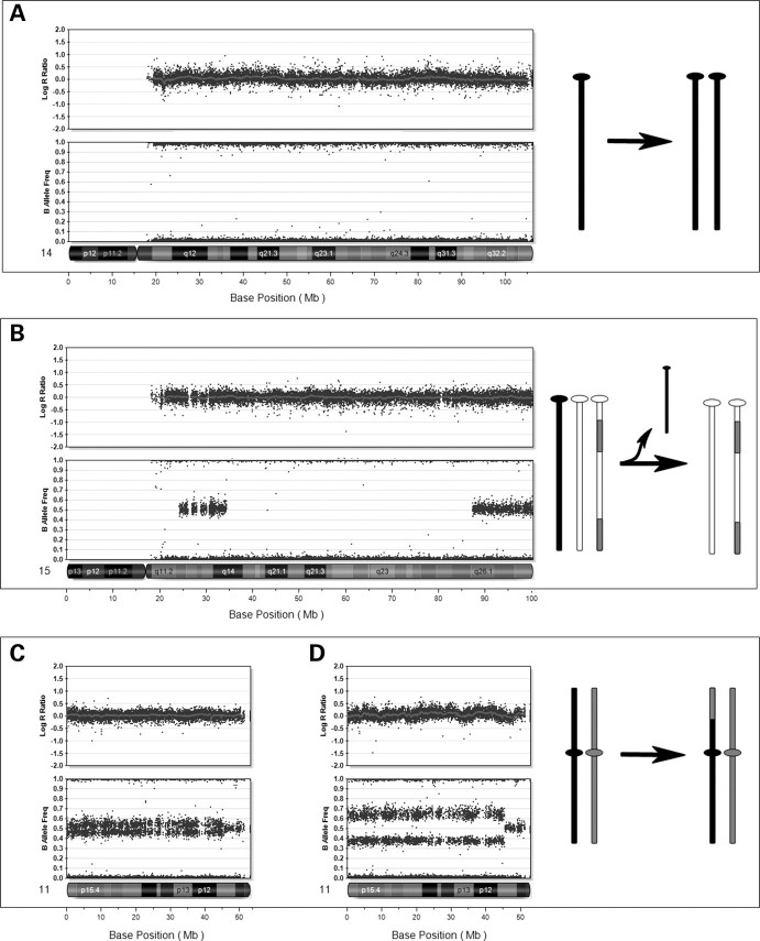
Mosaic aneuploidy and uniparental disomy (UPD) arise from mitotic or meiotic events. There are differences between these mechanisms in terms of (i) impact on embryonic development; (ii) co-occurrence of mosaic trisomy and UPD and (iii) potential recurrence risks. We used a genome-wide single nucleotide polymorphism (SNP) array to study patients with chromosome aneuploidy mosaicism, UPD and one individual with XX/XY chimerism to gain insight into the developmental mechanism and timing of these events. Sixteen cases of mosaic aneuploidy originated mitotically, and these included four rare trisomies and all of the monosomies, consistent with the influence of selective factors. Five trisomies arose meiotically, and three of the five had UPD in the disomic cells, confirming increased risk for UPD in the case of meiotic non-disjunction. Evidence for the meiotic origin of aneuploidy and UPD was seen in the patterns of recombination visible during analysis with 1-3 crossovers per chromosome. The mechanisms of formation of the UPD included trisomy rescue, with and without concomitant trisomy, monosomy rescue, and mitotic formation of a mosaic segmental UPD. UPD was also identified in an XX/XY chimeric individual, with one cell line having complete maternal UPD consistent with a parthenogenetic origin. Utilization of SNP arrays allows simultaneous evaluation of genomic alterations and insights into aneuploidy and UPD mechanisms. Differentiation of mitotic and meiotic origins for aneuploidy and UPD supports existence of selective factors against full trisomy of some chromosomes in the early embryo and provides data for estimation of recurrence and disease mechanisms.


Figures

Figure 1.
Figure 1.
Composite array results for mosaic deletions and duplications. This figure shows segments from different chromosomes illustrating mosaicism from 0–100%. For all figure parts, the percentages above the data indicate the level of mosaicism, with 0% representing a patient with normal copy number, and 100% representing a non-mosaic patient. (A) BeadStudio output for nine patients with varying levels of mosaicism for deletions involving autosomes. (B) BeadStudio output for seven patients with varying levels of mosaicism for trisomies. The pattern of B allele frequency indicates that the same two haplotypes present in the euploid cell line are also present in the triploid cell line at altered ratios. (C) BeadStudio output from seven patients with varying levels of mosaicism for trisomies. The additional B allele frequencies in the mosaic patients represent genotypes present in the trisomic cell line that are not present in the euploid cell line, suggesting a meiotic origin of the trisomy.
Figure 2.
Figure 2.
Mosaic monosomies. (A) BeadStudio output for patient no. 5 with a mosaic monosomy 7. Note the decreased log R ratio and altered B allele frequency. There is a somewhat lesser percent mosaicism around the centromere, which suggests that the deleted chromosome originated as a small pericentromeric marker, which was subsequently lost. (B) FISH confirmation of the monosomy 7 in interphase cells using a chromosome 7 centromere-specific probe. (C) Representation of the proposed mechanism, with the formation of a pericentromeric marker, which is subsequently lost to produce monosomy 7. (D) BeadStudio output for patient no. 4 with 45,X/47,XXX. (E) FISH confirmation of the parental origin of the X chromosomes. We used an X chromosome fosmid probe (G248P81417G5, labeled in red) within a known paternally inherited deletion within Xp22.3 indicating that the 45,X cell line contains the paternal X, while the 47,XXX cell line contains one paternal X and two non-deleted maternal X chromosomes. The X chromosome centromere is labeled in green. (F) Representation of origin of the 45,X/47,XXX showing mitotic non-disjunction.
Figure 3.
Figure 3.
Mosaic trisomies. (A) Mosaic trisomy 9 (20%) in patient no. 17, with no evidence for recombination suggesting a mitotic origin. On the right is a representation of the mitotic event. (B) Mosaic trisomy 14 (50%) in peripheral blood from patient no. 11, with a complex pattern of genotypes consistent with non-disjunction in meiosis I. There is evidence for two recombination sites at the points where genotype complexity changes. Illustration at right shows the distribution of genotypes resulting from meiotic recombination. (C) Mosaic trisomy 18 (10%) in peripheral blood of patient no. 14 with a genotype pattern consistent with non-disjunction in meiosis II. Evidence for two recombination sites are observed. Illustration at right shows regions of crossovers and resulting genotypes across the chromosome. (D) Mosaic trisomy 8 (40%) in patient no. 12 with a genotype pattern consistent with non-disjunction in meiosis I. Altered pattern near the telomere of the p-arm demonstrates UPD (isodisomy) for this region. This is illustrated in the figure on the right.
Figure 4.
Figure 4.
Uniparental disomy (UPD). In these cases, the log R ratio is consistent with normal copy number for all cases. (A) Complete isodisomy of chromosome 14 with loss of heterozygosity (LOH) for the entire chromosome in patient no. 22. This is consistent with the mechanism of monosomy rescue. (B) UPD of chromosome 15 in patient no. 29. Note the regions of LOH near the centromere and across the middle of the chromosome, which are interrupted by regions of heterozygosity, suggesting origin in meiosis II, with evidence of regions of recombination. (C) Segmental UPD of 11p11.2 to p-terminus in the DNA from cultured skin (10%) from patient no. 24. (D) Analysis of DNA from pancreatic tissue in patient no. 24, which had 30% mosaicism for the 11p LOH. This patient has a clinical diagnosis of focal hyperinsulinism.
Figure 5.
Figure 5.
Analysis of a chimeric individual (patient no. 30). (A) FISH analysis of a buccal sample using centromere probe for X and Y. (B) Cytogenetic analysis of cultured cells from hyper-pigmented skin revealed both XY and XX cells. (C) BeadStudio analysis of DNA from hyperpigmented tissue demonstrates that chromosomes 1 and 2 show altered B allele frequencies and a normal log R ratio. This altered B allele frequency was seen for all autosomes. (D) BeadStudio data from the X chromosome reveals only a single genotype at all loci. Note that the log R ratio reflects a 20% increase for the normal levels expected in a male and the B allele frequency of pseudoautosomal regions appears similar to that seen with the autosomes.

Similar articles

Cited by

References

    1. Hassold T.J., Jacobs P.A. Trisomy in man. Annu. Rev. Genet. 1984;18:69–97. - PubMed
    1. Hassold T., Hall H., Hunt P. The origin of human aneuploidy: where we have been, where we are going. Hum. Mol. Genet. 2007;16(Spec No. 2):R203–R208. - PubMed
    1. Kalousek D.K. Pathogenesis of chromosomal mosaicism and its effect on early human development. Am. J. Med. Genet. 2000;91:39–45. - PubMed
    1. Engel E. A fascination with chromosome rescue in uniparental disomy: Mendelian recessive outlaws and imprinting copyrights infringements. Eur. J. Hum. Genet. 2006;14:1158–1169. - PubMed
    1. Kotzot D. Complex and segmental uniparental disomy updated. J. Med. Genet. 2008;45:545–556. - PubMed

Publication types