. 2010 Apr 1;19(7):1153-64.
doi: 10.1093/hmg/ddp585. Epub 2010 Jan 6.

The snoRNA MBII-52 (SNORD 115) is processed into smaller RNAs and regulates alternative splicing

Affiliations

The snoRNA MBII-52 (SNORD 115) is processed into smaller RNAs and regulates alternative splicing

Shivendra Kishore et al. Hum Mol Genet. .

Abstract

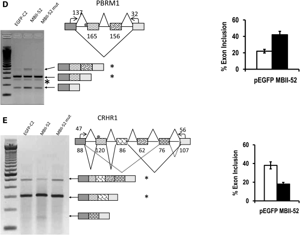
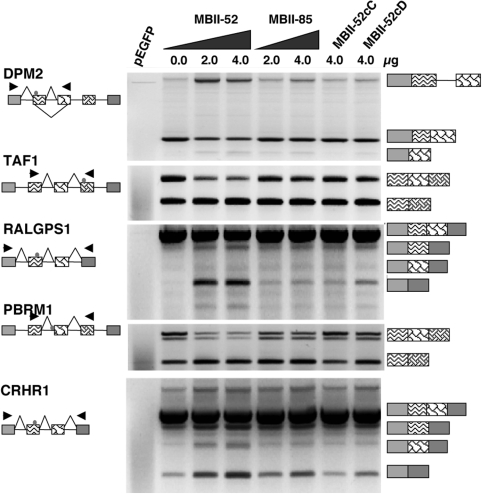
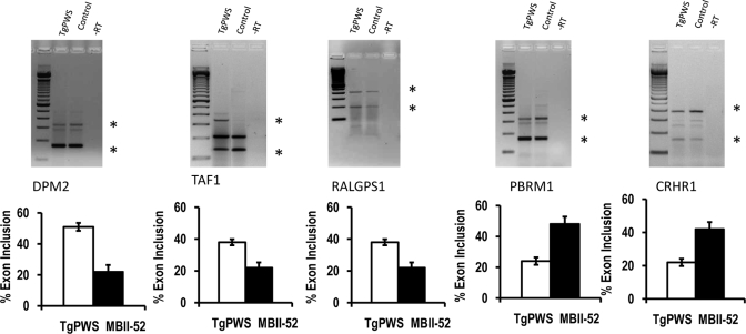
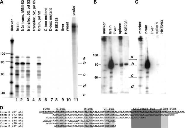
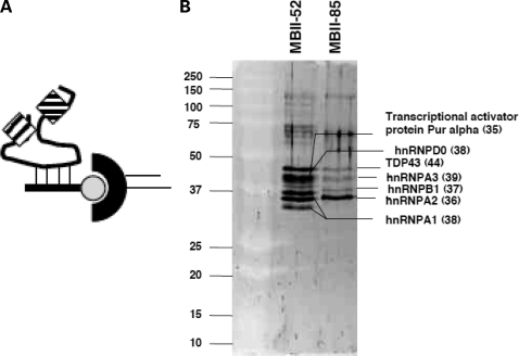
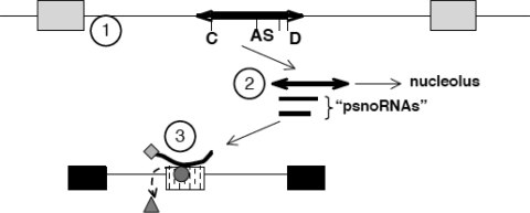
The loss of HBII-52 and related C/D box small nucleolar RNA (snoRNA) expression units have been implicated as a cause for the Prader-Willi syndrome (PWS). We recently found that the C/D box snoRNA HBII-52 changes the alternative splicing of the serotonin receptor 2C pre-mRNA, which is different from the traditional C/D box snoRNA function in non-mRNA methylation. Using bioinformatic predictions and experimental verification, we identified five pre-mRNAs (DPM2, TAF1, RALGPS1, PBRM1 and CRHR1) containing alternative exons that are regulated by MBII-52, the mouse homolog of HBII-52. Analysis of a single member of the MBII-52 cluster of snoRNAs by RNase protection and northern blot analysis shows that the MBII-52 expressing unit generates shorter RNAs that originate from the full-length MBII-52 snoRNA through additional processing steps. These novel RNAs associate with hnRNPs and not with proteins associated with canonical C/D box snoRNAs. Our data indicate that not a traditional C/D box snoRNA MBII-52, but a processed version lacking the snoRNA stem is the predominant MBII-52 RNA missing in PWS. This processed snoRNA functions in alternative splice-site selection. Its substitution could be a therapeutic principle for PWS.


Figures

Figure 1.
Figure 1.
MBII-52 changes the alternative splicing pattern of predicted targets. Computationally predicted MBII-52 target genes expressed in Neuro2A cells were analyzed by RT–PCR. Cells were transfected with 1 µg pEGFP-C2, 1 µg of the MBII-52 expressing construct pCMV/MBII-52 (MBII-52) (29) and 1 µg MBII-52 consensus box mutation, MBII-52 cm, (MBII-52 mut) expressing an antisense box mutation of MBII-52 (10). A representative ethidium bromide-stained agarose gel is shown. The adjacent diagram shows the part of the genes that was analyzed. Small arrows indicate the location of the primers used. The MBII-52 complementarity region is indicated by a dot. Numbers in boxes indicate the length of the exons and numbers next to PCR primers indicate the length of the amplified exon fragment. The structure of the PCR products is indicated by similar shading of exons in cDNA and genomic DNA. The statistical analysis of at least four independent experiments is shown on the right. Stars indicate the bands that were used for quantification. The sequences of the regulated exons are shown in Table 1.
Figure 1.
Figure 1.
MBII-52 changes the alternative splicing pattern of predicted targets. Computationally predicted MBII-52 target genes expressed in Neuro2A cells were analyzed by RT–PCR. Cells were transfected with 1 µg pEGFP-C2, 1 µg of the MBII-52 expressing construct pCMV/MBII-52 (MBII-52) (29) and 1 µg MBII-52 consensus box mutation, MBII-52 cm, (MBII-52 mut) expressing an antisense box mutation of MBII-52 (10). A representative ethidium bromide-stained agarose gel is shown. The adjacent diagram shows the part of the genes that was analyzed. Small arrows indicate the location of the primers used. The MBII-52 complementarity region is indicated by a dot. Numbers in boxes indicate the length of the exons and numbers next to PCR primers indicate the length of the amplified exon fragment. The structure of the PCR products is indicated by similar shading of exons in cDNA and genomic DNA. The statistical analysis of at least four independent experiments is shown on the right. Stars indicate the bands that were used for quantification. The sequences of the regulated exons are shown in Table 1.
Figure 2.
Figure 2.
Minigene analysis of MBII-52 target genes. The exons harboring the MBII-52 complementary region were subcloned into the exon trap vector pSpliceExpress. The structure of the resulting constructs pSE-RALGPS1, pSE-CRHR1, pSE-DPM2, pSE-PBRM1 and pSE-TAF1 as well as the location of the primers used for RT–PCR analysis is indicated on the left. pEGFP: only an expression construct for GFP is transfected. All other lanes contain 1 µg of pSE-reporter. MBII-52: cotransfection with 2 and 4 µg of MBII-52 expression construct, MBII-85: cotransfection with 2 and 4 µg of an MBII-85 expression construct, MBII52cC: cotransfection with 4 µg of a C-box mutant of MBII-52: MBII52cD: cotransfection with 4 µg of a D-box mutant of MBII-52. The structure of the products is shown schematically on the right, using the same shading scheme as in Figure 1. The usage of alternative exons indicated with a triangle was statistically evaluated. The comparison between MBII-52 and MBII-85 transfected cells showed statistically significant differences, the P-values of the Student's t-test were: DPM2: 0.001, TAF1: 0.023; RALGPS: 0.021; PBRM1: 0.076 and CRHR1: 0.002; (n = 4).
Figure 3.
Figure 3.
Comparison of RNA from TgPWS mice lacking MBII-52 expression and control littermates. Total brain samples from TgPWS mice lacking expression of the Prader–Willi critical region were compared with normal littermates expressing all the snoRNAs from the PWS critical region (control). Primers similar to Figure 1 were used. The structure of the gene products is indicated similar to Figures 1 and 2. Stars indicate the bands that were used for comparison. n = 6, other statistical evaluations: P = 0.093, <0.001, 0.0023, 0.001, 0.05 for DPM2, TAF1, RALGPS1, PBRM1 and CRHR1, respectively.
Figure 4.
Figure 4.
MBII-52 is processed into smaller RNAs. (A) RNase protection analysis using a probe detecting the MBII-52 copy used in transfection studies in Figures 1 and 2. Five microgram of the following total RNAs was hybridized to an MBII-52 antisense probe: (1): total mouse brain, (2): Neuro2A cells transfected with pCMV/MBII-52. Lanes (3–5) are protections from RNPs captured with oligonucleotides against the antisense box (Fig. 5). (3): Affinity captured RNA from Neuro2A cells expressing MBII-52 using a MBII-52 probe for pull down (pd), (4): affinity captured RNA from Neuro2A expressing MBII-52 using an MBII-85 probe (negative control) and (5): affinity captured RNA from brain using an MBII-52 probe for pull down. Lane 6: RNA from Neuro2A cells transfected with an expression construct containing a C-box mutant, (7): RNA from Neuro2A cells transfected with an expression construct containing a D-box mutant, (8): HEK293 cells non-transfected, (9, 10): RNA from liver and yeast. (11): Undigested probe. The marker is a 100 nt RNA base ladder. (B) Northern blot analysis of MBII-52. Fifteen microgram total RNA from brain, liver, spleen and HEK293 cells was separated on 15% polyacrylamide gels and probed with a 32P labeled probe for MBII-52. After stringent washing, cross-hybridizing bands in liver, spleen and HEK293 cells still remain. Exposure was overnight. (C) The filter from (B) was treated with RNase A/T1 and again washed. The cross-hybridizing bands disappear, but the signals corresponding to smaller bands remain. Exposure was for 3 days. (D) Sequences of the shorter RNAs. The stems and functional boxes are indicated. The clones are ordered according to their length. Form A corresponds to the published snoRNA MBII-52. Underlined nucleotides in forms C and D indicated predicted stems.
Figure 5.
Figure 5.
Analysis of proteins associated with MBII-52. (A) Experimental strategy: a biotinylated oligonucleotide is used to capture the snoRNP complex. The oligonucleotide is shown as a line, the complementarity is dashed, biotin is shown as a circle. The RNP complex (boxes) is isolated by streptavidin (half-circle)-capture from extracts expressing MBII-52 and washed in non-denaturing buffer. The extracts were prepared by transfecting MBII-52 expression constructs and performing Dignam mini-extracts. (B) Silver stain of a gel with affinity purified RNPs. MBII-52: the RNPs were isolated with a capture-oligonucleotide against MBII-52. MBII-85: the RNP was isolated with a capture oligonucleotide against MBII-85. Proteins were determined by mass spectrometry and are indicated. Sizes in kDa are shown in parentheses.
Figure 6.
Figure 6.
Model for MBII-52 action on RNA processing. (1) The PWS critical region contains snoRNAs (thick line) located in introns between non-coding exons (grey boxes). The snoRNA is characterized by a C box (C), D box (D) and an antisense box (AS), as well as stem-forming sequences (arrows). (2) This unit generates several RNAs, including the full-length snoRNA that shows its highest concentration in the nucleolus and Cajal bodies (35) as well as several shorter psnoRNAs (for processed snoRNAs). PsnoRNAs are present in the nucleoplasm where they associate with hnRNPs. (3) PsnoRNAs can change splice-site selection, most likely by binding to complementary sequences. We postulate that they either remove regulatory proteins from their targets (triangle) or bring in associated proteins to the exon recognition complex (diamond associated with the small RNA).

Similar articles

Cited by

References

    1. Pan Q., Shai O., Lee L.J., Frey B.J., Blencowe B.J. Deep surveying of alternative splicing complexity in the human transcriptome by high-throughput sequencing. Nat. Genet. 2008;40:1413–1415. - PubMed
    1. Wang E.T., Sandberg R., Luo S., Khrebtukova I., Zhang L., Mayr C., Kingsmore S.F., Schroth G.P., Burge C.B. Alternative isoform regulation in human tissue transcriptomes. Nature. 2008;456:470–476. - PMC - PubMed
    1. Stamm S., Ben-Ari S., Rafalska I., Tang Y., Zhang Z., Toiber D., Thanaraj T.A., Soreq H. Function of alternative splicing. Gene. 2005;344C:1–20. - PubMed
    1. Watkins N.J., Segault V., Charpentier B., Nottrott S., Fabrizio P., Bachi A., Wilm M., Rosbash M., Branlant C., Luhrmann R. A common core RNP structure shared between the small nucleoar box C/D RNPs and the spliceosomal U4 snRNP. Cell. 2000;103:457–466. - PubMed
    1. Matera A.G., Terns R.M., Terns M.P. Non-coding RNAs: lessons from the small nuclear and small nucleolar RNAs. Nat. Rev. Mol. Cell Biol. 2007;8:209–220. - PubMed

Publication types

Substances