. 2010 Mar 1;21(5):791-801.
doi: 10.1091/mbc.e09-08-0656. Epub 2010 Jan 6.

Intracellular targeting signals and lipid specificity determinants of the ALA/ALIS P4-ATPase complex reside in the catalytic ALA alpha-subunit

Affiliations

Intracellular targeting signals and lipid specificity determinants of the ALA/ALIS P4-ATPase complex reside in the catalytic ALA alpha-subunit

Rosa L López-Marqués et al. Mol Biol Cell. .

Abstract

Members of the P(4) subfamily of P-type ATPases are believed to catalyze flipping of phospholipids across cellular membranes, in this way contributing to vesicle biogenesis in the secretory and endocytic pathways. P(4)-ATPases form heteromeric complexes with Cdc50-like proteins, and it has been suggested that these act as beta-subunits in the P(4)-ATPase transport machinery. In this work, we investigated the role of Cdc50-like beta-subunits of P(4)-ATPases for targeting and function of P(4)-ATPase catalytic alpha-subunits. We show that the Arabidopsis P(4)-ATPases ALA2 and ALA3 gain functionality when coexpressed with any of three different ALIS Cdc50-like beta-subunits. However, the final cellular destination of P(4)-ATPases as well as their lipid substrate specificity are independent of the nature of the ALIS beta-subunit they were allowed to interact with.


Figures

Figure 1.
Figure 1.
ALA2 in combination with ALIS genes functionally complement the cold- and heavy metal–sensitive phenotype of a Δdrs2Δdnf1Δdnf2 yeast mutant. The triple yeast mutant Δdrs2Δdnf1Δdnf2 expressed ALA2 alone or in combination with ALIS genes under the control of a bidirectional galactose-inducible promoter. Yeast DRS2 and an empty vector were used as positive and negative controls, respectively. ALA2 was tagged with the hemagglutinin (HA) epitope and ALIS with the RGSH6 epitope. ALA2 expressed in concert with ALIS allows growth of the cold-sensitive yeast strain at 20°C, whereas the catalytically inactive mutant, ala2D381A, in combination with ALIS, did not support growth at 20°C. (A) Yeast cells were dropped on rich media at 30°C and at the restrictive temperature 20°C. (B) Yeast cells dropped on minimal media without metals or containing 200 μM CoCl2 or 2 mM ZnCl2. (C) Western blot analysis of membranes from yeast overexpressing HA:ALA2 or HA:ala2D381A in concert with RGSH6:ALIS proteins. Control, cells grown on glucose plates to repress expression; Induced, cells grown on galactose plates to induce overexpression; Empty, yeast cells transformed with an empty plasmid control; ala2, ala2D381A.
Figure 2.
Figure 2.
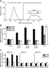
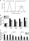
Coexpression of ALA2 and ALIS proteins complements the lipid uptake defect of the Δdrs2Δdnf1Δdnf2 yeast mutant. Yeast mutant cells expressing different protein combinations and wild-type cells were labeled with 1-palmitoyl-NBD lipids and then washed and analyzed by flow cytometry. Accumulation of NBD lipids was expressed as percentage of fluorescence intensity relative to control Δdrs2Δdnf1Δdnf2 mutant cells. (A) Coexpression of ALA2 and ALIS1 resulted in two populations of cells with low and high NBD-PS uptake, respectively. Representative histograms are shown. For quantitative analysis of lipid uptake the fluorescence intensity of the total population was determined. (B) ALA2 specifically promoted NBD-PS internalization in the presence of ALIS proteins. (C) The catalytically inactive mutant ala2D381A failed to promote NBD-PS internalization. (B and C) Results are averages ± SE from three independent experiments. One hundred percent corresponds to 45 ± 13 arbitrary units (NBD-PS), 31 ± 7 arbitrary units (NBD-PE), and 32 ± 6 arbitrary units (NBD-PC). WT, wild type; ala2, ala2D381A.
Figure 3.
Figure 3.
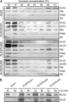
ALA2 reaches the plasma membrane when expressed in yeast. (A) PM-enriched membranes from yeast expressing HA:ALA2 or HA:ALA2 together with RGSH6:ALIS1, RGSH6:ALIS3, or RGSH6:ALIS5 were subjected to discontinuous sucrose density gradient fractionation. (B) Fractions corresponding to 30 and 48% sucrose were analyzed in parallel for each ALA2/ALIS combination to compare protein levels. Western blots were probed using the following antibodies: anti-Pma1p, PM; anti-Sed5p, Golgi apparatus; anti-Dpm1p, ER; anti-HA, ALA2; and anti-RGSH6, ALIS.
Figure 4.
Figure 4.
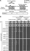
ALA2 as well as ALA3 in combination with ALIS proteins regulate lipid distribution in the PM of yeast. (A) Cytolytic, lipid-binding peptides allow probing of distribution of natural lipids. Wild-type yeast confines PS and PE in the cytoplasmic leaflet of the PM and is less sensitive to peptide-induced cytolysis compared with yeast mutants deficient in P4-ATPases. (B) Δdrs2Δdnf1Δdnf2 yeast cells were transformed with ALA2 and ALA3 alone or in combination with ALIS genes and dropped on plates containing PS-binding papuamide B or PE-binding duramycin. Yeast DRS2 and an empty vector were used as positive and negative controls, respectively. Control, nonexpressing cells; Induced, overexpressing yeast; No pep, no peptide included; WT, wild type; ala2, ala2D381A; ala3, ala3D413A.
Figure 5.
Figure 5.
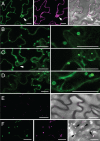
ALA2 leaves the ER and localizes to vesicular structures in the presence of ALIS. GFP:ALA2 was expressed in tobacco leaf epidermal cells alone or in the presence of ALIS. (A) When expressed alone, GFP:ALA2 colocalized with an ER-retained YFP:HDEL: left, GFP fluorescence; middle, YFP fluorescence; right, fluorescent signal overlay on the bright-field image. (B–D) On coexpression with an untagged ALIS, GFP:ALA2 was localized to vesicular structures: (B) coexpression with ALIS1; (C) coexpression with ALIS3; and (D) coexpression with ALIS5. In each case, two different magnifications are shown; (E) Prevacuolar compartment visualized by expression of the fusion protein BP-80:YFP containing the targeting signals for a plant homologue of the yeast receptor protein Vsp10p: left, YFP fluorescence; right, overlay of YFP signal on a bright-field image. (F) GFP:ALA2 expressed in the presence of ALIS colocalized with BP-80:YFP in aberrant prevacuolar compartment structures. Coexpression with ALIS3 is shown: left, GFP fluorescence; middle, YFP fluorescence; right, overlay of the fluorescent signals on the bright-field image. Arrows indicate the position of nuclei when visible. Bar, 25 μm.
Figure 6.
Figure 6.
Coexpression of ALA3 and ALIS proteins complements the lipid uptake defect of the Δdrs2Δdnf1Δdnf2 yeast mutant. Yeast mutant cells expressing different protein combinations and wild-type cells were labeled with 1-palmitoyl-NBD lipids and then washed and analyzed by flow cytometry. Accumulation of NBD lipids was expressed as percentage of fluorescence intensity relative to control Δdrs2Δdnf1Δdnf2 mutant cells. (A) Coexpression of ALA3 and ALIS1 resulted in cells displaying a broad distribution of NBD-PS uptake. Representative histograms are shown. For quantitative analysis of lipid uptake the fluorescence intensity of the total population was determined. (B) ALA3 facilitated the internalization of NBD-PS, -PE, and -PC in the presence of ALIS proteins; (C) The catalytically inactive mutant ala3D413A failed to promote NBD-lipid internalization. Results in B and C are averages ± SE from five independent experiments. One hundred percent corresponds to 51 ± 5 arbitrary units (NBD-PS), 40 ± 5 arbitrary units (NBD-PE), and 42 ± 4 arbitrary units (NBD-PC). WT, wild type; ala3, ala3D413A.
Figure 7.
Figure 7.
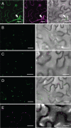
ALA3 leaves the ER and localizes to Golgi-like structures in the presence of ALIS. GFP:ALA3 was expressed in tobacco leaf epidermal cells alone or in the presence of ALIS. (A) When expressed alone, GFP:ALA3 colocalized with an ER-retained YFP:HDEL: left, GFP fluorescence; middle, YFP fluorescence; right, fluorescent signal overlay on the bright-field image. (B–D) On coexpression with untagged ALIS, GFP:ALA3 was localized to Golgi-like structures: (B) coexpression with ALIS1; (C) coexpression with ALIS3; and (D) coexpression with ALIS5. In each case: left GFP fluorescence; right, overlay (including autofluorescence, magenta) of GFP signal on a bright-field image. (E) Golgi bodies visualized by expression of a Golgi localized sialyl transferase fusion to YFP (ST:YFP). Arrows indicate the position of nuclei when visible. Bar, 25 μm.
Figure 8.
Figure 8.
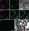
ALIS1 leaves the ER and localizes to different compartments in the presence of ALA2 and ALA3, respectively. A C-terminal fusion of ALIS1 to YFP was expressed in tobacco leaf epidermal cells alone or in combination with ALA2 and ALA3. (A) When expressed alone, ALIS1:YFP colocalized with an ER-retained GFP:HDEL: left, YFP fluorescence; middle, GFP fluorescence; right, overlay image of YFP and GFP fluorescent signals on the bright-field image. (B) When expressed with GFP:ALA2, ALIS1:YFP colocalized to vesicular structures: left, YFP fluorescence; middle, GFP fluorescence; right, overlay of YFP and GFP fluorescent signals. (C) On coexpression with an untagged ALA3, ALIS1:YFP was detected in Golgi-resembling structures: left, YFP fluorescence; right, overlay of YFP signal on the bright-field image. Arrows indicate the position of nuclei when visible. Bar, 25 μm.

Similar articles

Cited by

References

    1. Alder-Baerens N., Lisman Q., Luong L., Pomorski T., Holthuis J. C. Loss of P4 ATPases Drs2p and Dnf3p disrupts aminophospholipid transport and asymmetry in yeast post-Golgi secretory vesicles. Mol. Biol. Cell. 2006;17:1632–1642. - PMC - PubMed
    1. Axelsen K. B., Palmgren M. G. Evolution of substrate specificities in the P-type ATPase superfamily. J. Mol. Evol. 1998;46:84–101. - PubMed
    1. Axelsen K. B., Palmgren M. G. Inventory of the superfamily of P-type ion pumps in Arabidopsis. Plant Physiol. 2001;126:696–706. - PMC - PubMed
    1. Brandizzi F., Fricker M., Hawes C. A greener world: the revolution in plant bioimaging. Nat. Rev. Mol. Cell Biol. 2002;3:520–530. - PubMed
    1. Chantalat S., Park S. K., Hua Z., Liu K., Gobin R., Peyroche A., Rambourg A., Graham T. R., Jackson C. L. The Arf activator Gea2p and the P-type ATPase Drs2p interact at the Golgi in Saccharomyces cerevisiae. J. Cell Sci. 2004;117:711–722. - PubMed

Publication types