. 2010 Mar 1;21(5):767-77.
doi: 10.1091/mbc.e09-11-0967. Epub 2010 Jan 6.

Global up-regulation of microtubule dynamics and polarity reversal during regeneration of an axon from a dendrite

Affiliations

Global up-regulation of microtubule dynamics and polarity reversal during regeneration of an axon from a dendrite

Michelle C Stone et al. Mol Biol Cell. .

Abstract

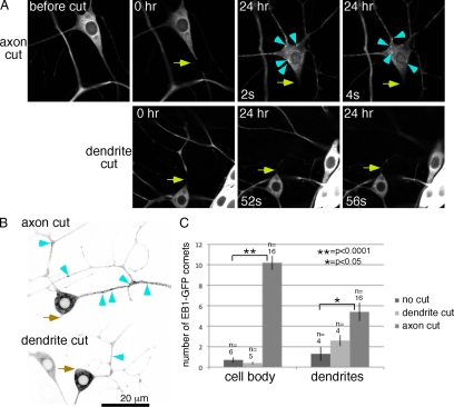
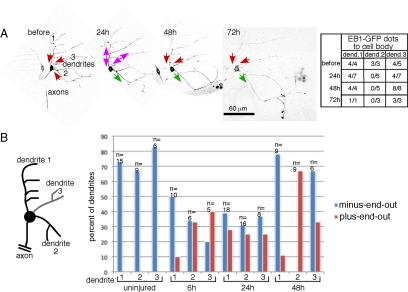
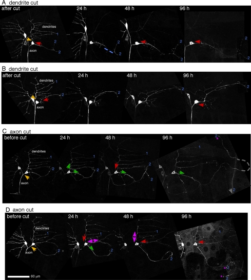
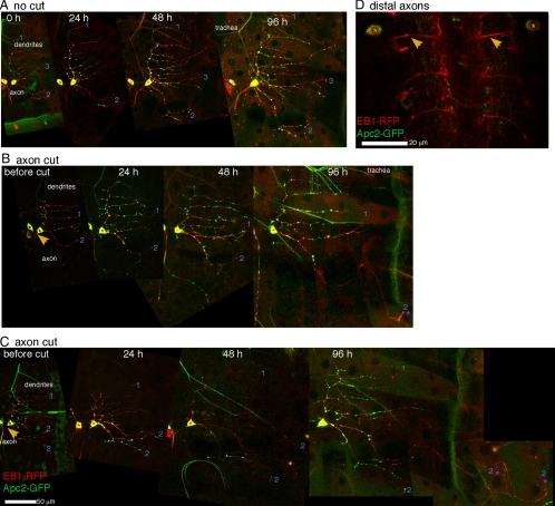
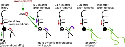
Axon regeneration is crucial for recovery after trauma to the nervous system. For neurons to recover from complete axon removal they must respecify a dendrite as an axon: a complete reversal of polarity. We show that Drosophila neurons in vivo can convert a dendrite to a regenerating axon and that this process involves rebuilding the entire neuronal microtubule cytoskeleton. Two major microtubule rearrangements are specifically induced by axon and not dendrite removal: 1) 10-fold up-regulation of the number of growing microtubules and 2) microtubule polarity reversal. After one dendrite reverses its microtubules, it initiates tip growth and takes on morphological and molecular characteristics of an axon. Only neurons with a single dendrite that reverses polarity are able to initiate tip growth, and normal microtubule plus-end dynamics are required to initiate this growth. In addition, we find that JNK signaling is required for both the up-regulation of microtubule dynamics and microtubule polarity reversal initiated by axon injury. We conclude that regulation of microtubule dynamics and polarity in response to JNK signaling is key to initiating regeneration of an axon from a dendrite.


Figures

Figure 1.
Figure 1.
The number of growing microtubules is up-regulated by axon, but not dendrite, severing. (A) Images of EB1-GFP in the ddaE neuron were acquired before, immediately after (0 h), and 24 h after axon (top row) or dendrite (bottom row) severing. Two frames are shown from each movie from the 24-h time point. Arrows indicate the site of UV laser-mediated severing. Arrowheads point out examples of EB1-GFP comets in the cell body. In all figures dorsal is up. (B) Panels from movies acquired 24 h after axon or dendrite severing are shown. Images were inverted for ease of identifying EB1-GFP comets in dendrites; examples are marked with arrowheads. (C) The number of EB1-GFP comets in the cell body or a region of dendrite 2 was counted in single frames from movies of uninjured neurons and from neurons 24 h after axon or dendrite severing. Three frames were averaged for each animal, error bars, SD of the average from all animals. n = number of animals scored (one neuron per animal). Unpaired t tests were used to determine whether the number of comets was significantly increased after dendrite or axon cutting. No significant difference between number of dots in the cell body before and after dendrite cutting was found. Significant differences were found for cell bodies and dendrites before and after axon cutting.
Figure 2.
Figure 2.
Axon injury induces orientation switching of dendritic microtubules. (A) Images of EB1-GFP in a single ddaE neuron at different times before or after axon injury. Several confocal images were projected to give a complete overview of the dendrite arbor. In zoomed in movies, the direction of EB1-GFP comet movement was scored. Comets moving toward the cell body represent minus-end-out microtubules and comets moving away from the cell body represent plus-end-out microtubules. The raw data are shown in the table and represented by arrows on the overview pictures. A green arrow indicates plus-end-out microtubule orientation, a red arrow indicates minus-end-out orientation and double arrow indicates mixed orientation. Movie 3 shows microtubule dynamics in this cell. (B) Microtubule orientation was quantitated in dendrites of uninjured neurons and neurons at different times after axon severing. Comets were scored in each dendrite as in A: EB1-GFP dots in the region of the dendrite between the cell body and first dendrite branch point were counted; dendrites with four or more comets were classified as plus-end-out if 75% or more comets moved away from the cell body and minus-end-out if 75% or more moved toward the cell body. The class in between was classified as mixed and is not shown explicitly in the table. n = number of dendrites classified for each time point.
Figure 3.
Figure 3.
Axon, but not dendrite, injury induces extensive tip growth from a dendrite. The axon or dendrite of the ddaE neuron was severed with a UV laser, and EB1-GFP was imaged at different time points. Overview images were compiled from movies of EB1-GFP, and microtubule orientation was scored as in Figure 2. Yellow arrows, site of laser severing; red arrows, minus-end-out microtubules; green arrows, plus-end-out microtubules; double arrows, mixed orientation. Stars label tips of processes that have extended by 96 h. Six of nine cells in which the axon was removed initiated tip growth and 0 of 5 in which a dendrite was removed initiated tip growth. The dendrites are numbered as in Figure 2. Movie frames were Z projected, maximum method, to show the entire dendritic tree. In some cases several frames of a movie had to be assembled next to one another to cover the complete area of the dendrites. The images were also rotated and placed on a black background so that the neuron would be seen in the same orientation at all time points. Scale is the same for all images.
Figure 4.
Figure 4.
Apc2-GFP is excluded from growing processes. Apc2-GFP and EB1-RFP were expressed in ddaE neurons. (A) Uninjured neurons were imaged over the same time course used in other experiments. At all times Apc2-GFP is seen in spots throughout the dendritic arbor. (B and C) Axon-severing experiments were performed as in Figure 3. In both cells shown tip growth is initiated from dendrite 2. Apc2-GFP is only found in the proximal region of this process at 96 h after axon removal, and this pattern was seen in a total of 12 of 12 neurons which initiated tip growth. (D) The ddaE neuron extends its axon from the body wall to the ventral ganglion. In live animals expressing Apc2-GFP and EB1-RFP in class I DA neurons, EB1-RFP can be seen in the distal axons that enter the ventral ganglion. Apc2-GFP is not seen in these axons. See Figure S4 for greyscale images of Apc2-GFP alone.
Figure 5.
Figure 5.
RNAi targeting msps blocks regeneration from a dendrite after axon removal. (A) The cell body and proximal dendrite of ddaE neurons expressing EB1-GFP and hairpin RNAs that target rtnl2 (control) or msps are shown. rtnl2 RNAi was used as a control as its loss has no known consequences in flies. EB1-GFP comets (red arrows) can be seen in control, but not msps RNAi neurons. (B) An axon-severing experiment as in Figure 3 was performed on a ddaE neuron expressing EB1-GFP and msps hairpin RNA. Cell shape (but not microtubule polarity, as no EB1-GFP comets were present) were tracked over time. No tip growth was observed (n = 5).
Figure 6.
Figure 6.
JNK is required for up-regulation of microtubule number and initiation of growth in response to axon removal. (A) ddaE neurons expressing EB1-GFP and mCD8-RFP (control), RNAi hairpins to target bsk, or bskDN, were imaged 24 h after axon removal. Numerous EB1-GFP comets (arrowheads) were seen in control neurons, and many fewer were seen is bskRNAi or bskDN neurons. (B) EB1-GFP comets in the cell body were quantitated 24 h after axon injury. Number of comets in individual frames of movies was counted as in Figure 1C. Genotypes of the larvae in order shown in the table were as follows: 1) UAS-Dicer2/UAS-mCD8-RFP; 221-Gal4, UAS-EB1-GFP/+; 2) UAS-Dicer2/UAS-bskRNAi; 221-Gal4, UAS-EB1-GFP/+; 3) UAS-mCD8-RFP/+; 221-Gal4, UAS-EB1-GFP/+; and 4) UAS-bskDN/+;; 221-Gal4; UAS-EB1-GFP. (C and D) A ddaE neuron expressing EB1-GFP and bskDN was tracked over time. The dendrite arbor of these cells retained the same shape over time as in controls (n = 6). In D, microtubule orientation was determine as in Figure 3, except that comets were quantitated throughout major dendrites because fewer comets were present. Red arrows, minus-end-out polarity (>75% of comets to the cell body); double arrows, mixed polarity (25–75% of comets to the cell body).
Figure 7.
Figure 7.
Reduction of JNK signaling affects microtubule polarity in uninjured neurons. Microtubule polarity was assayed by tracking direction of EB1-GFP comet movement in uninjured neurons. Comets were counted in main trunk of the comb dendrite (1), and in dendrite 2 (see Figure 2B). The percent of dots moving toward the cell body is shown for dendrites. The percent was calculated for each cell; error bars, SD. An unpaired t test was used to calculate the significance of the difference between wild type and bskDN. For axons, the percent of comets moving away from the cell body is shown.
Figure 8.
Figure 8.
Model for conversion of a dendrite to a regenerating axon after axon removal. Before axon injury, microtubules in dendrites have minus-end-out polarity (red arrows) and axonal microtubules are plus-end-out. Axon removal up-regulates JNK signaling, which switches microtubule polarity in dendrites, frequently resulting in mixed polarity (purple double arrows) and up-regulates the number of microtubules in the cell body (white circle) and throughout the dendrites. Over several days polarity resolves such that one dendrite takes on the axonal microtubule polarity and the rest return to minus-end-out polarity. After this point the process with axonal microtubule polarity initiates tip growth.

Similar articles

Cited by

References

    1. Adachi-Yamada T., Nakamura M., Irie K., Tomoyasu Y., Sano Y., Mori E., Goto S., Ueno N., Nishida Y., Matsumoto K. p38 mitogen-activated protein kinase can be involved in transforming growth factor beta superfamily signal transduction in Drosophila wing morphogenesis. Mol. Cell. Biol. 1999;19:2322–2329. - PMC - PubMed
    1. Ayaz D., Leyssen M., Koch M., Yan J., Srahna M., Sheeba V., Fogle K. J., Holmes T. C., Hassan B. A. Axonal injury and regeneration in the adult brain of Drosophila. J. Neurosci. 2008;28:6010–6021. - PMC - PubMed
    1. Baas P. W., Black M. M., Banker G. A. Changes in microtubule polarity orientation during the development of hippocampal neurons in culture. J. Cell Biol. 1989;109:3085–3094. - PMC - PubMed
    1. Baas P. W., Deitch J. S., Black M. M., Banker G. A. Polarity orientation of microtubules in hippocampal neurons: uniformity in the axon and nonuniformity in the dendrite. Proc. Natl. Acad. Sci. USA. 1988;85:8335–8339. - PMC - PubMed
    1. Bogoyevitch M. A., Kobe B. Uses for JNK: the many and varied substrates of the c-Jun N-terminal kinases. Microbiol. Mol. Biol. Rev. 2006;70:1061–1095. - PMC - PubMed

Publication types

Substances

LinkOut - more resources