. 2010 Mar 1;21(5):802-10.
doi: 10.1091/mbc.e09-09-0831. Epub 2010 Jan 6.

Numb independently antagonizes Sanpodo membrane targeting and Notch signaling in Drosophila sensory organ precursor cells

Affiliations

Numb independently antagonizes Sanpodo membrane targeting and Notch signaling in Drosophila sensory organ precursor cells

Xin Tong et al. Mol Biol Cell. .

Abstract

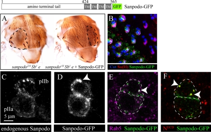
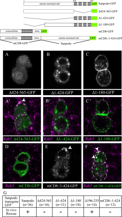
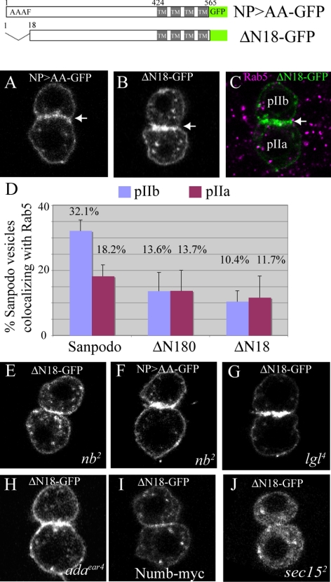
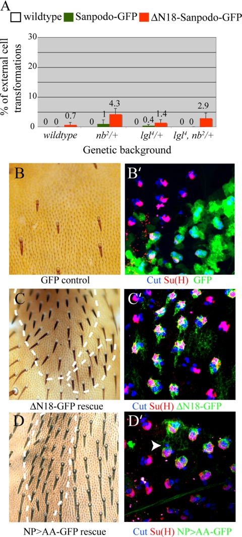
In Drosophila, mitotic neural progenitor cells asymmetrically segregate the cell fate determinant Numb in order to block Notch signaling in only one of the two daughter cells. Sanpodo, a membrane protein required for Notch signaling in asymmetrically dividing cells, is sequestered from the plasma membrane to intracellular vesicles in a Numb-dependent way after neural progenitor cell mitosis. However, the significance of Numb-dependent Sanpodo regulation is unclear. In this study, we conducted a structure-function analysis to identify the determinants of Sanpodo targeting in vivo. We identified an NPAF motif in the amino-terminal cytoplasmic tail of Sanpodo, which is conserved among insect Sanpodo homologues. The Sanpodo NPAF motif is predicted to bind directly to the Numb phosphotyrosine-binding domain and is critical for Numb binding in vitro. Deletion or mutation of the NPAF motif results in accumulation of Sanpodo at the plasma membrane in Numb-positive cells in vivo. Genetic analysis of Sanpodo NPAF mutants shows that Numb-dependent Sanpodo endocytic targeting can be uncoupled from Notch signaling regulation. Our findings demonstrate that Sanpodo contains an evolutionarily conserved motif that has been linked to Numb-dependent regulation in vertebrates and further support the model that Numb regulates Notch signaling independently of Sanpodo membrane trafficking in neural progenitor cells.


Figures

Figure 1.
Figure 1.
Analysis of Sanpodo-GFP function and localization in vivo. Schematic of wild-type Sanpodo transgene tagged with GFP at the C-terminus (TM, transmembrane). (A) Sanpodo-GFP fully restores the wild-type bristle pattern in sanpodoC55 mutant clones, which typically show extensive balding. (B) Clusters of four to five Cut-positive (blue) cells have a single Cut-positive, Su(H)-positive cell (magenta) in both wild-type and sanpodoC55 MARCM mutant clones expressing Sanpodo-GFP (green; white dashed line delineates the clone border). (C) Immunocytochemistry of endogenous Sanpodo in the pIIa and pIIb cell (here and in all subsequent figures pIIb cells are at the top, and pIIa cells are at the bottom). (D) Ten minutes after SOP cell mitotic exit, Sanpodo-GFP localizes to small puncta and to the membrane in the pIIa cell, whereas in the pIIb cell, Sanpodo-GFP localizes to large puncta (white arrowhead). (E and F) Sanpodo-GFP (green) colocalizes with Rab5 (magenta, E) and NotchECD (red, F) in pIIb cells. Genotypes: (A and B) yw, ubx-FLP; sca-Gal4, UAS-sanpodo-GFP/+; FRT82B sanpodoC55 Sb1 e/FRT82B tub-Gal80. Similar results were obtained with a rescue of FRT82B sanpodoG104 e clones. (C) yw (D–F) yw; neur-Gal4/UAS-sanpodo-GFP.
Figure 2.
Figure 2.
Dynamic regulation of Sanpodo-GFP membrane targeting during asymmetric cell division. (A) Sanpodo-GFP localizes to the pIIa/pIIb cell interface within ten minutes of completion of cytokinesis and is targeted to anterior vesicles in the pIIb cells. (B and D) In nb2, lgl4, and α-adaear4 mutants, Sanpodo-GFP is enriched at the membrane during mitosis and accumulates strongly at the pIIa/pIIb cell membrane after cytokinesis. (E and F) Under conditions of numb-myc overexpression, or in sec152 mutants, Sanpodo is depleted from the plasma membrane of the mitotic SOP and the newly formed pIIa and pIIb cells. Genotypes: (A) yw/+; neur-Gal4/UAS-sanpodo-GFP, yw, ubx-FLP/+; (B) y+ nb2 ck FRT40A, or (C) lgl4 FRT40A or (D) adaear4 FRT40A/tub-Gal80 FRT40A; neur-Gal4, UAS-sanpodo-GFP/+, (E) yw/+; neur-Gal4, UAS-sanpodo-GFP/UAS-numb-myc, and (F) yw, ubx-FLP/+; sca-Gal4, UAS-sanpodo-GFP/+; FRT82B sec152/FRT82B tub-Gal80.
Figure 3.
Figure 3.
The Sanpodo amino-terminal tail is sufficient for endocytic targeting in vivo. Schematic of Sanpodo truncation mutants and mCD8-GFP, and the mCD8-Sanpodo amino-terminal tail-GFP chimera (mCD8::1-424-GFP). (A–C) Deletion of the Sanpodo transmembrane domains (Δ424-565, A and A′), N-terminal tail (Δ1-424, B and B′), or the N-terminus (Δ1-180, C and C′), result in failure to target the transgene to endosomes in pIIb cells. (D) mCD8-GFP (green) is localized to the plasma membrane and intracellular membrane compartments in pIIa and pIIb cells, but is excluded from the cell nucleus and Rab5 early endosomes. (E) mCD8:: 1-424-GFP localizes to large puncta in pIIb cells following asymmetric cell division (white arrowhead). (F′) mCD8::1-424-GFP puncta colocalize with Rab5 in pIIb cells (white arrowheads). *(A–C and E) Live cells; (A′–C′, D and F) fixed cells; GFP-tagged transgenes in green and Rab5 in magenta. (G) Rescue experiments testing the ability of mutant transgenes to restore external cell fates in sanpodoC55 mutant clones (n = number of flies of each genotype scored for rescue).
Figure 4.
Figure 4.
The conserved Sanpodo NPAF motif regulates Numb binding. (A) The evolutionary relationship of species of the Superorder Endopterygota and the predicted amino acid sequence of the region containing the NPAF within the amino-terminus (perfectly conserved amino acids labeled with magenta blocks) and the alignment of the Sanpodo peptide with the NAK, ApoER2, and APP peptides. (B) A structural model of the cleft formed by the Numb PTB domain (cyan ribbon) binding to the Sanpodo NPAF motif (yellow backbone) based on the crystal structure of Disabled1 bound to ApoER2. Predicted hydrogen bonds are indicated (magenta lines), and the conserved asparagine residue (N) forms hydrogen bonds with the Numb PTB domain and the Sanpodo peptide backbone. (C) Amino acid substitution mutations (NP>AA and N>E) in the NPAF motif abrogate the Sanpodo amino-terminal tail binding to Numb-Myc in an immunoprecipitation assay.
Figure 5.
Figure 5.
The Sanpodo NPAF is required for endocytic targeting in vivo. (A–C) Sanpodo mutants with either point mutations in the NPAF motif (NP>AA) or the NPAF motif removed (ΔN18) localize to the plasma membrane (white arrow) and small puncta in pIIa and pIIb cells and do not colocalize with early endosomes (labeled by Rab5, magenta) in pIIb cells. (D) Graph representing the decrease in percentage of puncta colocalizing with the early endosome marker Rab5 in Sanpodo-Δ1-180-GFP and Sanpodo ΔN18-GFP mutants in the pIIb cell compared with wild-type Sanpodo. Colocalized puncta were visually identified from individual planes of apical-basal z stacks through pIIa/pIIb cell pairs at a0.5-μm interval. Error bars, SEM. (E–I) Sanpodo NPAF mutant (NP>AA or ΔN18) localization in nb2, lgl4, and α-adaear4 mutants or under conditions of numb-myc overexpression (I) is similar to Sanpodo-NPAF mutant localization in wild-type cells shown in A and B. (J) In sec152 mutants, Sanpodo-NPAF mutant is depleted from the plasma membrane of the mitotic SOP and the newly formed pIIa and pIIb cells. Genotypes: yw; neur-Gal4/UAS-sanpodo-NP>AA-GFP (A), yw; neur-Gal4/UAS-sanpodoN18-GFP (B and C), yw, ubx-FLP/+; y+ nb2 ck FRT40A/CyO (E and F), or lgl4 FRT40A/CyO (G), or adaear4 FRT40A/CyO (H) were crossed to yw; tub-Gal80 FRT40A; neur-Gal4, UAS-sanpodo-NP>AA-GFP/TM6 (F) or yw; tub-Gal80 FRT40A; neur-Gal4,UAS-sanpodoN18-GFP (E, G, and H), yw; neur-Gal4, UAS-sanpodoN18-GFP/UAS-numb-myc (I), and yw, ubx-FLP/+; sca-Gal4, UAS-sanpodoN18-GFP/+; FRT82B sec152/FRT82B tub-Gal80 (J).
Figure 6.
Figure 6.
The Sanpodo NPAF motif is not required Sanpodo function in external sensory organ lineage cells. (A) Overexpression of either wild-type or ΔN18 Sanpodo mutant transgenes show low frequencies of external cell fate transformations (i.e., extra sockets) in sensory organs of the head, even in backgrounds where numb and/or lgl dose is lowered. (BD) Both Sanpodo ΔN18-GFP and Sanpodo NP>AA-GFP completely restore wild-type cell fates to all external sensory organs in sanpodoC55 Sb e MARCM clones, whereas GFP alone does not. sanpodo mutant ES organs are marked by Sb/Sb bristles and approximate clone boundaries are marked by dashed lines in C and D. GFP, Sanpodo ΔN18-GFP, and Sanpodo NP>AA-GFP (green) mark the sanpodo mutant ES organ cells in B′, C′, and D′. External organ lineage cells are labeled with Cut (blue) and socket cells with Su(H) (red) antibodies. Cell fate transformations, such as twinned bristles, are observed in rescued flies at a low frequency (<2%, not shown). Genotypes: sca-Gal4, or sca-Gal4, UAS-sanpodo-GFP, or sca-Gal4, UAS-sanpodoN18-GFP were crossed to the following: (A) yw, nb2/CyO or lgl4/CyO, or lgl4, nb2/CyO; (B and B′) yw, ubx-FLP/+; sca-Gal4/UAS-EGFP; (C and C′) UAS-sanpodoN18-GFP; or (D and D′) UAS-sanpodo-NP>AA-GFP or FRT82B sanpodoC55 Sb1 e/FRT82B tub-Gal80; similar results were obtained with a rescue of FRT82B sanpodoG104 e clones.

Similar articles

Cited by

References

    1. Berdnik D., Torok T., Gonzalez-Gaitan M., Knoblich J. A. The endocytic protein alpha-Adaptin is required for numb-mediated asymmetric cell division in Drosophila. Dev. Cell. 2002;3:221–231. - PubMed
    1. Borg J. P., Ooi J., Levy E., Margolis B. The phosphotyrosine interaction domains of X11 and FE65 bind to distinct sites on the YENPTY motif of amyloid precursor protein. Mol. Cell. Biol. 1996;16:6229–6241. - PMC - PubMed
    1. Brand A. H., Perrimon N. Targeted gene expression as a means of altering cell fates and generating dominant phenotypes. Development. 1993;118:401–415. - PubMed
    1. Calderwood D. A., Fujioka Y., de Pereda J. M., Garcia-Alvarez B., Nakamoto T., Margolis B., McGlade C. J., Liddington R. C., Ginsberg M. H. Integrin beta cytoplasmic domain interactions with phosphotyrosine-binding domains: a structural prototype for diversity in integrin signaling. Proc. Natl. Acad. Sci. USA. 2003;100:2272–2277. - PMC - PubMed
    1. Canutescu A. A., Shelenkov A. A., Dunbrack R. L., Jr A graph-theory algorithm for rapid protein side-chain prediction. Protein Sci. 2003;12:2001–2014. - PMC - PubMed

Publication types

LinkOut - more resources