. 2010 Mar 1;21(5):753-66.
doi: 10.1091/mbc.e09-07-0555. Epub 2010 Jan 6.

Structural mutants of the spindle pole body cause distinct alteration of cytoplasmic microtubules and nuclear dynamics in multinucleated hyphae

Affiliations

Structural mutants of the spindle pole body cause distinct alteration of cytoplasmic microtubules and nuclear dynamics in multinucleated hyphae

Claudia Lang et al. Mol Biol Cell. .

Abstract

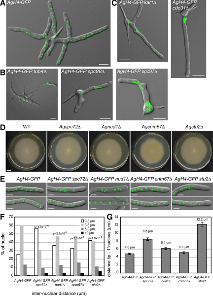
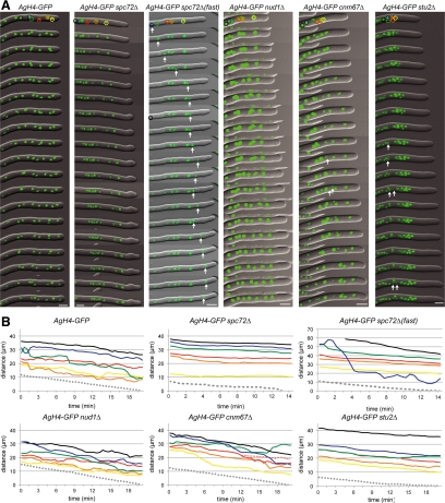
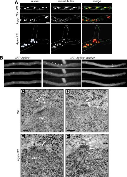
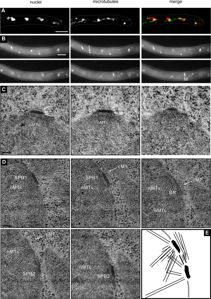
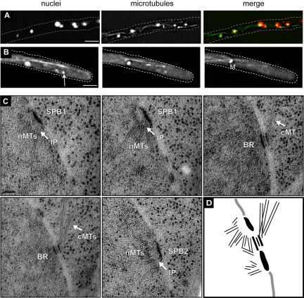
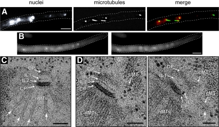
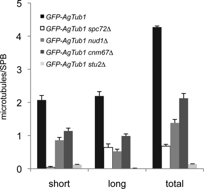
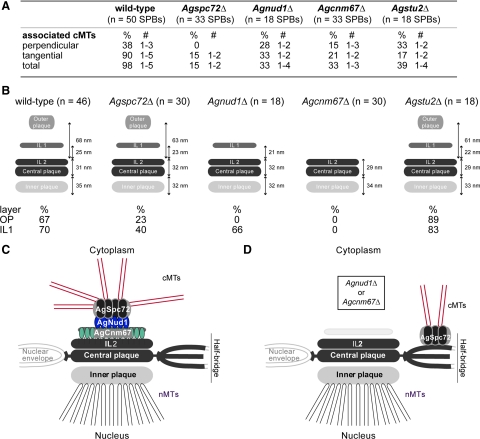
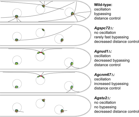
In the multinucleate fungus Ashbya gossypii, cytoplasmic microtubules (cMTs) emerge from the spindle pole body outer plaque (OP) in perpendicular and tangential directions. To elucidate the role of cMTs in forward/backward movements (oscillations) and bypassing of nuclei, we constructed mutants potentially affecting cMT nucleation or stability. Hyphae lacking the OP components AgSpc72, AgNud1, AgCnm67, or the microtubule-stabilizing factor AgStu2 grew like wild- type but showed substantial alterations in the number, length, and/or nucleation sites of cMTs. These mutants differently influenced nuclear oscillation and bypassing. In Agspc72Delta, only long cMTs were observed, which emanate tangentially from reduced OPs; nuclei mainly moved with the cytoplasmic stream but some performed rapid bypassing. Agnud1Delta and Agcnm67Delta lack OPs; short and long cMTs emerged from the spindle pole body bridge/half-bridge structures, explaining nuclear oscillation and bypassing in these mutants. In Agstu2Delta only very short cMTs emanated from structurally intact OPs; all nuclei moved with the cytoplasmic stream. Therefore, long tangential cMTs promote nuclear bypassing and short cMTs are important for nuclear oscillation. Our electron microscopy ultrastructural analysis also indicated that assembly of the OP occurs in a stepwise manner, starting with AgCnm67, followed by AgNud1 and lastly AgSpc72.


Figures

Figure 1.
Figure 1.
Deletion of A. gossypii SPB components. Overlays of DIC image and AgH4-GFP signals from cells grown for ∼12 h at 30°C. (A) A typical wild-type mycelium at this stage contains multiple branches and 50–100 nuclei. (B) Terminal phenotype of mutants lacking components of the γ-tubulin complex. (C) Terminal phenotype of mutants lacking half-bridge components. Some mutants shown in B and C arrest as small mycelium with up to 12 branches containing a few nuclei and did not arrest as germlings with one nucleus or two nuclei. This is due to a maternal effect, where remnants of the wild-type protein are packed into mutant spores (Gladfelter et al., 2006). In A. gossypii, targeted deletions are performed with young mycelium containing ∼20 haploid nuclei that initially generate heterokaryotic strains: mixtures of nuclei carrying either the wild type or the deletion allele. On starvation, haploid mononucleate spores are produced in the heterokaryotic hyphae, and a substantial part of the mutant spores contain wild-type (maternal) protein, which can account for early growth and nuclear division in otherwise fatal deletions. Bars, 10 μm. (D) Radial growth of wild-type and deletion mutants on solid medium during 7 d of incubation at 30°C. Bar, 1 cm. (E) Overlays of DIC image and AgH4-GFP signals from hyphae showing nuclear distributions in wild-type and mutant strains. Bars, 5 μm. (F) Distribution of distances between adjacent nuclei was plotted using distance measurements between the nuclei in the first 50 μm of at least 20 different hyphae for each strain (n > 200 for each strain). The center of the GFP signal was used as the central point of the nucleus. Confidence values (p) for the χ2 test were calculated for each data set between wild-type and mutant distributions. (G) Distance between the hyphal tip and the first nucleus was determined. Error bars represent the SE of the mean, n > 60 for each strain.
Figure 2.
Figure 2.
Time-lapse analysis of nuclear migration in different mutants. (A) Overlays of DIC and AgH4-GFP signals from time-lapse video imaging of wild-type (Supplemental Movie S1), Agspc72Δ (Supplemental Movie S2), Agnud1Δ (Supplemental Movie S4), Agcnm67Δ (Supplemental Movie S5), and Agstu2Δ (Supplemental Movie S6) hyphae. Images were captured every 30 s, and 1-min interval frames are shown. Migration of the first six nuclei were tracked and are shown in the schematic. In Agspc72Δ(fast) (Supplemental Movie S3), images from each 30-s time point are shown for the first 7 min followed by 1-min interval frames; the rapidly moving nucleus is indicated by an arrow. Nuclei undergoing mitosis are indicated by arrows in Agcnm67Δ and Agstu2Δ. Bars, 5 μm. (B) Positions of the first six nuclei in each hyphae and the hyphal tip (gray dotted line) were tracked throughout each time course and are plotted. In wild-type, the nuclei were observed to undergo bypassing and oscillations. In Agspc72Δ(fast), one nucleus (blue) moves rapidly toward the tip, thereby bypassing four other nuclei in <5 min and traveling distances up to 8.9 μm within a 30-s interval (1.5–2 min). The other nuclei in this hypha as well as in the other Agspc72Δ and Agstu2Δ mutants move toward the tip with the cytoplasmic stream without undergoing any bypassing or oscillation.
Figure 3.
Figure 3.
Nuclear movement, cMTs and SPB structure in Agspc72Δ. (A) Wild-type and Agspc72Δ mutants were stained with Hoechst to visualize DNA and anti-α-tubulin antibodies to detect microtubules. In the bottom image of Agspc72Δ, a cluster of nuclei can be seen at a branch site. These hyphae lack short cMTs, whereas long cMTs that extend along the growth axis are still present. A detached microtubule can be seen in the upper tip region. Bars, 5 μm. (B) Representative, deconvolved images of a Z-stack of a wild-type and two Agspc72Δ hyphae expressing GFP-AgTUB1. The complete stacks are available as Supplemental Movies S7, S8, and S9. Long and short cMTs can be seen emerging from bright foci that represent the SPBs in wild-type, but Agspc72Δ SPBs often lack associated cMTs. Arrowheads point to a long cMT in Agspc72Δ, which may facilitate bypassing. An arrow indicates an anaphase spindle. Bar, 5 μm. (C) EM image of SPB-attached microtubules in wild-type hyphae. Bar, 100 nm. The SPB is associated with nuclear microtubules (nMTs) at the IP and a tangential and perpendicular microtubule at its OP (arrows). (D) SPB structure in wild-type. Bar, 100 nm. A central plaque (CP) plus IL1 and IL2 are marked by arrows. A small amount of amorphous material could also be detected above IL1 and is part of the outer plaque (OP) (Lang et al., 2010). (E) EM image of SPB-attached microtubules in Agspc72Δ. Bar, 100 nm. The SPB is associated with nMTs at the IP and a tangential microtubule close to the central plaque (arrow). In this and other thin sections, we never observed perpendicular microtubules. (F) SPB structure in Agspc72Δ. Bars, 100 nm. A CP plus IL1 and IL2 are marked by arrows. In some cases, a small amount of amorphous material could also be detected above IL1, which could be part of the OP. However, the size of this OP remnant was substantially reduced compared with a wild-type OP (Lang et al., 2010).
Figure 4.
Figure 4.
SPBs of Agnud1Δ lack an OP and nucleate cMTs from the bridge. (A) Agnud1Δ mutants were stained with Hoechst to visualize DNA and anti-α-tubulin antibodies to detect microtubules. Long cMTs that extend along the growth axis are still present and also a few short cMTs were detected. Bar, 5 μm. (B) Representative, deconvolved images of a Z-stack from Agnud1Δ hypha expressing GFP-AgTUB1. The complete stack is available as Supplemental Movie S10. Long and short cMTs can be seen emerging form both sides of a mitotic spindle (M) and from bright foci that represent the SPBs. An arrow points to a SPB lacking cMTs. Bar, 5 μm. (C and D) EM images of Agnud1Δ mutants. Bars, 100 nm. (C) Serial section images of a single SPB in an Agnud1Δ mutant. The central plaque (CP) and IL1 and IL2 were observed, whereas the outer plaque could not be detected in any section. (D) Serial section images of duplicated side-by-side SPBs (SPB1 and SPB2) connected by a bridge (BR) in the Agnud1Δ mutant. Nuclear microtubules (nMTs) as well as two cMTs can be seen. The cMTs seem to emerge from the bridge region. (E) Schematic summarizing the five serial sections images of D.
Figure 5.
Figure 5.
cMT nucleation and SPB structure in Agcnm67Δ hyphae. (A) Agcnm67Δ hypha stained with Hoechst to visualize DNA and anti-α-tubulin antibodies to detect microtubules. Long cMTs that extend along the growth axis are present and a few short cMTs were detected. Bar, 5 μm. (B) Representative, deconvolved images of a Z-stack of an Agcnm67Δ hypha expressing GFP-AgTub1. The complete stack is available as Supplemental Movie S11. Long and short cMTs can be seen emerging form both sides of a mitotic spindle (M) and from bright foci that represent the SPBs. An arrow points to a SPB lacking cMTs. Bar, 5 μm. (C) Serial sections of duplicated side-by-side SPBs (SPB1 and SPB2) connected by a bridge (BR), which nucleates three cMTs in an Agcnm67Δ mutant. Nuclear microtubules (nMTs) are formed at the SPB IP. Images were aligned using AutoAligner to produce the schematic in D. Bars, 100 nm.
Figure 6.
Figure 6.
Microtubule stability and SPB structure in Agstu2Δ mutants. (A) Agstu2Δ hypha stained with Hoechst to visualize DNA and anti-α-tubulin antibodies to detect microtubules. Only short cMTs were detected. Bar, 5 μm. (B) Representative, deconvolved images of a Z-stack of an Agstu2Δ hypha expressing GFP-AgTUB1. The complete stack is available as Supplemental Movie S12. (C) EM image of a SPB from Agstu2Δ showing OP, IL2, IL1, and central plaque (CP) layers as well as nuclear microtubules (nMTs). (D) Serial sections of an Agstu2Δ SPB also show the layered SPB structure and nMTs as in A. In addition, three very short cMTs are visible, which have both perpendicular and tangential orientations with the SPB. Arrows point to “flared” microtubule ends frequently seen by EM in Agstu2Δ mutants. Bars, 100 nm.
Figure 7.
Figure 7.
Quantitation of short and long cMTs in hyphae expressing GFP-AgTUB1. The number of short (≤5-μm) and long (>5-μm) cMTs per SPB was quantitated in wild-type and mutant hyphae expressing GFP-AgTUB1, and average values observed for both classes of microtubules are presented along with a combined total. Error bars indicate SE of the mean (n = 77, 39, 46, 92, and 58 SPBs for GFP-AgTUB1, GFP-AgTUB1 spc72Δ, GFP-AgTUB1 nud1Δ, GFP-AgTUB1 cnm67Δ, and GFP-AgTUB1 stu2Δ, respectively). Note that only a minor fraction of the long cMTs in GFP-AgTUB1 spc72Δ and GFP-AgTUB1 nud1Δ are >12 μm, whereas in GFP-AgTUB1 cnm67Δ mutants, a substantial fraction is >12 μm.
Figure 8.
Figure 8.
Comparison of A. gosspyii wild-type and mutant SPB structure and cMT nucleation based on EM analysis. (A) Quantitation of cMTs that emerge per SPB in wild-type and mutants based on EM analysis. (B) Schematics of wild-type and mutant SPBs depicting the observed layers and averaged distances between the layers, based on measurements shown in Supplemental Table S3. The percentage of SPBs in which the OP and IL1 were seen is indicated below. (C) Model of the A. gossypii SPB based on SPB morphology we observed in different deletion mutants. The layered SPB of A. gossypii is composed of AgCnm67 in IL1, which recruits AgNud1. Next, AgSpc72 binds to form the OP and tether the γ-tubulin complex that nucleates cMTs. (D) In the absence of AgCNM67 or AgNUD1, cMTs are formed from the bridge region (Brachat et al., 1998; Gruneberg et al., 2000; Hoepfner et al., 2000). Studies in budding yeast lead us to hypothesize that AgSpc72 and the γ-tubulin complex relocalize here (Knop and Schiebel, 1998), at least in these mutants. In Agnud1Δ, IL1 remains, whereas it is lost in Agcnm67Δ.
Figure 9.
Figure 9.
Model of the cytoplasmic sides of SPBs and attached cMTs in wild-type and mutant hyphae of A. gossypii. The four deletion mutants presented here have altered cMT arrays compared with wild-type, and these alterations can explain most of the observed changes in nuclear mobility and in distance control to the hyphal tip assuming that microtubules can exert pulling forces upon cortical contacts, which may be transient, e.g., causing oscillations, or more stable, e.g., for nuclear bypassing driven by long microtubules. See Discussion for details.

Similar articles

Cited by

References

    1. Adams I. R., Kilmartin J. V. Localization of core spindle pole body (SPB) components during SPB duplication in Saccharomyces cerevisiae. J. Cell Biol. 1999;145:809–823. - PMC - PubMed
    1. Al-Bassam J., van Breugel M., Harrison S. C., Hyman A. Stu2p binds tubulin and undergoes an open-to-closed conformational change. J. Cell Biol. 2006;172:1009–1022. - PMC - PubMed
    1. Ayad-Durieux Y., Knechtle P., Goff S., Dietrich F., Philippsen P. A PAK-like protein kinase is required for maturation of young hyphae and septation in the filamentous ascomycete Ashbya gossypii. J. Cell Sci. 2000;113:4563–4575. - PubMed
    1. Brachat A., Kilmartin J. V., Wach A., Philippsen P. Saccharomyces cerevisiae cells with defective spindle pole body outer plaques accomplish nuclear migration via half-bridge-organized microtubules. Mol. Biol. Cell. 1998;9:977–991. - PMC - PubMed
    1. Brachat S., Dietrich F. S., Voegeli S., Zhang Z., Stuart L., Lerch A., Gates K., Gaffney T., Philippsen P. Reinvestigation of the Saccharomyces cerevisiae genome annotation by comparison to the genome of a related fungus: Ashbya gossypii. Genome Biol. 2003;4:R45. - PMC - PubMed

Publication types

Substances

LinkOut - more resources