. 2010 Mar 1;21(5):725-38.
doi: 10.1091/mbc.e09-04-0268. Epub 2010 Jan 6.

Fate specification and tissue-specific cell cycle control of the Caenorhabditis elegans intestine

Affiliations

Fate specification and tissue-specific cell cycle control of the Caenorhabditis elegans intestine

Alexandra Segref et al. Mol Biol Cell. .

Abstract

Coordination between cell fate specification and cell cycle control in multicellular organisms is essential to regulate cell numbers in tissues and organs during development, and its failure may lead to oncogenesis. In mammalian cells, as part of a general cell cycle checkpoint mechanism, the F-box protein beta-transducin repeat-containing protein (beta-TrCP) and the Skp1/Cul1/F-box complex control the periodic cell cycle fluctuations in abundance of the CDC25A and B phosphatases. Here, we find that the Caenorhabditis elegans beta-TrCP orthologue LIN-23 regulates a progressive decline of CDC-25.1 abundance over several embryonic cell cycles and specifies cell number of one tissue, the embryonic intestine. The negative regulation of CDC-25.1 abundance by LIN-23 may be developmentally controlled because CDC-25.1 accumulates over time within the developing germline, where LIN-23 is also present. Concurrent with the destabilization of CDC-25.1, LIN-23 displays a spatially dynamic behavior in the embryo, periodically entering a nuclear compartment where CDC-25.1 is abundant.


Figures

Figure 1.
Figure 1.
C. elegans excess endoderm phenotypes. (A) Left, GFP fluorescence of JR1838 carrying the elt-2::GFP transgene at the comma stage or in early arrested embryos after control (ctrl), lin-23, cul-1, gsk-3, or apr-1 RNAi or in IA592 lin-23::FLAG-TY (see Materials and Methods for genotype). Right, corresponding Nomarski counterparts. Bar, 10 μm. (B) lin-23(RNAi) triggers predominantly hyperplasia of the intestine under mild RNAi conditions. Top, phenotypes of strain JR1838 carrying elt-2::GFP (int.), JR667 wIs51 carrying a seam cell GFP reporter (seam), and IA105 carrying the hypodermal reporter dpy-7::GFP (hyp.) in control or lin-23(RNAi) embryos at the threefold stage. Bar, 10 μm. Bottom, quantification of GFP-positive cells (mean ± SD, number of embryos from two independent experiments in parentheses): intestinal cells (int.): control, 20.0 ± 1.0 (40); lin-23(RNAi), 32.7 ± 6.8 (58); seam cells: control, 19.4 ± 1.2 (81) and lin-23(RNAi), 18.3 ± 2.8 (79); and hypodermal cells (hyp.): control, 65.3 ± 4.6 (42) and lin-23(RNAi), 62.3 ± 7.4 (45).
Figure 2.
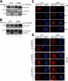
Figure 2.
RNAi of lin-23 and cul-1 but not gsk-3 mimic CDC-25.1(S46F) protein increase in all embryonic blast cells. (A) Extracts derived from JR1838 (WT, lanes 1 and 2) or IA530 cdc-25.1(ij48) (S46F, lanes 3 and 4) embryos after control (−) or lin-23(RNAi) (+) were applied to SDS-PAGE followed by Western blotting against CDC-25.1, LIN-23, or β-actin as loading control. (B) Essentially as in A, but RNAi was performed against gsk-3 followed by Western blotting against CDC-25.1, GSK3β, and β-actin. (C) Indirect immunostaining of CDC-25.1 (red panels) with corresponding DAPI counterparts (blue panels) in JR1838 (WT) or IA530 cdc-25.1(ij48) (S46F) at approximately the 4, 30, 60, or 200 cell stage. Bar, 10 μm. The images shown are representative. Images like these were used in the quantification of intensity of CDC-25.1 immunofluorescence; details of how this was performed are given in the legend to Supplemental Figure 1. (D) Indirect immunostaining of CDC-25.1 (red panels) with corresponding DAPI images (blue channels) in JR1838 (WT) or IA530 cdc-25.1(ij48) (S46F) after control, lin-23, cul-1, or gsk-3 RNAi. Bars, 10 μm.
Figure 3.
Figure 3.
CDC-25.1(S46) is crucial for LIN-23 interaction in C. elegans embryos. (A) Immunoprecipitation of CDC-25.1 from C. elegans wild-type strain JR1838 (WT, lanes 1, 3, and 4) compared with IA530 cdc-25.1(ij48) (S46F, lanes 2, 5, and 6). Samples were precipitated with an anti-CDC-25.1 antibody (lanes 3 and 5) and as control rabbit IgG (lanes 4 and 6). Three percent (9 μg of total protein) of the total fraction were applied to SDS-PAGE for input (lanes 1 and 2) and 30% of the eluates (lanes 3–6), followed by Western blotting and sequential probing with anti-ubiquitin, anti-LIN-23, and anti-CDC-25.1 antibodies. Small inset, weak exposure of input for anti-LIN-23 to show equal protein loading. A typical result obtained from two independent experiments is shown. (B) Comparison of CDC-25.1 protein levels from L4-stage hermaphrodites, mainly expressing CDC-25.1 in the germline (lanes 1 and 2), with the protein levels in the embryo (lanes 3 and 4). Similar amounts of total proteins derived from extracts of JR1838 (WT) and IA530 cdc-25.1(ij48) (S46F) were applied to SDS-PAGE, followed by Western blotting with anti-CDC-25.1 and anti-β-ACTIN antibodies, result was obtained from two independent experiments. (C) Immunoprecipitation of CDC-25.1 with the anti-CDC-25.1 antibody (lanes 1 and 3) or as control rabbit IgG (lanes 2 and 4) from extracts derived from L4-stage hermaphrodites (lanes 1 and 2) or embryos (lanes 3 and 4). Eluates were applied to SDS-PAGE followed by Western blotting against LIN-23 and CDC-25.1 (similar to A, two independent experiments). (D) Immunoprecipitation of LAP-tagged CDC-25.1 from embryos. Embryos containing transgenically expressed CDC-25.1::LAP (strain IA535) or as a control GFP::LacZ (strain JR 1838) expressed in intestinal cells were immunoprecipitated with anti S-tag resin. We loaded 1.25% of the total fraction on SDS-PAGE for the input and flow-through (lanes 1–4) and 33% of the total for the eluates (5–6). Western blots were probed with anti-GFP antibodies (top) or anti-LIN-23 antibodies (bottom).
Figure 4.
Figure 4.
Subcellular distribution of LIN-23 in early embryonic blastomeres and of LIN-23 and CDC-25.1 in adult germline. (A–E) Indirect immunostaining of LIN-23 in wild type (A–D) or lin-23(RNAi)–treated embryos (E) by using an anti-LIN-23 antibody (red panels) with corresponding DAPI counterparts (blue panels). Early two-cell embryo (A), late two-cell embryo (B), four-cell embryo (C and E), and 28-cell embryo (D). Nuclear localization of LIN-23 indicated by white arrows in the blastomere AB (B) and ABa and Abp (C). Bar, 10 μm. (F) Indirect immunostaining of LIN-23 in the adult germline of the wild-type strain N2 (green panels) with corresponding DAPI counterparts (blue panels). The position of oocyte nuclei are marked with white arrows and the turn of the gonad labeled with T. The proximal and distal arms of the gonad are labeled and both shown are from the same intact isolated gonad. Bar, 10 μm. (G–H) Indirect immunostaining of LIN-23 in the adult germline of the wild-type strain N2 (green panels) with corresponding DAPI counterparts (blue panels). The section shown is from approximately the middle of the distal arm. The nuclei are packed around the outside of the distal arm, as seen in the top focal plane (G). The core of the distal arm does not contain nuclei, but has strong LIN-23 staining (H). Bar, 10 μm. (I) Indirect immunostaining of CDC-25.1 in the adult germline of the wild-type strain N2 (green panels) with corresponding DAPI counterparts (blue panels). The position of oocyte nuclei are marked with white arrows and the turn of the gonad labeled with T. The proximal and distal arms of the gonad are labeled and are shown in the same orientation as that for F. Bar, 10 μm. (J) Indirect immunostaining of LIN-23 in the adult germline of the wild-type strain N2 (green panels) with corresponding DAPI counterparts (blue panels). The section shown is from approximately the middle of the distal arm similar to the section shown in G, but at higher magnification. Bar, 10 μm. (K) Indirect immunostaining of CDC-25.1 in the adult germline of the wild-type strain N2 (green panels) with corresponding DAPI counterparts (blue panels). The section shown is from approximately the middle of the distal arm similar to the section shown in G, but at higher magnification. Bar, 10 μm.
Figure 5.
Figure 5.
Maternally supplied LIN-23 controls intestinal cell proliferation in the embryo. (A) Offspring derived from C. elegans strain IA565 carrying the intestinal-specific GFP marker npa-1::GFP::LacZ (see Materials and Methods for genotype) was analyzed for the numbers of intestinal nuclei in lin-23(e1883) null [lin-23(−/−)], lin-23(e1883) heterozygotes [lin-23(+/−)], or lin-23 wild type [lin-23 (+/+)] hermaphrodites. We were able to confirm the genotype of larvae and embryos using PCR and a restriction site polymorphism between the mutant and wild type. (B) Western blot analysis of LIN-23 protein levels of whole animal lysates of strains analyzed in the top panels. C, cross-reacting band detected with the anti-LIN-23 antibody. A typical result is shown from two independent experiments and several comparative protein loadings.
Figure 6.
Figure 6.
The lin-23::FLAG-TY allele rescues germline function of a lin-23 null mutant but causes embryonic defects. (A) Relative percentage of viable F1 embryos produced by the C. elegans strains N2 Bristol (WT), IA589 lin-23(e1883) null mutant carrying the lin-23::FLAG-TY allele (lin-23::FLAG-TY), and the lin-23(e1883) null mutant (derived from CB3514). For strain genotypes, see Materials and Methods. Mean ± SD, 100 ± 10.4% (WT, three independent experiments), 129 ± 16.3% (lin-23:: FLAG-TY), and 0% (no viable embryos) [lin-23(e1883)], six independent experiments for both. (B) Percentage of hatch rate of N2 Bristol (WT) and IA589 (lin-23::FLAG-TY). Mean ± SD (number of embryos in parentheses): 100 ± 2.5% (525) (WT, three experiments), 28.7 ± 15% (1350) (lin-23::FLAG-TY, six experiments). (C) Comparison of CDC-25.1 stability in lin-23 and lin-23::FLAG-TY embryos. Equal amount of total protein from embryonic extracts of JR1838 (WT, LIN-23, lane 1), IA530 cdc-25.1(ij48) (S46F, LIN-23, lane 2), IA592 (WT, LIN-23::FLAG-TY, lane 3), and IA593 (S46F, LIN-23::FLAG-TY, lane 4) were applied on SDS-polyacrylamide gel and blotted against CDC-25.1, LIN-23, and β-actin. The cause for the low LIN-23::FLAG-TY level in the CDC-25.1(S46F) background is currently unknown. (D) Immunoprecipitation of CDC-25.1 (lanes 3 and 5) compared with control IgG (lanes 4 and 6) from embryonic extracts derived of the strains JR1838 (WT, lanes 1, 3, and 4) or IA592 (+TAG, lanes 2, 5, and 6) a lin-23 null strain carrying the lin-23::FLAG-TY allele (see Materials and Methods). Specific copurification of endogenous LIN-23 is observed to endogenous CDC-25.1 but LIN-23::FLAG-TY results in weaker binding to CDC-25.1 (compare lanes 5 and 3). Samples were precipitated with the CDC-25.1 antibody and 3 and 30% of the total fraction applied to SDS-PAGE for input (lanes 1 and 2) and eluates (lanes 3–6), respectively, followed by Western blotting against LIN-23 or CDC-25.1. The presence of the tag in LIN-23 promotes an up-shift of 2.4 kDa in SDS-PAGE.
Figure 7.
Figure 7.
Cell lineage analysis of the E, MS, and C blastomeres. Lineages are derived from wild type or cdc-25(ij48) embryos with or without lin-23 or gsk-3(RNAi), as indicated. The wild-type C blastomere generates ectodermal and mesodermal cells; only Cp is shown. Lineages expressing the intestinal marker elt-2::GFP are depicted in green; wild-type MS lineage is in red; wild-type C is in pink; and undetermined but nonintestinal fates in blue. Vertical lines, time between cell divisions; time, minutes post first cleavage. Lineages were followed up to the time indicated by the ends of vertical lines. Lineage analysis was performed on the following: three embryos each for lin-23 or gsk-3 RNAi in the wild-type background and gsk-3(RNAi) in the cdc-25.1(ij48) background and two embryos for lin-23(RNAi) in the cdc-25.1(ij48) background. For the effect of lin-23(RNAi) on the E lineage, those shown are typical, but there is some variability with RNAi. For gsk-3(RNAi) on the E lineage, in four embryos analyzed the lineage derived from Ep adopted the intestinal fate, in one embryo the lineage derived from Epr adopted the intestinal fate, and in one embryo the E lineage was wild type. For gsk-3(RNAi) on the C lineage, all six embryos analyzed behaved as shown with Cp adopting the intestinal fate. For lin-23(RNAi) on the C lineage, ectopic intestinal fate from Cp was observed in two embryos (one of each genotype) and three were wild type. In all cases, the point of specification of the endodermal fate was determined by tracing back to a common ancestor, those cells that at the end of the lineage recordings are determined to express the elt-2::GFP marker.

References

    1. Ashcroft N., Golden A. CDC-25.1 regulates germline proliferation in Caenorhabditis elegans. Genesis. 2002;33:1–7. - PubMed
    1. Ashcroft N. R., Srayko M., Kosinski M. E., Mains P. E., Golden A. RNA-Mediated interference of a cdc25 homolog in Caenorhabditis elegans results in defects in the embryonic cortical membrane, meiosis, and mitosis. Dev. Biol. 1999;206:15–32. - PubMed
    1. Bao Z., Zhao Z., Boyle T. J., Murray J. I., Waterston R. H. Control of cell cycle timing during C. elegans embryogenesis. Dev. Biol. 2008;318:65–72. - PMC - PubMed
    1. Bei Y., Hogan J., Berkowitz L. A., Soto M., Rocheleau C. E., Pang K. M., Collins J., Mello C. C. SRC-1 and Wnt signaling act together to specify endoderm and to control cleavage orientation in early C. elegans embryos. Dev. Cell. 2002;3:113–125. - PubMed
    1. Bischoff M., Schnabel R. A posterior centre establishes and maintains polarity of the Caenorhabditis elegans embryo by a Wnt-dependent relay mechanism. PLoS Biol. 2006;4:e396. - PMC - PubMed

Publication types

LinkOut - more resources