. 2010 Jan;8(1):35-45.
doi: 10.1158/1541-7786.MCR-09-0220. Epub 2010 Jan 6.

Sunitinib induces apoptosis and growth arrest of medulloblastoma tumor cells by inhibiting STAT3 and AKT signaling pathways

Affiliations

Sunitinib induces apoptosis and growth arrest of medulloblastoma tumor cells by inhibiting STAT3 and AKT signaling pathways

Fan Yang et al. Mol Cancer Res. 2010 Jan.

Abstract

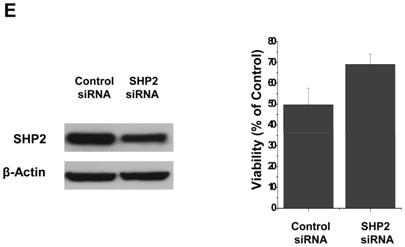
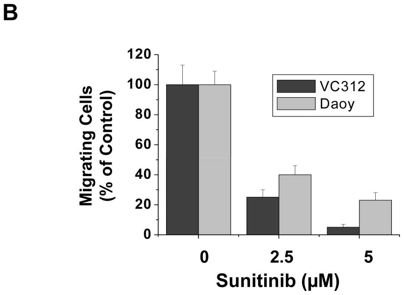
Medulloblastomas are the most frequent malignant brain tumors in children. Sunitinib is an oral multitargeted tyrosine kinase inhibitor used in clinical trials as an antiangiogenic agent for cancer therapy. In this report, we show that sunitinib induced apoptosis and inhibited cell proliferation of both a short-term primary culture (VC312) and an established cell line (Daoy) of human medulloblastomas. Sunitinib treatment resulted in the activation of caspase-3 and cleavage of poly(ADP-ribose) polymerase and upregulation of proapoptotic genes, Bak and Bim, and inhibited the expression of survivin, an antiapoptotic protein. Sunitinib treatment also downregulated cyclin E, cyclin D2, and cyclin D3 and upregulated p21Cip1, all of which are involved in regulating cell cycle. In addition, it inhibited phosphorylation of signal transducer and activator of transcription 3 (STAT3) and AKT (protein kinase B) in the tumor cells. Dephosphorylation of STAT3 (Tyr(705)) induced by sunitinib was helped by a reduction in activities of Janus-activated kinase 2 and Src. Additionally, sodium vanadate, an inhibitor of protein tyrosine phosphatases, partially blocked the inhibition of phosphorylated STAT3 by sunitinib. Loss of phosphorylated AKT after sunitinib treatment was accompanied by decreased phosphorylation of downstream proteins glycogen synthase kinase-3beta and mammalian target of rapamycin. Expression of a constitutively activated STAT3 mutant or myristoylated AKT partially blocked the effects of sunitinib in these tumor cells. Sunitinib also inhibited the migration of medulloblastoma tumor cells in vitro. These findings suggest the potential use of sunitinib for the treatment of pediatric medulloblastomas.


Conflict of interest statement

Disclosure of Potential Conflicts of Interest

No potential conflicts of interest were disclosed.

Figures

Figure 1
Figure 1
Sunitinib induced tumor cell apoptosis and changes in expression of pro-apoptotic and anti-apoptotic genes in the primary culture (VC312) of medulloblastoma. (A) Tumor cells were treated with sunitinib at indicated concentrations for 24h and 48 h, and Annexin V-APC positive apoptotic cells were determined by flow cytometry. (B) Expression of full-length or cleaved caspase-3 and PARP were analyzed by immunoblottings with total cell lysates after 24 h sunitinib treatment. Anti-β-actin monoclonal antibody was used as a loading control. (C) Effects of sunitinib on the expression of pro-apoptotic proteins, Bim, Bak, Bax, Puma and Noxa or anti-apoptotic proteins, survivin, Mcl-1, Bcl-xL and Bcl-2 were also evaluated by immunoblottings. (D) Sunitinib (2.5 μM) inhibited expression of survivin mRNA after 24 h treatment, which was determined by real-time PCR (left panel). Transient transfection of human survivin expression vector partially decreased the effect of sunitinib on the cell viability (right panel). (E) Sunitinib increased the levels of Bak and Bim mRNA and pre-treatment of AcD, a transcriptional inhibitor, blocked the effects of sunitinib in VC312 cells. Data shown are the mRNA ratio of Bak or Bim to GAPDH and determined by real-time quantitive PCR.
Figure 1
Figure 1
Sunitinib induced tumor cell apoptosis and changes in expression of pro-apoptotic and anti-apoptotic genes in the primary culture (VC312) of medulloblastoma. (A) Tumor cells were treated with sunitinib at indicated concentrations for 24h and 48 h, and Annexin V-APC positive apoptotic cells were determined by flow cytometry. (B) Expression of full-length or cleaved caspase-3 and PARP were analyzed by immunoblottings with total cell lysates after 24 h sunitinib treatment. Anti-β-actin monoclonal antibody was used as a loading control. (C) Effects of sunitinib on the expression of pro-apoptotic proteins, Bim, Bak, Bax, Puma and Noxa or anti-apoptotic proteins, survivin, Mcl-1, Bcl-xL and Bcl-2 were also evaluated by immunoblottings. (D) Sunitinib (2.5 μM) inhibited expression of survivin mRNA after 24 h treatment, which was determined by real-time PCR (left panel). Transient transfection of human survivin expression vector partially decreased the effect of sunitinib on the cell viability (right panel). (E) Sunitinib increased the levels of Bak and Bim mRNA and pre-treatment of AcD, a transcriptional inhibitor, blocked the effects of sunitinib in VC312 cells. Data shown are the mRNA ratio of Bak or Bim to GAPDH and determined by real-time quantitive PCR.
Figure 2
Figure 2
Sunitinib inhibited tumor cell proliferation and activities of STAT3 and AKT in VC312 cells. (A) Cells were treated with 0, 1.25, 2.5, or 5 μM sunitinib for 24 h and 48 h and cell proliferation was evaluated by MTS assay. (B) Expression of cyclin E, D1, D2, D3, p21Cip1 and p27Kip1 which are important for cell cycle was evaluated by immunoblotings after 24 h sunitinib treatment. (C) Immunoblotting analyses showed effects of sunitinib on total or phosphorylated STAT3, AKT and MAPK(44/42) after 4 h treatment. (D) Inhibition of STAT3 (Tyr705) and AKT (Ser473) phosphorylation were quickly induced by sunitinib after 5 μM sunitinib treatment. (E) Pre-incubation of sunitinib (5 μM) for 20 min inhibited the phosphorylation of STAT3 (Tyr705) induced by IL-6 (10 ng/ml). Anti-β-actin monoclonal antibody was used as a loading control.
Figure 2
Figure 2
Sunitinib inhibited tumor cell proliferation and activities of STAT3 and AKT in VC312 cells. (A) Cells were treated with 0, 1.25, 2.5, or 5 μM sunitinib for 24 h and 48 h and cell proliferation was evaluated by MTS assay. (B) Expression of cyclin E, D1, D2, D3, p21Cip1 and p27Kip1 which are important for cell cycle was evaluated by immunoblotings after 24 h sunitinib treatment. (C) Immunoblotting analyses showed effects of sunitinib on total or phosphorylated STAT3, AKT and MAPK(44/42) after 4 h treatment. (D) Inhibition of STAT3 (Tyr705) and AKT (Ser473) phosphorylation were quickly induced by sunitinib after 5 μM sunitinib treatment. (E) Pre-incubation of sunitinib (5 μM) for 20 min inhibited the phosphorylation of STAT3 (Tyr705) induced by IL-6 (10 ng/ml). Anti-β-actin monoclonal antibody was used as a loading control.
Figure 3
Figure 3
Inhibition of phosphorylated STAT3 induced by sunitinib may involve in multiple mechanisms in VC312 cells. (A) A key role of STAT3 in sunitinib induced anti-tumor effects. VC312 cells were stably transfected with a constitutively activated STAT3 mutant, pSTAT3-C, and the success of transfection was confirmed by immunobloting assay with flag-antibody (top panel). VC312 cells transfected with pSTAT3-C or vector only were treated with 2.5 μM sunitinib for 48 h and cell proliferation was evaluated by MTS assay. Untransfected cells (Untran) treated in the same way as transfected cells were used as positive control. (B) Sunitinib inhibited phosphorylated JAK2 and Src, upstream activators of STAT3. Total and phosphorylated JAK2 and Src were determined by immunoblotting analyses after 5 μM sunitinib treatment at indicated times. (C) Protein tyrosine phosphatases may be involved in de-phosphorylation of STAT3 induced by sunitinib. Sodium vanadate (Na3VO4) reverses the de-phosphorylation of STAT3 (Tyr705) induced by sunitinib. Pre-treatment with 0.5 mM or 1 mM sodium vanadate, a general inhibitor of protein tyrosine phosphatases, inhibited the de-phosphorylation induced by sunitinib (5 μM). (D) Expression of total or phosphorylated SHP2 after sunitinib treatment. Anti-β-actin monoclonal antibody was used as a loading control. (E) Transfection of SHP2 siRNA partially decreased SHP2 expression (left panel) and the effect of sunitinib (2.5 μM) on cell viability after 24 h treatment.
Figure 3
Figure 3
Inhibition of phosphorylated STAT3 induced by sunitinib may involve in multiple mechanisms in VC312 cells. (A) A key role of STAT3 in sunitinib induced anti-tumor effects. VC312 cells were stably transfected with a constitutively activated STAT3 mutant, pSTAT3-C, and the success of transfection was confirmed by immunobloting assay with flag-antibody (top panel). VC312 cells transfected with pSTAT3-C or vector only were treated with 2.5 μM sunitinib for 48 h and cell proliferation was evaluated by MTS assay. Untransfected cells (Untran) treated in the same way as transfected cells were used as positive control. (B) Sunitinib inhibited phosphorylated JAK2 and Src, upstream activators of STAT3. Total and phosphorylated JAK2 and Src were determined by immunoblotting analyses after 5 μM sunitinib treatment at indicated times. (C) Protein tyrosine phosphatases may be involved in de-phosphorylation of STAT3 induced by sunitinib. Sodium vanadate (Na3VO4) reverses the de-phosphorylation of STAT3 (Tyr705) induced by sunitinib. Pre-treatment with 0.5 mM or 1 mM sodium vanadate, a general inhibitor of protein tyrosine phosphatases, inhibited the de-phosphorylation induced by sunitinib (5 μM). (D) Expression of total or phosphorylated SHP2 after sunitinib treatment. Anti-β-actin monoclonal antibody was used as a loading control. (E) Transfection of SHP2 siRNA partially decreased SHP2 expression (left panel) and the effect of sunitinib (2.5 μM) on cell viability after 24 h treatment.
Figure 4
Figure 4
Sunitinib inhibits phosphorylation of GSK-3β and mTOR and expression of activated AKT partially reversed effect of sunitinib on VC312 cells. (A) Effects of sunitinib on the expression of total or phosphorylated GSK-3β and mTOR were detected by immunoblotting analyses with specific antibodies. (B) VC312 cells were transiently transfected with a constitutively activated AKT expressing plasmid for 24h, and then cells were treated with 1.25 μM sunitinib for 48 h. Cell proliferation was evaluated by MTS assay. (C) Effects of expressing activated STAT3 and AKT on regulatory proteins for cell cycle and apoptosis. VC312 cells contained stable transfeted pSTAT3-C were transiently transfected with pAKT-C for 24 h, followed by treating with 5 μM sunitinib for another 24 h. Immunoblotting analyses were performed with specific antibodies.
Figure 4
Figure 4
Sunitinib inhibits phosphorylation of GSK-3β and mTOR and expression of activated AKT partially reversed effect of sunitinib on VC312 cells. (A) Effects of sunitinib on the expression of total or phosphorylated GSK-3β and mTOR were detected by immunoblotting analyses with specific antibodies. (B) VC312 cells were transiently transfected with a constitutively activated AKT expressing plasmid for 24h, and then cells were treated with 1.25 μM sunitinib for 48 h. Cell proliferation was evaluated by MTS assay. (C) Effects of expressing activated STAT3 and AKT on regulatory proteins for cell cycle and apoptosis. VC312 cells contained stable transfeted pSTAT3-C were transiently transfected with pAKT-C for 24 h, followed by treating with 5 μM sunitinib for another 24 h. Immunoblotting analyses were performed with specific antibodies.
Figure 5
Figure 5
Sunitinib inhibits cell proliferation and induces apoptosis in an established cell line (Daoy) of medulloblastoma. (A) and (B) Induction of apoptosis and inhibition of cell growth after sunitinib treatment were determined by Annexin V-APC staining and MTS assay, respectively. (C) Expression of full-length or cleaved caspase-3 and PARP was analyzed by immunoblottings. (D) Effects of sunitinib on total or phosphorylated STAT3, AKT, MAK(44/42) after 4 h sunitinib treatment.
Figure 5
Figure 5
Sunitinib inhibits cell proliferation and induces apoptosis in an established cell line (Daoy) of medulloblastoma. (A) and (B) Induction of apoptosis and inhibition of cell growth after sunitinib treatment were determined by Annexin V-APC staining and MTS assay, respectively. (C) Expression of full-length or cleaved caspase-3 and PARP was analyzed by immunoblottings. (D) Effects of sunitinib on total or phosphorylated STAT3, AKT, MAK(44/42) after 4 h sunitinib treatment.
Figure 6
Figure 6
Sunitinib inhibits the migration of human medulloblastoma cells. VC312 and Daoy cells were plated in 6-well plates and next day a single scratch was made in the confluent monolayer, followed by sunitinib or vehicle (DMSO) treatment for 24 h. (A) Each scratch was photographed after 24 h treatment. (B) The data represent the average and SD from two independent experiments in duplicate.
Figure 6
Figure 6
Sunitinib inhibits the migration of human medulloblastoma cells. VC312 and Daoy cells were plated in 6-well plates and next day a single scratch was made in the confluent monolayer, followed by sunitinib or vehicle (DMSO) treatment for 24 h. (A) Each scratch was photographed after 24 h treatment. (B) The data represent the average and SD from two independent experiments in duplicate.

References

    1. Rossi A, Caracciolo V, Russo G, Reiss K, Giordano A. Medulloblastoma: from molecular pathology to therapy. Clin Cancer Res. 2008;14:971–976. - PMC - PubMed
    1. McNeil DE, Cote TR, Clegg L, Rorke LB. Incidence and trends in pediatric malignancies medulloblastoma/primitive neuroectodermal tumor: a SEER update. Surveillance Epidemiology and End Results. Med Pediatr Oncol. 2002;39:190–194. - PubMed
    1. Zurawel RH, Chiappa SA, Allen C, Raffel C. Sporadic medulloblastomas contain oncogenic beta-catenin mutations. Cancer Res. 1998;58:896–899. - PubMed
    1. Hartmann W, Digon-Sontgerath B, Koch A, Waha A, Endl E, Dani I, et al. Phosphatidylinositol 3′-kinase/AKT signaling is activated in medulloblastoma cell proliferation and is associated with reduced expression of PTEN. Clin Cancer Res. 2006;12:3019–3027. - PubMed
    1. Yu H, Jove R. The STATs of cancer--new molecular targets come of age. Nat Rev Cancer. 2004;4:97–105. - PubMed

Publication types

MeSH terms