. 2010 Mar;84(6):2972-82.
doi: 10.1128/JVI.02151-09. Epub 2010 Jan 6.

Biochemical and structural characterization of cathepsin L-processed Ebola virus glycoprotein: implications for viral entry and immunogenicity

Affiliations

Biochemical and structural characterization of cathepsin L-processed Ebola virus glycoprotein: implications for viral entry and immunogenicity

Chantelle L Hood et al. J Virol. 2010 Mar.

Abstract

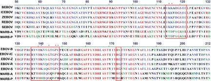
Ebola virus (EBOV) cellular attachment and entry is initiated by the envelope glycoprotein (GP) on the virion surface. Entry of this virus is pH dependent and associated with the cleavage of GP by proteases, including cathepsin L (CatL) and/or CatB, in the endosome or cell membrane. Here, we characterize the product of CatL cleavage of Zaire EBOV GP (ZEBOV-GP) and evaluate its relevance to entry. A stabilized recombinant form of the EBOV GP trimer was generated using a trimerization domain linked to a cleavable histidine tag. This trimer was purified to homogeneity and cleaved with CatL. Characterization of the trimeric product by N-terminal sequencing and mass spectrometry revealed three cleavage fragments, with masses of 23, 19, and 4 kDa. Structure-assisted modeling of the cathepsin L-cleaved ZEBOV-GP revealed that cleavage removes a glycosylated glycan cap and mucin-like domain (MUC domain) and exposes the conserved core residues implicated in receptor binding. The CatL-cleaved ZEBOV-GP intermediate bound with high affinity to a neutralizing antibody, KZ52, and also elicited neutralizing antibodies, supporting the notion that the processed intermediate is required for viral entry. Together, these data suggest that CatL cleavage of EBOV GP exposes its receptor-binding domain, thereby facilitating access to a putative cellular receptor in steps that lead to membrane fusion.


Figures

FIG. 1.
FIG. 1.
Characterization of recombinant full-length ZEBOV-GP. (A) Schematic of EBOV GP plasmid construct containing the GP1 region (orange; residues 31 to 500), with the RBD (dark-dotted orange; residues 54 to 201) and the MUC domain (white-dotted orange; residues 309 to 500), the GP2 region (green; residues 501 to 647), signal sequence (red; residues 1 to 30) and foldon domain (blue). The furin cleavage site is shown directly prior to GP2 at residue 500, and the thrombin cleavage site is shown directly prior to the His tag. N-linked glycan sites are shown as “Y,” and disulfide bonds are shown in red as “S”. (B) His affinity-purified EBOV GP was thrombin digested and resolved by gel filtration size-exclusion chromatography. The approximate peak size was 600 kDa. Molecular mass standards shown are as follows: thyroglobulin, 669 kDa; ferritin, 440 kDa; aldolase, 158 kDa; ovalbumin, 43 kDa; and ribonuclease A, 13.7 kDa. (C) Gel filtration peak fractions were separated by SDS-PAGE and Coomassie stained (left) or transferred to nitrocellulose membrane and Western blotted with an Ebola-specific antibody detecting both GP1 and GP2 (middle) and show GP1 and GP2 at approximately 120 and 25 kDa, respectively. Peak fractions were also analyzed by Native Blue gel electrophoresis to confirm that the protein was trimeric (right) and showed an approximately 600-kDa GP1:GP2 trimer protein. (D) Dynamic light scattering analysis of full-length recombinant ZEBOV-GP. Molecular weight (MW; in thousands) and polydispersity percent (Pd%) of peak fractions following thrombin digestion and size exclusion chromatography on a Superose6 column.
FIG. 2.
FIG. 2.
Characterization of CatL-cleaved EBOV GP. (A) Thrombin-digested EBOV GP was cleaved with CatL and resolved on a Superdex200 column. Molecular mass standards shown are the same as for Fig. 1B. The approximate size was 150 kDa by dynamic light scatter analysis. (B) The reduced protein size is shown by SDS-PAGE Coomassie staining (left) and Western blotting using an Ebola virus-specific antibody (right). (C) The three N terminals resulting from CatL cleavage of ZEBOV-GP are shown in boldface and red type, with the GP1 sequences in orange and GP2 in green. N-linked glycan sites (black) and residues important for virus entry (blue) are also shown. The estimated C termini were calculated based on mass analyses of reduced and nonreduced CatL cleavage reactions and dynamic light scatter (shown as trimer) to confirm the protein trimeric state and mass. (D) Dynamic light scattering analysis of recombinant CatL-processed ZEBOV-GP. Molecular weight (MW; in thousands) and polydispersity percent (Pd%) of peak fractions following cleavage and size exclusion chromatography on a Superdex200 column.
FIG. 3.
FIG. 3.
Mapping and modeling of the ZEBOV-GP CatL cleavage site. (A) Schematic of the EBOV GP sequence aligned with secondary structure features. Alpha-helices and beta-strands are numbered and shown in gray (or outlined in a black dotted line for tentative assignment), and the three N termini at resides 31, 201, and 501 are shown in boldface and red. The N-terminal CatL cleavage site in GP1 is shown at residue 201 (red arrow) and the C-terminal site approximately at residue 222 (dotted red arrow), while the C-terminal of GP2 extends to residue 647 (dotted red arrow). The disordered loop between beta-strands 13 and 14 is shown by a black dotted line. The furin cleavage site is depicted by a black arrow at residue 502. GP1 (orange), GP2 (green), N-glycans (black), and residues important for entry (blue) are labeled as in Fig. 2. The region deleted upon CatL cleavage, including the entire MUC domain, is highlighted in blue. (B) The modeled CatL-cleaved monomer structure is shown based on the recently reported structure and Protein Data Bank file number 3CSY (10). Beta-strands and alpha-helices of the head region are numbered, with GP1 and GP2 residues shown in orange and green, respectively. The first GP1 CatL cleavage site is shown in the disordered loop between β13 and β14, with the latter beta-strand being covalently associated to beta-strand 9. This model assumes there is no conformational change following processing by CatL. (C) The EBOV GP trimer structure (upper panel) (10) is shown by ribbons with a transparent surface representation and red lines depicting the CatL cleavage site in the β13-14 loop. As reported in reference , GP1 (shades of orange) forms a chalice, while GP2 (shades of green) girdles the base of GP1. The modeled CatL-cleaved EBOV trimer structure (lower panel) (based on reference and using the same color scheme as the upper panel) shows the removal of the glycan cap while maintaining the GP2 and the intact fusion peptide, and it also assumes that no conformational change results from the cleavage process. All graphic representations were produced with PyMOL.
FIG. 4.
FIG. 4.
Ability of CatL-cleaved trimeric EBOV-GP ZEBOV-GP to elicit neutralizing antibodies and bind KZ52. (A) The neutralization by antisera from groups of five mice immunized and boosted at weeks 0, 4, and 8 with 20 μg of either CatL-cleaved or uncleaved ZEBOV-GP in 50 μl PBS and an equal volume of Ribi adjuvant was analyzed by incubating the mouse sera (collected 10 days after each vaccination) with an ZEBOV-GP pseudotyped lentiviral reporter vector expressing luciferase. The neutralization activity was calculated based on the luciferase activity relative to values reported for preimmune mouse sera. CatL-cleaved ZEBOV-GP induced the highest titer of neutralizing antibody response, while the uncleaved ZEBOV-GP was 3-fold less potent. (B) Mouse sera from CatL-cleaved ZEBOV-GP mice were analyzed for neutralizing activity against all five EBOV-GP strains individually pseudotyped or a VSV control with lentiviral reporter vectors expressing luciferase. Matched Zaire GP and Ivory Coast GP pseudotyped viruses showed the highest neutralization, while Sudan GP, Bundibugyo GP, and Reston GP pseudotyped viruses showed limited neutralization. The VSV control showed no neutralization. (C) Surface plasmon resonance analysis of CatL-cleaved ZEBOV-GP (upper panel) and SEBOV-GP (lower panel) showed that KZ52 binds both CatL-cleaved EBOV GP strains with a binding affinity of 1.5 nM and 63 nM, respectively.
FIG. 5.
FIG. 5.
Filovirus GPs receptor-binding domain sequence alignment. EBOV-GP species Bundibugyo (BEBOV), Ivory Coast (ICEBOV), Zaire (ZEBOV), Reston (REBOV), and Sudan (SEBOV) and Marburg virus strains Angola (MARB-A) and Popp (MARB-P) were sequence aligned in the receptor-binding domain. Residues in red are conserved among all filoviruses, and the six conserved core residues are marked with an asterisk. Residues that are located in close surface proximity to the six conserved residues are boxed in red.
FIG. 6.
FIG. 6.
Receptor-binding residues modeled on CatL-cleaved EBOV GP trimer structure. (A) Surface representation of the ZEBOV-GP trimer structure (as reported in reference 10) depicting N-glycan sites in the head region (red) (side view; left panel) and residues important for virus entry (blue) (top view; right panel). GP1 is shown in shades of orange and GP2 in shades of green. (B) The surface-modeled CatL-cleaved EBOV-GP trimer structure (based on reference 10) reveals the complete removal of all N-linked glycans (red) from the head region surface (side view; left panel) and exposes the conserved core of the RBD and critical residues for virus entry (blue) (top view; right panel). The approximately 20-Å by 15-Å footprint reported by Lee et al., which contains residues important for virus entry, is indicated in the black circled regions of the top view (right panel). (C) Using a ribbon diagram and transparent surface rendering, the model of CatL-cleaved ZEBOV-GP complexed to the heavy (blue) and light (purple) chains of KZ52 depicts unobstructed binding of KZ52 to the processed EBOV GP and supports the surface plasmon resonance binding data. The color scheme of EBOV GP is the same as in Fig. 5. All graphic representations were produced with PyMOL.
FIG. 7.
FIG. 7.
Schematic model of ZEBOV-GP entry and fusion events. Attachment and binding of the virion (purple) to the host cell membrane is the first step required for EBOV-GP virus entry and replication, followed by endocytosis of the virion into vesicles. The critical step of virus entry is CatL/CatB cleavage at acidic pH 5 to 6, which acts to prime the GP for subsequent membrane fusion. Virus on the cell surface may also encounter factors that are present in CatL-containing endosomes. A recent study implicated α5β1 integrins in the regulation of double chain (DC) CatB and L expression. Our results indicate that CatL processing alone is insufficient to trigger the conformational change and suggest that CatB and/or additional cellular factors are required to initiate the conformational change leading to the final membrane fusion step and release of the nucleocapsid into the cytoplasm.

References

    1. Alvarez, C. P., F. Lasala, J. Carrillo, O. Muñiz, A. L. Corbí, and R. Delgado. 2002. C-type lectins DC-SIGN and L-SIGN mediate cellular entry by Ebola virus in cis and in trans. J. Virol. 76:6841-6844. - PMC - PubMed
    1. Brindley, M. A., L. Hughes, A. Ruiz, P. B. McCray, Jr., A. Sanchez, D. A. Sanders, and W. Maury. 2007. Ebola virus glycoprotein 1: identification of residues important for binding and postbinding events. J. Virol. 81:7702-7709. - PMC - PubMed
    1. Cadene, M., and B. T. Chait. 2000. A robust, detergent-friendly method for mass spectrometric analysis of integral membrane proteins. Anal. Chem. 72:5655-5658. - PubMed
    1. Chan, S. Y., C. J. Empig, F. J. Welte, R. F. Speck, A. Schmaljohn, J. F. Kreisberg, and M. A. Goldsmith. 2001. Folate receptor-α is a cofactor for cellular entry by Marburg and Ebola viruses. Cell 106:117-126. - PubMed
    1. Chandran, K., N. J. Sullivan, U. Felbor, S. P. Whelan, and J. M. Cunningham. 2005. Endosomal proteolysis of the Ebola virus glycoprotein is necessary for infection. Science 308:1643-1645. - PMC - PubMed

Publication types

MeSH terms