. 2010 Mar;84(6):2859-70.
doi: 10.1128/JVI.02053-09. Epub 2010 Jan 6.

Functional analysis of RNA structures present at the 3' extremity of the murine norovirus genome: the variable polypyrimidine tract plays a role in viral virulence

Affiliations

Functional analysis of RNA structures present at the 3' extremity of the murine norovirus genome: the variable polypyrimidine tract plays a role in viral virulence

Dalan Bailey et al. J Virol. 2010 Mar.

Erratum in

  • J Virol. 2010 Oct;84(20):10943. Reese, Jivan [corrected to Rees, Jivan]

Abstract

Interactions of host cell factors with RNA sequences and structures in the genomes of positive-strand RNA viruses play various roles in the life cycles of these viruses. Our understanding of the functional RNA elements present in norovirus genomes to date has been limited largely to in vitro analysis. However, we recently used reverse genetics to identify evolutionarily conserved RNA structures and sequences required for norovirus replication. We have now undertaken a more detailed analysis of RNA structures present at the 3' extremity of the murine norovirus (MNV) genome. Biochemical data indicate the presence of three stable stem-loops, including two in the untranslated region, and a single-stranded polypyrimidine tract [p(Y)] of variable length between MNV isolates, within the terminal stem-loop structure. The well-characterized host cell pyrimidine binding proteins PTB and PCBP bound the 3'-untranslated region via an interaction with this variable sequence. Viruses lacking the p(Y) tract were viable both in cell culture and upon mouse infection, demonstrating that this interaction was not essential for virus replication. However, competition analysis with wild-type MNV in cell culture indicated that the loss of the p(Y) tract was associated with a fitness cost. Furthermore, a p(Y)-deleted mutant showed a reduction in virulence in the STAT1(-/-) mouse model, highlighting the role of RNA structures in norovirus pathogenesis. This work highlights how, like with other positive-strand RNA viruses, RNA structures present at the termini of the norovirus genome play important roles in virus replication and virulence.


Figures
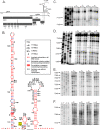
FIG. 1.
FIG. 1.
The murine norovirus genome contains three 3′-terminal stem-loop RNA structures. (A) Schematic representation of murine norovirus genome, highlighting the four predicted open reading frames and the mature replicase proteins produced from ORF1. (B) Predicted RNA secondary structure of the 3′ end of the MNV genome. The positions of the RNase cleavage sites, as determined by limited RNase digestion followed by primer extension, are highlighted on the bioinformatically predicted structure for the MNV 3′ end. The genetic sequence variation of 38 published murine norovirus 3′-end sequences is also provided, highlighting those bases which either are invariant, show signs of covariation, vary but maintain base pairing, or vary without maintaining base pairing. ClustalW analysis was performed using all available murine norovirus sequences in the NCBI database (details available on request). The stop codon of the VP2 coding sequence in SL1 is also highlighted. (C to F) In vitro-transcribed RNA encompassing SL1, -2, and -3, with a poly(A) tail of 27 nucleotides in length, was subjected to limited RNase digestion with dilutions of RNases and to subsequent primer extension analysis. A sequencing ladder obtained using the same primer allows the identification of the RNase cleavage sites. Analysis was performed a minimum of three times, and one representative gel is shown. Nucleotide positions are numbered according to their positions in the murine norovirus genome. Note that data are shown for regions containing RNase cleavage sites only.
FIG. 2.
FIG. 2.
Murine norovirus 3′-end mutants. Schematic representation of the MNV 3′ end, highlighting the various deletion and point mutants under study. The diagram at the top left highlights the nucleotide positions of the various stem-loops, SL1 to SL3, and the GA-rich sequence in SL2 deleted in the corresponding mutants. The positions of the nucleotide alterations in SL2 ssm, SL3 ssm, and SL3 ssm R are highlighted with filled circles or squares, with the corresponding changes introduced shown offset in the open circles or squares. The predicted secondary structures of SL3 in the SL3 ssm NR mutant, with the deleted sequence boxed, in SL3 GNRA, and in SL3 AAAA are also shown. Note that for the purposes of this figure, the base pairing predicted to occur between the poly(A) tail and the MNV 3′ UTR has been omitted. In addition, the schematics for SL2 ssm, SL3 ssm, SL3 ssm R, and SL3 ssm NR are for illustrative purposes only, since the predicted secondary structure is significantly modified by the introduced mutations.
FIG. 3.
FIG. 3.
PCBP2 and PTB binding to the murine norovirus 3′ end. EMSAs demonstrated the ability of recombinant PCBP2 and PTB to interact with the wild-type MNV 3′ end (A and D, respectively) or the SL3 GNRA 3′ end (B and E, respectively). Quantification of the corresponding EMSA products is expressed as the percentage of probe in any complex with either recombinant PCBP2 (C) or PTB (F). The positions of the free probe (P) and various RNA-protein complexes (C, CI, CII, and CIII) are also highlighted.
FIG. 4.
FIG. 4.
Biochemical footprinting confirmation of PTB binding site. Polyacrylamide gel analysis was performed on primer extension products obtained from transcripts digested with the single-strand-specific RNase T2 in the presence of PCBP2 (A) or GST, GST-PTB, or buffer alone (B). A sequencing ladder was run alongside, obtained using the same primer, to highlight the sequence protected by PCBP2 and PTB binding (boxed). (C) Schematic representation of SL3 highlighting the PCBP2 and PTB binding site identified by footprinting analysis.
FIG. 5.
FIG. 5.
Viral growth kinetics of wild-type and SL3 GNRA murine noroviruses in tissue culture. Single-cycle (A) and multicycle (B) growth kinetics of wild-type virus and the virus lacking the 3′ p(Y) tract (SL3 GNRA) were determined with the murine macrophage cell line RAW 264.7. Cells were infected at multiplicities of infection of 4 and 0.01 (A and B, respectively), and samples were taken at various time points postinfection. The yield of infectious virus was determined by TCID50 titration in RAW264.7 cells and expressed as TCID50/ml. Infections were performed in triplicate, and the averages were plotted along with standard deviations. Viral protein synthesis was also examined by preparing protein extracts and subsequently analyzing them by Western blotting of 10 μg of protein for the viral polymerase (NS7), viral capsid (VP1), or an endogenous host cell protein (GAPDH). Note that the absence of GAPDH at 12 h postinfection during the single-cycle growth curve analysis was the likely result of the effect of virus infection on host cell protein synthesis.
FIG. 6.
FIG. 6.
Competition analysis of wild-type and SL3 GNRA murine noroviruses. Sequencing chromatograms were obtained for the virus population obtained after 1 to 5 passages of a 1:1 mix of wild-type and SL3 GNRA viruses in RAW 264.7 cells. Cells were infected at an approximate multiplicity of infection of 0.01 for 48 h before dilution and passage onto an additional monolayer. For virus sequence analysis, the virus population isolated at each passage was used to infect a fresh monolayer at a high multiplicity of infection (>3) for ∼16 h before viral RNA was extracted from the cells and sequenced by reverse transcription-PCR.
FIG. 7.
FIG. 7.
Viral growth kinetics of virulent wild-type and SL3 GNRA viruses in primary bone-derived murine macrophage cells. Sequence-verified stocks of wild-type or SL3 GNRA murine norovirus in the CW1.P1 backbone (see the text for more details) were used to infected either RAW 264.7 cells or primary bone-derived macrophage cells at a multiplicity of infection of 0.01 (based on viral titration in RAW 264.7 cells). Infections were performed in triplicate, and samples were taken at various times postinfection prior to TCID50 titration in RAW 264.7 cells. The average titers were plotted as TCID50/ml, with the standard deviations also shown.
FIG. 8.
FIG. 8.
Virulence analysis of wild-type and SL3 GNRA murine noroviruses in STAT1−/− mice. (A) Age- and sex-matched groups of STAT1−/− mice were orally inoculated with 1,000 TCID50 of sequence-verified, partially purified wild-type or SL3 GNRA murine norovirus recovered in the virulent backbone (see the text for details), and their weight was monitored daily. Control mice were inoculated with a lysate from cells prepared in a similar manner to that for the virus stocks (see Materials and Methods for further details). ***, P < 0.001; *, P < 0.05 by two-way ANOVA with Bonferroni posttests. (B) qPCR with tissues; (C) qPCR with feces. The numbers of viral genome copies were determined by quantitative real-time reverse transcription-PCR. RNAs were extracted from numerous tissues at 5 days postinfection and from feces at 3 and 5 days postinfection, and the numbers of genome copies per μg of total RNA were determined as described in Materials and Methods. The means are shown, with standard errors highlighted. The significant difference recorded at day 3 postinfection was observed using a standard t test.

References

    1. Bailey, D., L. B. Thackray, and I. G. Goodfellow. 2008. A single amino acid substitution in the murine norovirus capsid protein is sufficient for attenuation in vivo. J. Virol. 82:7725-7728. - PMC - PubMed
    1. Belliot, G., S. V. Sosnovtsev, K. O. Chang, P. McPhie, and K. Y. Green. 2008. Nucleotidylylation of the VPg protein of a human norovirus by its proteinase-polymerase precursor protein. Virology 374:33-49. - PMC - PubMed
    1. Buchholz, U. J., S. Finke, and K.-K. Conzelmann. 1999. Generation of bovine respiratory syncytial virus (BRSV) from cDNA: BRSV NS2 is not essential for virus replication in tissue culture, and the human RSV leader region acts as a functional BRSV genome promoter. J. Virol. 73:251-259. - PMC - PubMed
    1. Chachu, K. A., A. D. LoBue, D. W. Strong, R. S. Baric, and H. W. Virgin. 2008. Immune mechanisms responsible for vaccination against and clearance of mucosal and lymphatic norovirus infection. PLoS Pathog. 4:e1000236. - PMC - PubMed
    1. Chang, K.-O., S. V. Sosnovtsev, G. Belliot, A. D. King, and K. Y. Green. 2006. Stable expression of a Norwalk virus RNA replicon in a human hepatoma cell line. Virology 353:463-473. - PubMed

Publication types

MeSH terms