

Adenovirus type 5 E1A and E6 proteins of low-risk cutaneous beta-human papillomaviruses suppress cell transformation through interaction with FOXK1/K2 transcription factors
- PMID: 20053746
- PMCID: PMC2826030
- DOI: 10.1128/JVI.02119-09
Adenovirus type 5 E1A and E6 proteins of low-risk cutaneous beta-human papillomaviruses suppress cell transformation through interaction with FOXK1/K2 transcription factors
Abstract
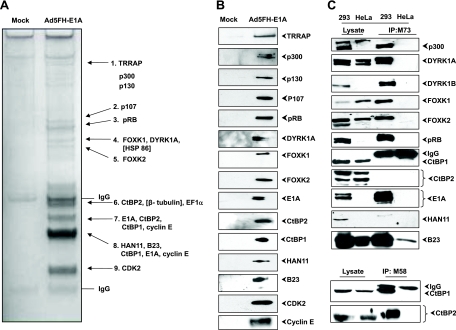
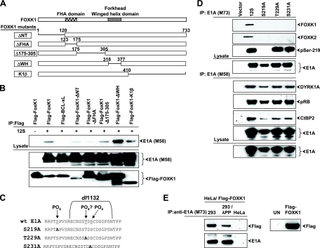
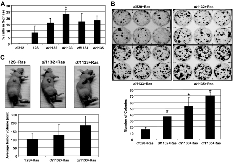
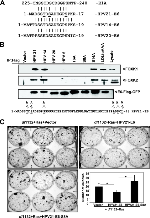
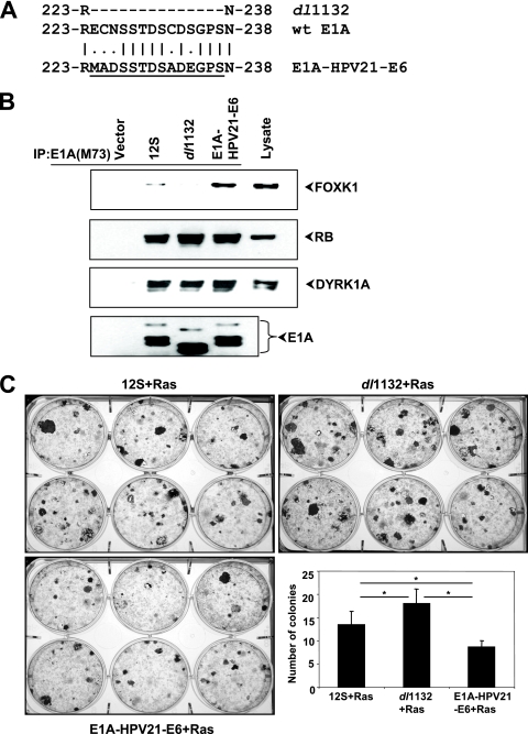
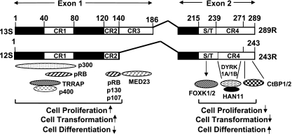
The adenovirus (Adv) oncoprotein E1A stimulates cell proliferation and inhibits differentiation. These activities are primarily linked to the N-terminal region (exon 1) of E1A, which interacts with multiple cellular protein complexes. The C terminus (exon 2) of E1A antagonizes these processes, mediated in part through interaction with C-terminal binding proteins 1 and 2 (CtBP1/2). To identify additional cellular E1A targets that are involved in the modulation of E1A C-terminus-mediated activities, we undertook tandem affinity purification of E1A-associated proteins. Through mass spectrometric analysis, we identified several known E1A-interacting proteins as well as novel E1A targets, such as the forkhead transcription factors, FOXK1/K2. We identified a Ser/Thr-containing sequence motif in E1A that mediated interaction with FOXK1/K2. We demonstrated that the E6 proteins of two beta-human papillomaviruses (HPV14 and HPV21) associated with epidermodysplasia verruciformis also interacted with FOXK1/K2 through a motif similar to that of E1A. The E1A mutants deficient in interaction with FOXK1/K2 induced enhanced cell proliferation and oncogenic transformation. The hypertransforming activity of the mutant E1A was suppressed by HPV21 E6. An E1A-E6 chimeric protein containing the Ser/Thr domain of the E6 protein in E1A interacted efficiently with FOXK1/K2 and inhibited cell transformation. Our results suggest that targeting FOXK1/K2 may be a common mechanism for certain beta-HPVs and Adv5. E1A exon 2 mutants deficient in interaction with the dual-specificity kinases DYRK1A/1B and their cofactor HAN11 also induced increased cell proliferation and transformation. Our results suggest that the E1A C-terminal region may suppress cell proliferation and oncogenic transformation through interaction with three different cellular protein complexes: FOXK1/K2, DYRK(1A/1B)/HAN11, and CtBP1/2.
Figures

Similar articles
-
Functional similarity between E6 proteins of cutaneous human papillomaviruses and the adenovirus E1A tumor-restraining module.J Virol. 2013 Jul;87(13):7781-6. doi: 10.1128/JVI.00037-13. Epub 2013 May 1. J Virol. 2013. PMID: 23637414 Free PMC article.
-
Interaction of CtBP with adenovirus E1A suppresses immortalization of primary epithelial cells and enhances virus replication during productive infection.Virology. 2013 Sep 1;443(2):313-20. doi: 10.1016/j.virol.2013.05.018. Epub 2013 Jun 5. Virology. 2013. PMID: 23747199 Free PMC article.
-
The adaptor protein DCAF7 mediates the interaction of the adenovirus E1A oncoprotein with the protein kinases DYRK1A and HIPK2.Sci Rep. 2016 Jun 16;6:28241. doi: 10.1038/srep28241. Sci Rep. 2016. PMID: 27307198 Free PMC article.
-
The C-terminal region of E1A: a molecular tool for cellular cartography.Biochem Cell Biol. 2012 Apr;90(2):153-63. doi: 10.1139/o11-080. Epub 2012 Jan 31. Biochem Cell Biol. 2012. PMID: 22292450 Review.
-
Modulation of oncogenic transformation by the human adenovirus E1A C-terminal region.Curr Top Microbiol Immunol. 2004;273:139-61. doi: 10.1007/978-3-662-05599-1_5. Curr Top Microbiol Immunol. 2004. PMID: 14674601 Review.
Cited by
-
The forkhead transcription factor FOXK2 acts as a chromatin targeting factor for the BAP1-containing histone deubiquitinase complex.Nucleic Acids Res. 2014 Jun;42(10):6232-42. doi: 10.1093/nar/gku274. Epub 2014 Apr 19. Nucleic Acids Res. 2014. PMID: 24748658 Free PMC article.
-
NHEJ pathway is involved in post-integrational DNA repair due to Ku70 binding to HIV-1 integrase.Retrovirology. 2019 Nov 6;16(1):30. doi: 10.1186/s12977-019-0492-z. Retrovirology. 2019. PMID: 31690330 Free PMC article.
-
The forkhead transcription factor FOXK2 promotes AP-1-mediated transcriptional regulation.Mol Cell Biol. 2012 Jan;32(2):385-98. doi: 10.1128/MCB.05504-11. Epub 2011 Nov 14. Mol Cell Biol. 2012. PMID: 22083952 Free PMC article.
-
Insights from the protein interaction Universe of the multifunctional "Goldilocks" kinase DYRK1A.Front Cell Dev Biol. 2023 Oct 12;11:1277537. doi: 10.3389/fcell.2023.1277537. eCollection 2023. Front Cell Dev Biol. 2023. PMID: 37900285 Free PMC article. Review.
-
The Influence of E1A C-Terminus on Adenovirus Replicative Cycle.Viruses. 2017 Dec 19;9(12):387. doi: 10.3390/v9120387. Viruses. 2017. PMID: 29257057 Free PMC article.
References
-
- Anderson, R. D., R. E. Haskell, H. Xia, B. J. Roessler, and B. L. Davidson. 2000. A simple method for the rapid generation of recombinant adenovirus vectors. Gene Ther. 7:1034-1038. - PubMed
-
- Arany, Z., D. Newsome, E. Oldread, D. M. Livingston, and R. Eckner. 1995. A family of transcriptional adaptor proteins targeted by the E1A oncoprotein. Nature 374:81-84. - PubMed
-
- Arron, J. R., M. M. Winslow, A. Polleri, C. P. Chang, H. Wu, X. Gao, J. R. Neilson, L. Chen, J. J. Heit, S. K. Kim, N. Yamasaki, T. Miyakawa, U. Francke, I. A. Graef, and G. R. Crabtree. 2006. NFAT dysregulation by increased dosage of DSCR1 and DYRK1A on chromosome 21. Nature 441:595-600. - PubMed
-
- Avvakumov, N., A. E. Kajon, R. C. Hoeben, and J. S. Mymryk. 2004. Comprehensive sequence analysis of the E1A proteins of human and simian adenoviruses. Virology 329:477-492. - PubMed
-
- Bahler, J. 2005. Cell-cycle control of gene expression in budding and fission yeast. Annu. Rev. Genet 39:69-94. - PubMed
Publication types
MeSH terms
Substances
Grants and funding
LinkOut - more resources
Full Text Sources
