. 2010 Mar;84(6):2983-95.
doi: 10.1128/JVI.01805-09. Epub 2010 Jan 6.

Prophylactic administration of bacterially derived immunomodulators improves the outcome of influenza virus infection in a murine model

Affiliations

Prophylactic administration of bacterially derived immunomodulators improves the outcome of influenza virus infection in a murine model

Elizabeth B Norton et al. J Virol. 2010 Mar.

Abstract

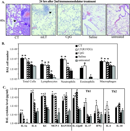
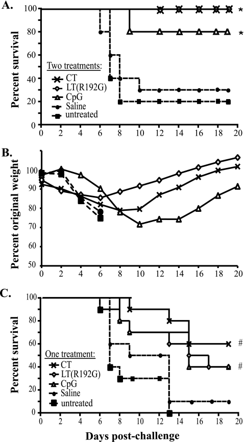
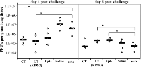
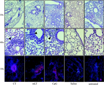
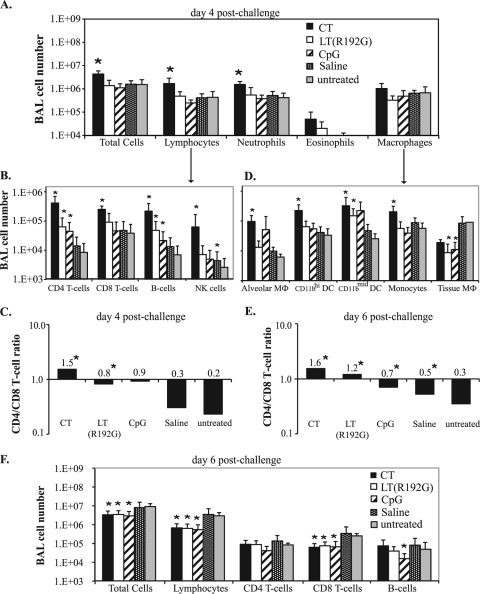
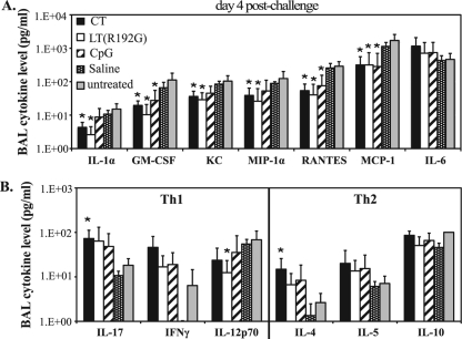
Prophylactic or therapeutic immunomodulation is an antigen-independent strategy that induces nonspecific immune system activation, thereby enhancing host defense to disease. In this study, we investigated the effect of prophylactic immunomodulation on the outcome of influenza virus infection using three bacterially derived immune-enhancing agents known for promoting distinct immunological profiles. BALB/c mice were treated nasally with either cholera toxin (CT), a mutant form of the CT-related Escherichia coli heat-labile enterotoxin designated LT(R192G), or CpG oligodeoxynucleotide. Mice were subsequently challenged with a lethal dose of influenza A/PR/8/34 virus 24 h after the last immunomodulation treatment and either monitored for survival or sacrificed postchallenge for viral and immunological analysis. Treatment with the three immunomodulators prevented or delayed mortality and weight loss, but only CT and LT(R192G) significantly reduced initial lung viral loads as measured by plaque assay. Analysis performed 4 days postinfection indicated that prophylactic treatments with CT, LT(R192G), or CpG resulted in significantly increased numbers of CD4 T cells, B cells, and dendritic cells and altered costimulatory marker expression in the airways of infected mice, coinciding with reduced expression of pulmonary chemokines and the appearance of inducible bronchus-associated lymphoid tissue-like structures in the lungs. Collectively, these results suggest that, despite different immunomodulatory mechanisms, CT, LT(R192G), and CpG induce an initial inflammatory process and enhance the immune response to primary influenza virus challenge while preventing potentially damaging chemokine expression. These studies provide insight into the immunological parameters and immune modulation strategies that have the potential to enhance the nonspecific host response to influenza virus infection.


Figures

FIG. 1.
FIG. 1.
Nasal immunomodulator treatments alter the steady-state immune status in the lung. BALB/c mice were nasally treated twice, 1 week apart, with 5 μg of CT, LT(R192G), or CpG. Controls received saline or were untreated. Mice were sacrificed 24 h later and samples collected for cellular, cytokine, and histological analyses. (A) Representative H&E-stained lung sections. Magnification, ×40. Arrows indicate areas with inflammatory infiltrates and inflamed septae. (B) Recovered BAL cells were characterized for the number of immune system cells, based on hemacytometer cell counts and cytospin/Hema3 differentiation analysis. (C) BAL inflammatory cytokine and chemokine levels, including Th1/Th2 cytokines, were detected using a Bioplex cytokine assay. Bars represent standard deviations of the means (n ≥ 4), with significance values indicated with an asterisk for P values of <0.05 by Mann-Whitney U test.
FIG. 2.
FIG. 2.
CT, LT(R192G), and CpG improve survival and differentially affect weight loss after influenza virus challenge. Groups of BALB/c mice (n ≥ 10) were untreated or nasally treated twice with saline or with 5 μg of immunomodulator [CT, LT(R192G), or CpG]. Twenty-four hours after the last treatment mice were infected with influenza A/PR/8/34 virus and monitored for survival (A) or weight loss (B). Data shown are group averages. Significance values indicated by asterisks represent log-rank test results for survival for CT, LT(R192G), and CpG treatment groups compared to untreated mice (P < 0.001, P < 0.001, and P = 0.002, respectively). Significance values for percent original weight loss are described in the text. (C) Alternatively, mice were infected 24 h after a single immunomodulator treatment and monitored for survival. Significance values (indicated by #) represent log-rank test results for survival for CT, LT(R192G), and CpG groups compared to untreated mice (P < 0.001, P = 0.006, and P = 0.001, respectively).
FIG. 3.
FIG. 3.
Immunomodulators alter viral burden in mouse lungs during influenza virus infection. Groups of BALB/c mice (n = 5) untreated or nasally treated twice with CT, LT(R192G), CpG, or saline were challenged with influenza A/PR/8/34 virus and sacrificed 4 or 6 days postchallenge. Recovered lung tissue was weighed and homogenized. Virus levels in lung homogenates were detected by plaque assay in MDCK cells. Values for individual animals (diamonds) and group medians (lines) are represented. Significance values (indicated by *) for Mann-Whitney U test were P = 0.008, P = 0.032, P = 0.036, and P = 0.009 for day 4 CT, day 4 LT(R192G), day 6 LT(R192G), and day 6 CpG, respectively. untx, untreated.
FIG. 4.
FIG. 4.
Immunomodulator treatment induces cellular aggregates in influenza virus-infected mouse lungs, suggestive of iBALT. Groups of BALB/c mice (n = 5) were nasally treated and infected as explained in the text. Recovered lung tissue on day 4 postchallenge was H&E stained (magnification for rows indicated on left margin); cellular aggregates are also indicated (arrows). Other lung sections were immunofluorescently stained with B220 (red), CD4 (green), and DAPI (blue) (10× magnification). Pictures are representative of group images.
FIG. 5.
FIG. 5.
Immunomodulator treatments alter airway cell composition during influenza virus infection. Groups of BALB/c mice (n = 5) were nasally treated and infected as explained in the text. Recovered BAL cells on day 4 or 6 postchallenge were characterized based on hemacytometer cell counts and cytospin/Hema3 differentiation analysis (A and F) and/or flow cytometry analysis of lymphocyte subtypes (B and F), APC subtypes (D), and CD4/CD8 T-cell ratios (C and E). Bars represent group means ± standard deviations, with significance values indicated by asterisks (P < 0.05, by Mann-Whitney U test, comparing treated to untreated groups).
FIG. 6.
FIG. 6.
Immunomodulator treatments decrease potentially damaging chemokine levels during influenza virus infection. Groups of BALB/c mice (n = 5) were nasally treated [twice with CT, LT(R192G), CpG, or saline] or left untreated, challenged 24 h later with influenza A/PR/8/34 virus, and sacrificed 4 days postchallenge. Supernatants from BAL samples were analyzed with a mouse cytokine/chemokine 23 Bio-plex array to detect levels of inflammatory cytokines and chemokines (A) and Th1/Th2 cytokines (B). Bars represent means ± standard deviations, with significance values indicated (*, P < 0.05 by Mann-Whitney U test).

Similar articles

Cited by

References

    1. Anonymous. 2008. Influenza fact sheet no. 211. World Health Organization, Geneva, Switzerland.
    1. Bagley, K. C., S. F. Abdelwahab, R. G. Tuskan, and G. K. Lewis. 2006. Cholera toxin indirectly activates human monocyte-derived dendritic cells in vitro through the production of soluble factors, including prostaglandin E(2) and nitric oxide. Clin. Vaccine Immunol. 13:106-115. - PMC - PubMed
    1. Banerjee, B., K. J. Kelly, J. N. Fink, J. D. Henderson, Jr., N. K. Bansal, and V. P. Kurup. 2004. Modulation of airway inflammation by immunostimulatory CpG oligodeoxynucleotides in a murine model of allergic aspergillosis. Infect. Immun. 72:6087-6094. - PMC - PubMed
    1. Bowman, C. C., and J. D. Clements. 2001. Differential biological and adjuvant activities of cholera toxin and Escherichia coli heat-labile enterotoxin hybrids. Infect. Immun. 69:1528-1535. - PMC - PubMed
    1. Bromander, A., J. Holmgren, and N. Lycke. 1991. Cholera toxin stimulates IL-1 production and enhances antigen presentation by macrophages in vitro. J. Immunol. 146:2908-2914. - PubMed

Publication types

MeSH terms