

Overexpression of EpCAM in uterine serous papillary carcinoma: implications for EpCAM-specific immunotherapy with human monoclonal antibody adecatumumab (MT201)
- PMID: 20053761
- PMCID: PMC2806489
- DOI: 10.1158/1535-7163.MCT-09-0675
Overexpression of EpCAM in uterine serous papillary carcinoma: implications for EpCAM-specific immunotherapy with human monoclonal antibody adecatumumab (MT201)
Abstract
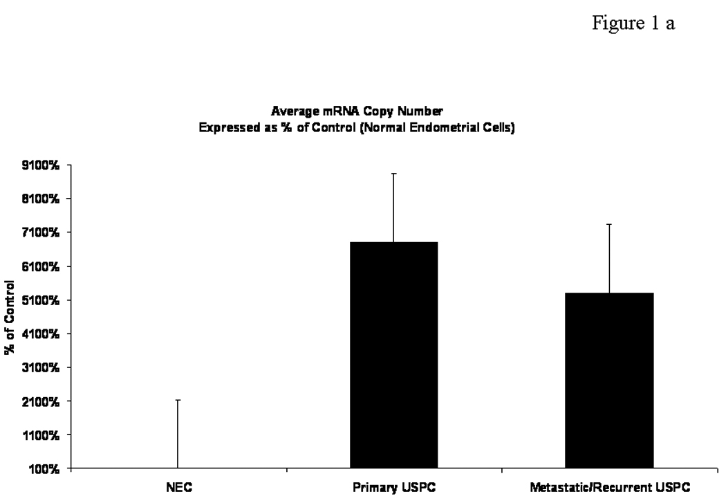
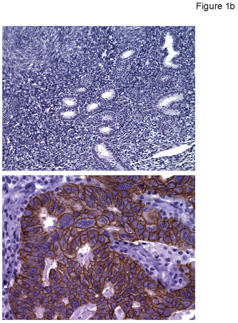
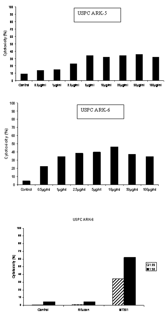
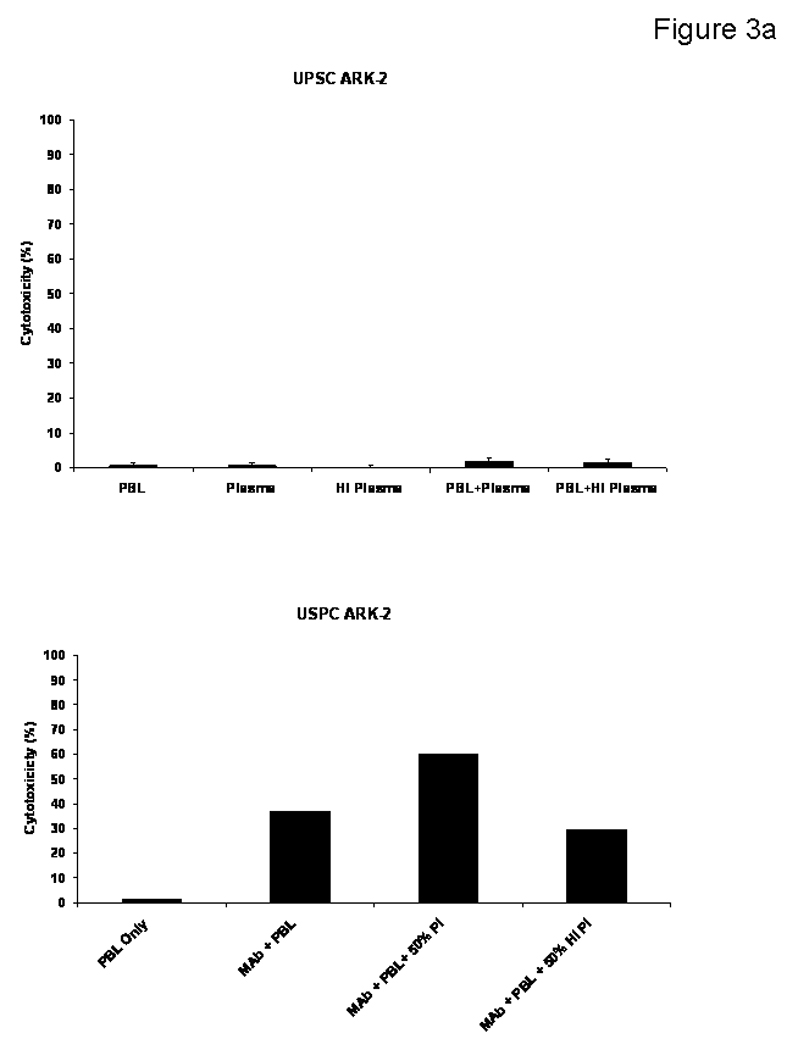
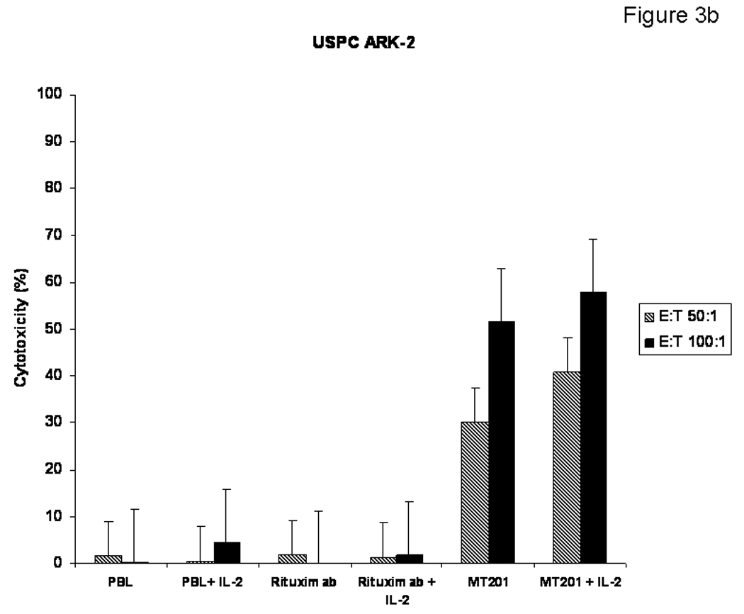
We evaluated the expression of epithelial cell adhesion molecule (EpCAM) and the potential of MT201 (adecatumumab), a human monoclonal antibody against EpCAM, in uterine serous papillary carcinoma (USPC). EpCAM expression was evaluated by real-time PCR and immunohistochemistry in a total of 56 USPC fresh-frozen biopsies and paraffin-embedded tissues. EpCAM surface expression was also evaluated by flow cytometry and immunohistochemistry in six USPC cell lines. Sensitivity to MT201 antibody-dependent cellular cytotoxicity and complement-dependent cytotoxicity was tested against a panel of primary USPC cell lines expressing different levels of EpCAM in standard 5-h (51)Cr release assays. EpCAM transcript was significantly overexpressed in fresh-frozen USPC when compared with normal endometrial cells (NEC). Median (minimum-maximum) copy number was 943.8 (31.5-1568.3) in tumor samples versus 12.9 (1.0-37.0) in NEC (P < 0.001). By immunohistochemistry, EpCAM expression was found in 96% (26 out of 27) of USPC samples with significantly higher expression compared with NECs (P < 0.001). High surface expression of EpCAM was found in 83% (five out of six) of the USPC cell lines tested by flow cytometry. EpCAM-positive cell lines were found highly sensitive to MT201-mediated antibody-dependent cellular cytotoxicity in vitro, whereas primary USPC cell lines were resistant to natural killer cell-dependent cytotoxicity. Human plasma IgG did not significantly inhibit MT201-mediated cytotoxicity against USPC. EpCAM is highly expressed in uterine serous carcinoma at mRNA and protein levels, and primary USPC are highly sensitivity to MT201-mediated cytotoxicity. MT201 might represent a novel therapeutic strategy in patients harboring advanced/recurrent or metastatic USPC refractory to standard treatment modalities.
Conflict of interest statement
Figures
Similar articles
-
Primary cervical carcinoma cell lines overexpress epithelial cell adhesion molecule (EpCAM) and are highly sensitive to immunotherapy with MT201, a fully human monoclonal anti-EpCAM antibody.Int J Gynecol Cancer. 2010 Dec;20(9):1440-7. doi: 10.1111/IGC.0b013e3181fb18a1. Int J Gynecol Cancer. 2010. PMID: 21370592 Free PMC article.
-
Uterine serous papillary carcinomas overexpress human trophoblast-cell-surface marker (Trop-2) and are highly sensitive to immunotherapy with hRS7, a humanized anti-Trop-2 monoclonal antibody.Cancer. 2011 Jul 15;117(14):3163-72. doi: 10.1002/cncr.25891. Epub 2011 Jan 18. Cancer. 2011. PMID: 21246534 Free PMC article.
-
High-grade, chemotherapy-resistant ovarian carcinomas overexpress epithelial cell adhesion molecule (EpCAM) and are highly sensitive to immunotherapy with MT201, a fully human monoclonal anti-EpCAM antibody.Am J Obstet Gynecol. 2010 Dec;203(6):582.e1-7. doi: 10.1016/j.ajog.2010.07.041. Epub 2010 Sep 25. Am J Obstet Gynecol. 2010. PMID: 20870202 Free PMC article.
-
Adecatumumab: an anti-EpCAM monoclonal antibody, from the bench to the bedside.Expert Opin Biol Ther. 2010 Jun;10(6):951-8. doi: 10.1517/14712598.2010.482098. Expert Opin Biol Ther. 2010. PMID: 20426706 Review.
-
EpCAM: A new therapeutic target for an old cancer antigen.Cancer Biol Ther. 2003 Jul-Aug;2(4):320-6. doi: 10.4161/cbt.2.4.451. Cancer Biol Ther. 2003. PMID: 14508099 Review.
Cited by
-
An Ep-ICD based index is a marker of aggressiveness and poor prognosis in thyroid carcinoma.PLoS One. 2012;7(9):e42893. doi: 10.1371/journal.pone.0042893. Epub 2012 Sep 25. PLoS One. 2012. PMID: 23049733 Free PMC article.
-
Eradication of Established Tumors by Chemically Self-Assembled Nanoring Labeled T Cells.ACS Nano. 2018 Jul 24;12(7):6563-6576. doi: 10.1021/acsnano.8b01308. Epub 2018 Jun 4. ACS Nano. 2018. PMID: 29792808 Free PMC article.
-
Epithelial cell adhesion molecule regulates tumor initiation and tumorigenesis via activating reprogramming factors and epithelial-mesenchymal transition gene expression in colon cancer.J Biol Chem. 2012 Nov 16;287(47):39449-59. doi: 10.1074/jbc.M112.386235. Epub 2012 Sep 18. J Biol Chem. 2012. PMID: 22989882 Free PMC article.
-
Expression of αV-integrins in uterine serous papillary carcinomas; implications for targeted therapy with intetumumab (CNTO 95), a fully human antagonist anti-αV-integrin antibody.Int J Gynecol Cancer. 2011 Aug;21(6):1084-90. doi: 10.1097/IGC.0b013e3182187324. Int J Gynecol Cancer. 2011. PMID: 21633302 Free PMC article.
-
Overexpression of EpCAM and Trop2 in pituitary adenomas.Int J Clin Exp Pathol. 2014 Oct 15;7(11):7907-14. eCollection 2014. Int J Clin Exp Pathol. 2014. PMID: 25550831 Free PMC article.
References
-
- Jemal A, Siegel R, Ward E, et al. Cancer statistics, 2008. Vol. 58. CA: a Cancer Journal for Clinicians; 2008. pp. 71–96. - PubMed
-
- Bohkman JV. Two pathogenetic types of endometrial carcinoma. Gynecol Oncol. 1983;15:10–17. - PubMed
-
- Sherman ME, Bitterman P, Rosenshein NB, et al. Uterine serous carcinoma. A morphologically diverse neoplasm with unifying clinicopathological features. Am J Surg Pathol. 1992;16:600–610. - PubMed
-
- Carcangiu ML, Chambers JT. Uterine papillary serous carcinoma: a study on 108 cases with emphasis on prognostic significance of associated endometrioid carcinoma, absence of invasion, and concomitant ovarian cancer. Gynecol Oncol. 1992;47:298–305. - PubMed
Publication types
MeSH terms
Substances
Grants and funding
LinkOut - more resources
Full Text Sources
Other Literature Sources
Medical
Miscellaneous
