. 2010 Jan;9(1):67-78.
doi: 10.1158/1535-7163.MCT-09-0734. Epub 2010 Jan 6.

Glioblastoma cancer-initiating cells inhibit T-cell proliferation and effector responses by the signal transducers and activators of transcription 3 pathway

Affiliations

Glioblastoma cancer-initiating cells inhibit T-cell proliferation and effector responses by the signal transducers and activators of transcription 3 pathway

Jun Wei et al. Mol Cancer Ther. 2010 Jan.

Abstract

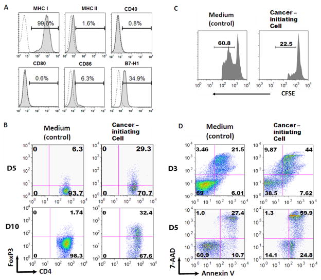
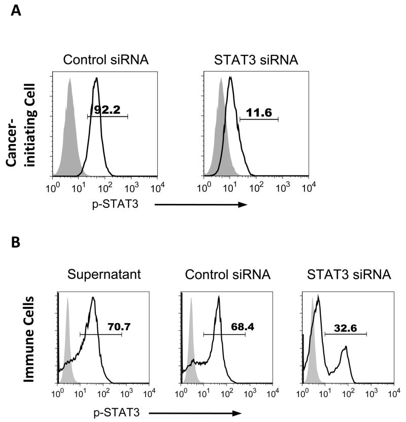
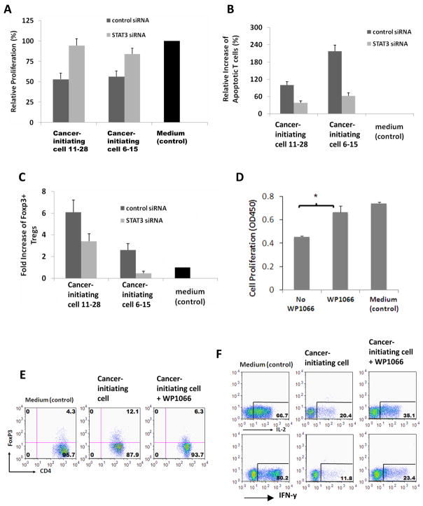
Glioblastoma multiforme (GBM) is a lethal cancer that responds poorly to radiotherapy and chemotherapy. Glioma cancer-initiating cells have been shown to recapitulate the characteristic features of GBM and mediate chemotherapy and radiation resistance. However, it is unknown whether the cancer-initiating cells contribute to the profound immune suppression in GBM patients. Recent studies have found that the activated form of signal transducer and activator of transcription 3 (STAT3) is a key mediator in GBM immunosuppression. We isolated and generated CD133+ cancer-initiating single colonies from GBM patients and investigated their immune-suppressive properties. We found that the cancer-initiating cells inhibited T-cell proliferation and activation, induced regulatory T cells, and triggered T-cell apoptosis. The STAT3 pathway is constitutively active in these clones and the immunosuppressive properties were markedly diminished when the STAT3 pathway was blocked in the cancer-initiating cells. These findings indicate that cancer-initiating cells contribute to the immune evasion of GBM and that blockade of the STAT3 pathway has therapeutic potential.


Figures

Figure 1
Figure 1. Characterization of human glioma-associated cancer-initiating cells
A. A representative image of neurospheres from one glioma-associated cancer-initiating cell is shown. B. The glioma-associated cancer-initiating cells were surface stained with antibodies to CD133, CD34 and CD45. Representative FACS histogram plots for CD133+ cells are shown for target staining (shaded line) with associated isotype controls (gray line). Percentages of the positive populations are shown. C. After 7 days of culture in differentiating medium, the glioma-associated cancer-initiating cells differentiated into GFAP+ astroglial lineage cells, MAP2+ neuronal lineage cells, and GalC+ oligodendroglial lineage cells (magnification X 40) indicating the glioma-associated cancer-initiating cells have multi-potent differentiation potential. D. A representative image of a glioma-associated cancer-initiating cell xenografted into the frontal lobe of a nude mouse. The tumor that developed from the glioma-associated cancer-initiating cell caused enlargement of the brain and were diffusely infiltrative (arrow on right), including into white matter tracts such as the corpus callosum (arrow on left).
Figure 2
Figure 2. Glioma-associated cancer-initiating cells are immunosuppressive of human T cells
A. Immune surface phenotype of a representative glioma-associated cancer-initiating cell. The glioma-associated cancer-initiating cells were surface stained with antibodies to MHC I, MHC II, CD40, CD80, CD86, and B7-H1. Representative FACS histogram plots for one glioma-associated cancer-initiating cell are shown for target staining (solid line) with associated isotype controls (dotted line). Percentages of the positive populations are shown. B. The supernatants from the glioma-associated cancer-initiating cells induce an increase in the number of CD4+FoxP3+ Tregs on both day 5 and 10. Representative FACS plots are shown. C. The glioma-associated cancer-initiating cell induced FoxP3+ Tregs suppress T cell proliferation. T cells that were treated with glioma-associated cancer-initiating cell supernatants were harvested, co-cultured for 3 days with autologous PBMCs (labeled with CFSE, responder cells) at a 1:1 ratio in the presence of soluble anti-CD3 and subsequently analyzed via FACScan. The number above the line in each histogram represents proliferating responder cells. D. The glioma-associated cancer-initiating cell supernatants induce T cell apoptosis after 3 days of exposure to the supernatants. Similar data was obtained after 5 days of exposure. After culturing with the glioma-associated cancer-initiating cell supernatants, T cells were stimulated with anti-CD3/CD28 and stained with 7-AAD and Annexin V. Compared to medium alone (control), the glioma-associated cancer-initiating cells enhanced T cell apoptosis.
Figure 3
Figure 3. Glioma-associated cancer-initiating cells express p-STAT3 and induce p-STAT3 expression in human immune cells
A. STAT3 siRNA treated glioma-associated cancer-initiating cells express lower p-STAT3 compared to control siRNA. B. Glioma-associated cancer-initiating cell supernatants enhance the expression of p-STAT3 in PBMCs, and the supernatants from STAT3 siRNA treated cancer-initiating cells reduce p-STAT3 level in PBMCs. p-STAT3 expression in normal donor PBMCs was measured by intracellular p-STAT3 (pY705) staining via flow cytometry after 3 days of co-culture with cancer-initiating cell supernatants in the presence of anti-CD3/CD28 stimulation. Representative histograms are shown from three independent experiments. Black line: p-STAT3; Gray shade: isotype control.
Figure 4
Figure 4. Immunosuppression mediated by glioma-associated cancer-initiating cells is reversed with p-STAT3 inhibition
A. Two glioma-associated cancer-initiating cell lines were transfected with STAT3 siRNA or control siRNA. After 3 days, the glioma-associated cancer-initiating cells were harvested for p-STAT3 staining, and conditioned media were collected for conducting T cell immune function assays. Inhibition of T cell proliferation mediated by glioma-associated cancer-initiating cells is reversed with STAT3 siRNA inhibition. Proliferation of T cells from normal donor was measured with cell division of CFSE-labeled T cells via flow cytometry after 5 days of culture. B. siRNA treated glioma-associated cancer-initiating cells reduces T cell apoptosis. Cultured T cells on day 5 from (A) were analyzed for apoptosis. C. siRNA treated glioma-associated cancer-initiating cells reduces the generation of FoxP3+ Tregs. Cultured T cells on day 5 from (A) were stained for CD4 and FoxP3, and FACS data were converted into bar graphs showing fold change in the percentage of FoxP3+ Tregs versus medium alone (control; set at baseline of 1). In A–C, media only served as negative controls represented by black bars. The results are averages from three independent experiments, with error bars demonstrating standard deviation. P < 0.05 for all control and STAT3 siRNA comparisons. D. Similar to the STAT-3 siRNA, the inhibition of T cell proliferation mediated by glioma-associated cancer-initiating cells is reversed by WP1066. Proliferation of the T cells was measured by CCK-8 staining after culturing the T cells with the supernatants from the cancer-initiating cells or the cancer-initiating cells treated with WP1066 for 4 days. The results are averages from three independent experiments. *P <0.05. E. Treatment of glioma-associated cancer-initiating cells with WP1066 reduces the generation of FoxP3+ Tregs. F. Inhibition of the pro-inflammatory cytokine IL-2 and IFN-y by the T cells in the presence of supernatant from glioma-associated cancer-initiating cells is partially reversed by WP1066. Intracellular cytokine staining of CD3+ T cells from (D) for IL-2 and IFN-y was performed on day 4. In E and F, FACS plots from one representative glioma-associated cancer-initiating cell experiment were shown but similar results were obtained with glioma-associated cancer-initiating cells from three other patients.

Similar articles

Cited by

References

    1. Kurpad SN, Zhao XG, Wikstrand CJ, Batra SK, McLendon RE, Bigner DD. Tumor antigens in astrocytic gliomas [Review] Glia. 1995 Nov;15(3):244–56. - PubMed
    1. Rosenberg SA, Yang JC, Restifo NP. Cancer immunotherapy: moving beyond current vaccines. Nat Med. 2004 Sep;10(9):909–15. - PMC - PubMed
    1. Dey M, Hussain SF, Heimberger AB. The role of glioma microenvironment in immune modulation: potential targets for intervention. Lett Drug Des Discov. 2006;3(7):443–51.
    1. Hickey WF, Hsu BL, Kimura H. T-lymphocyte entry into the central nervous system. J Neurosci Res. 1991 Feb;28(2):254–60. - PubMed
    1. Morford LA, Elliott LH, Carlson SL, Brooks WH, Roszman TL. T cell receptor-mediated signaling is defective in T cells obtained from patients with primary intracranial tumors. J Immunol. 1997 Nov 1;159(9):4415–25. - PubMed

Publication types

MeSH terms