. 2010 Mar;298(3):F617-24.
doi: 10.1152/ajprenal.00636.2009. Epub 2010 Jan 6.

CREB trans-activation of disruptor of telomeric silencing-1 mediates forskolin inhibition of CTGF transcription in mesangial cells

Affiliations

CREB trans-activation of disruptor of telomeric silencing-1 mediates forskolin inhibition of CTGF transcription in mesangial cells

Zhiyuan Yu et al. Am J Physiol Renal Physiol. 2010 Mar.

Abstract

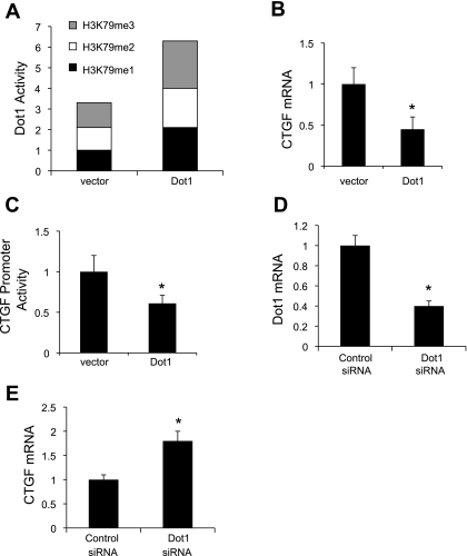
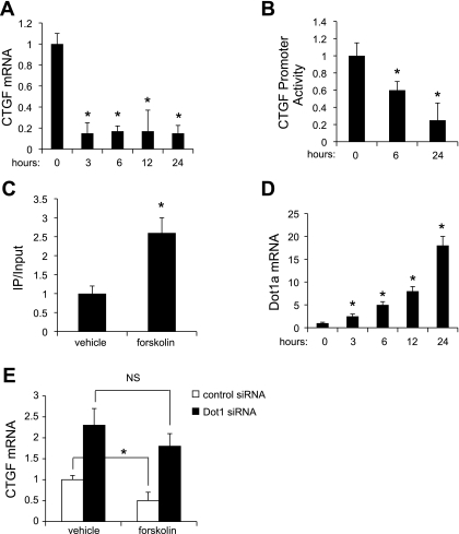
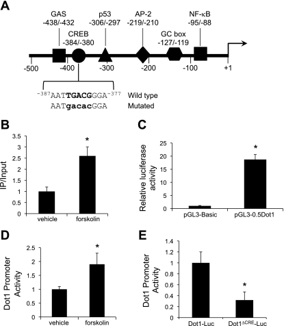
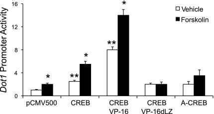
Connective tissue growth factor (CTGF) participates in diverse fibrotic processes including glomerulosclerosis. The adenylyl cyclase agonist forskolin inhibits CTGF expression in mesangial cells by unclear mechanisms. We recently reported that the histone H3K79 methyltransferase disruptor of telomeric silencing-1 (Dot1) suppresses CTGF gene expression in collecting duct cells (J Clin Invest 117: 773-783, 2007) and HEK 293 cells (J Biol Chem In press). In the present study, we characterized the involvement of Dot1 in mediating the inhibitory effect of forskolin on CTGF transcription in mouse mesangial cells. Overexpression of Dot1 or treatment with forskolin dramatically suppressed basal CTGF mRNA levels and CTGF promoter-luciferase activity, while hypermethylating H3K79 in chromatin associated with the CTGF promoter. siRNA knockdown of Dot1 abrogated the inhibitory effect of forskolin on CTGF mRNA expression. Analysis of the Dot1 promoter sequence identified a CREB response element (CRE) at -384/-380. Overexpression of CREB enhanced forskolin-stimulated Dot1 promoter activity. A constitutively active CREB mutant (CREB-VP16) strongly induced Dot1 promoter-luciferase activity, whereas overexpression of CREBdLZ-VP16, which lacks the CREB DNA-binding domain, abolished this activation. Mutation of the -384/-380 CRE resulted in 70% lower levels of Dot1 promoter activity. ChIP assays confirmed CREB binding to the Dot1 promoter in chromatin. We conclude that forskolin stimulates CREB-mediated trans-activation of the Dot1 gene, which leads to hypermethylation of histone H3K79 at the CTGF promoter, and inhibition of CTGF transcription. These data are the first to describe regulation of the Dot1 gene, and disclose a complex network of genetic and epigenetic controls on CTGF transcription.


Figures

Fig. 1.
Fig. 1.
Dot1 represses endogenous connective tissue growth factor (CTGF) mRNA expression and basal CTGF promoter activity in mouse mesangial cells (MMCs). A: overexpressed Dot1 increases H3K79 methylation states in acid extracts of histone proteins. MMCs were transiently transfected with pEGFPC3 (vector) or pEGFPC3-mDot1 (Dot1). Acid extracts of histone proteins were then prepared and assayed for H3K79 methylation states with a fluorometric assay as described in materials and methods. The H3K79me1 value for vector-transected cells was set at 1, and the other values were normalized to it; n = 3. B: overexpressed Dot1 downregulates basal CTGF mRNA expression and promoter activity in mesangial cells. MMCs were transiently transfected with pEGFPC3 (vector) or pEGFPC3-mDot1 (Dot1) as in A, and total RNA was prepared for analysis of CTGF mRNA levels by quantitative RT-PCR. *P < 0.05 vs. vector, n = 4. C: luciferase assay demonstrating that Dot1 overexpression suppresses expression of a stably incorporated CTGF promoter-luciferase construct in MMCs. A mesangial cell line harboring stably transfected pGL3Zeocin-3.8CTGF was transiently transfected with pEGFPC3 (vector) or pEGFPC3-mDot1 (Dot1). Twenty-four hours later, cell lysates were prepared, and the firefly luciferase activity of each sample was normalized to its protein content to generate the “CTGF promoter activity.” The relative luciferase activity of the vector-transfected cells was designated as 1 and utilized to determine the relative level and the significance of the other samples. *P < 0.05 vs. vector-transfected controls, n = 4. D: mRNA analysis showing siRNA knockdown of Dot1 expression in MMCs. MMCs were treated with scrambled control siRNA or Dot1-specific siRNA and then subjected to qRT-PCR analysis. *P < 0.05 vs. scrambled siRNA-transfected controls, n = 4. E: mRNA analysis showing that siRNA knockdown of Dot1 upregulates expression of endogenous CTGF mRNA expression in MMCs. MMCs were treated with scrambled control siRNA or Dot1-specific siRNA and then subjected to qRT-PCR analysis of endogenous CTGF mRNA expression. *P < 0.05 vs. scrambled siRNA, n = 3.
Fig. 2.
Fig. 2.
Forskolin inhibits CTGF mRNA expression and promoter activity as it increases H3K79me3 methylation associated with chromatin at the CTGF 5′-flanking region and Dot1 mRNA expression. A: mesangial cells were treated with vehicle or forskolin (1 μM) for the indicated number of hours, and total RNA was harvested for analysis of CTGF and β-actin mRNAs from the same sample levels by quantitative RT-PCR. The CTGF mRNA value was normalized to that of β-actin, which was invariant under the 2 conditions. *P < 0.05 vs. vehicle-treated cells, n = 4. B: luciferase assay demonstrating that forskolin inhibits the activity of the stably incorporated CTGF promoter-luciferase construct in a time-dependent manner. *P < 0.05 vs. vehicle-treated cells, n = 3. C: ChIP-qPCR analysis of H3K79me3 occupancy at the CTGF 5′-flanking region. Mesangial cells were treated with vehicle or forskolin (1 μM) for 24 h. ChIP assays were then performed with antibody directed against H3K79me3 or nonimmune IgG followed by qPCR with a primer set designed to amplify −3491 to −3070 of the CTGF gene. The value for the final amplicon/input DNA with anti-H3K79me3 from the vehicle-treated cells was set as 1, and the value from the forskolin-treated cells was normalized to it. Values are means ± SE of 3 separate experiments, each performed in triplicate. D: Dot1 mRNA levels were measured in mesangial cells treated with forskolin (1 μM) for the indicated number of hours by quantitative RT-PCR. The levels were normalized to that of β-actin, which was invariant under the 2 conditions. *P < 0.05 vs. vehicle-treated cells, n = 4. E: Dot1 knockdown abrogates the ability of forskolin to inhibit CTGF mRNA expression. Mesangial cells were transfected with scrambled control siRNA or Dot1-specific siRNA. The transfected cells were treated with vehicle or forskolin (1 μM) for 24 h, RNA was then harvested, and quantitative RT-PCR was used to determine CTGF mRNA levels. *P < 0.05 vs. vehicle-treated cells, n = 5. NS, not significant.
Fig. 3.
Fig. 3.
A: map of the proximal 5′-flanking region of the murine Dot1 gene. Consensus sites for the binding of selected transcription factors are indicated. Sequence of the −384/−380 region and the mutated sequence (mutations in lower case letters) used for trans-activation assays are also indicated. B: ChIP-qPCR analysis of CRE-binding protein (CREB) occupancy at the Dot1 proximal promoter. MMCs were treated with vehicle or forskolin (1 μM) for 24 h. ChIP assays were then performed with antibody directed against CREB or nonimmune IgG followed by qPCR with a primer set designed to amplify −582 to −162 of the Dot1 gene. The value for the final amplicon/input DNA with anti-CREB from the vehicle-treated cells was set as 1, and the value from the forskolin-treated cells was normalized to it. Values are means ± SE of 3 separate experiments, each performed in triplicate. C: luciferase assay showing functional promoter activity of the transiently transfected pGL3-0.5Dot1 construct. pGL3-Basic or pGL3-0.5Dot1 was cotransfected with pRL-SV40 into MMCs, and firefly and Renilla luciferase activities were measured in triplicate for each sample. Firefly luciferase activity was normalized to that of Renilla luciferase activity to generate “Dot1 promoter activity.” *P < 0.05 vs. pGL3-Basic-transfected cells, n = 3. D: luciferase activity showing forskolin (1 μM for 24 h) induction of pGL3-0.5Dot1. MMCs were cotransfected with pGL3-0.5Dot1 and pRL-SV40 in MMCs, and the cells were treated with vehicle or forskolin (1 μM) for 24 h. Firefly luciferase activity was normalized to that of Renilla luciferase activity to generate “Dot1 promoter activity,” measured in triplicate for each sample. *P < 0.05 vs. vehicle-treated cells, n = 3. E: functional analysis of the −384/−380 CRE in the proximal promoter of the murine Dot1 gene. Core nucleotides of the CRE in the pGL3-0.5Dot1 were mutated by point mutagenesis to create pGL3-0.5Dot1Δ−384/−380 (Dot1Δ−384/−380-Luc) as described in materials and methods. The construct or pGL3-0.5Dot1 was cotransfected with pRL-SV40 into MMCs, and firefly and Renilla luciferase activities were measured in triplicate for each sample. Firefly luciferase activity was normalized to that of Renilla luciferase activity to generate Dot1 promoter activity. Error bars indicate ±SE. *P ≤ 0.05 vs. pGL3-0.5Dot1 (Dot1-Luc), n = 3.
Fig. 4.
Fig. 4.
CREB trans-activates the proximal promoter of the murine Dot1 gene in MMCs. pGL3-0.5Dot1 reporter construct and pRL-SV40 were cotransfected with the expression vector for CREB, a constitutively active CREB mutant (CREB-VP16), CREBdLZ-VP16, the CREB dominant-negative mutant A-CREB, or an insertless mammalian expression vector containing the cytomegalovirus promoter CMV500. Twenty-four hours after transfection, cell lysates were prepared and firefly and Renilla luciferase activities in lysates of the cells were assayed. Firefly luciferase activity was normalized to Renilla luciferase activity. The bar graph plots the luciferase activities in the various transfection conditions. Error bars indicate ±SE. *P ≤ 0.05 vs. vehicle-treated pCMV500-transfected cells; **P ≤ 0.05 vs. forskolin-treated pCMV500-transfected cells, n = 3.

Similar articles

Cited by

References

    1. Ahn S, Olive M, Aggarwal S, Krylov D, Ginty DD, Vinson C. A dominant-negative inhibitor of CREB reveals that it is a general mediator of stimulus-dependent transcription of c-fos. Mol Cell Biol 18: 967–977, 1998 - PMC - PubMed
    1. Barry ER, Krueger W, Jakuba CM, Veilleux E, Ambrosi DJ, Nelson CE, Rasmussen TP. ES cell cycle progression and differentiation require the action of the histone methyltransferase Dot1L. Stem Cells 27: 1538–1547, 2009 - PMC - PubMed
    1. Black SA, Jr, Palamakumbura AH, Stan M, Trackman PC. Tissue-specific mechanisms for CCN2/CTGF persistence in fibrotic gingiva: interactions between cAMP and MAPK signaling pathways, and prostaglandin E2-EP3 receptor mediated activation of the c-JUN N-terminal kinase. J Biol Chem 282: 15416–15429, 2007 - PMC - PubMed
    1. Blom IE, van Dijk AJ, Wieten L, Duran K, Ito Y, Kleij L, deNichilo M, Rabelink TJ, Weening JJ, Aten J, Goldschmeding R. In vitro evidence for differential involvement of CTGF, TGFbeta, and PDGF-BB in mesangial response to injury. Nephrol Dial Transplant 16: 1139–1148, 2001 - PubMed
    1. Burns WC, Kantharidis P, Thomas MC. The role of tubular epithelial-mesenchymal transition in progressive kidney disease. Cells Tissues Organs 185: 222–231, 2007 - PubMed

Publication types

MeSH terms

LinkOut - more resources