

C-arm CT measurement of cerebral blood volume in ischemic stroke: an experimental study in canines
- PMID: 20053809
- PMCID: PMC7963973
- DOI: 10.3174/ajnr.A1851
C-arm CT measurement of cerebral blood volume in ischemic stroke: an experimental study in canines
Abstract
Background and purpose: CBV is a key parameter in distinguishing penumbra from ischemic core. The purpose of this study was to compare CBV measurements acquired with standard PCT with ones obtained with C-arm CT in a canine stroke model.
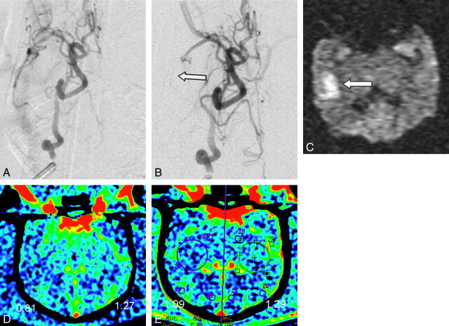
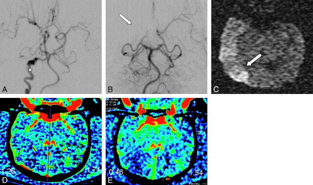
Materials and methods: Under an institutionally approved protocol, unilateral MCA strokes were created in 10 canines. Four hours later, DWI was used to confirm the presence of an infarct. CBV maps acquired with PCT were compared with ones acquired by using C-arm CT. Three experienced observers, blinded to the technique used for acquisition, evaluated the CBV maps.
Results: An ischemic stroke was achieved in 9 of the 10 animals. Areas of reduced CBV were detected in 70%-75% of the PCT studies and in 83%-87% of the C-arm CT examinations, with false-positives in 1.7% and 3.3%, respectively. False-negatives were found in 25% of the PCT and 12.2% of the C-arm CT studies. In all studies, there was a significant difference between the absolute CBV values in normal and abnormal tissue (P < .005) and no significant difference between PCT and C-arm CT CBV values in either the normal or the abnormal parenchyma (P > .05).
Conclusions: CBV measurements made with C-arm CT compare well with ones made with PCT. While further work is required both to fully validate the technique and to define its ultimate clinical value, it appears that it offers a feasible method for assessing CBV in the angiography suite.
Figures
References
-
- Lloyd-Jones D, Adams R, Carnethon M, et al. . Heart disease and stroke statistics: 2009 update—a report from the American Heart Association Statistics Committee and Stroke Statistics Subcommittee. Circulation 2009;119:e21–181. Epub 2008 Dec 15 - PubMed
-
- Schaefer PW, Barak ER, Kamalian S, et al. . Quantitative assessment of core/penumbra mismatch in acute stroke: CT and MR perfusion imaging are strongly correlated when sufficient brain volume is imaged. Stroke 2008;39:2986–92 - PubMed
-
- Tan JC, Dillon WP, Liu S, et al. . Systematic comparison of perfusion-CT and CT-angiography in acute stroke patients. Ann Neurol 2007;61:533–43 - PubMed
-
- Parsons MW, Pepper EM, Bateman GA, et al. . Identification of the penumbra and infarct core on hyperacute noncontrast and perfusion CT. Neurology 2007;68:730–36 - PubMed
Publication types
MeSH terms
LinkOut - more resources
Full Text Sources
Medical