Comparative Study
. 2010 Jan 6;30(1):99-109.
doi: 10.1523/JNEUROSCI.4305-09.2010.

Selective T-type calcium channel block in thalamic neurons reveals channel redundancy and physiological impact of I(T)window

Affiliations
Comparative Study

Selective T-type calcium channel block in thalamic neurons reveals channel redundancy and physiological impact of I(T)window

Fanny M Dreyfus et al. J Neurosci. .

Abstract

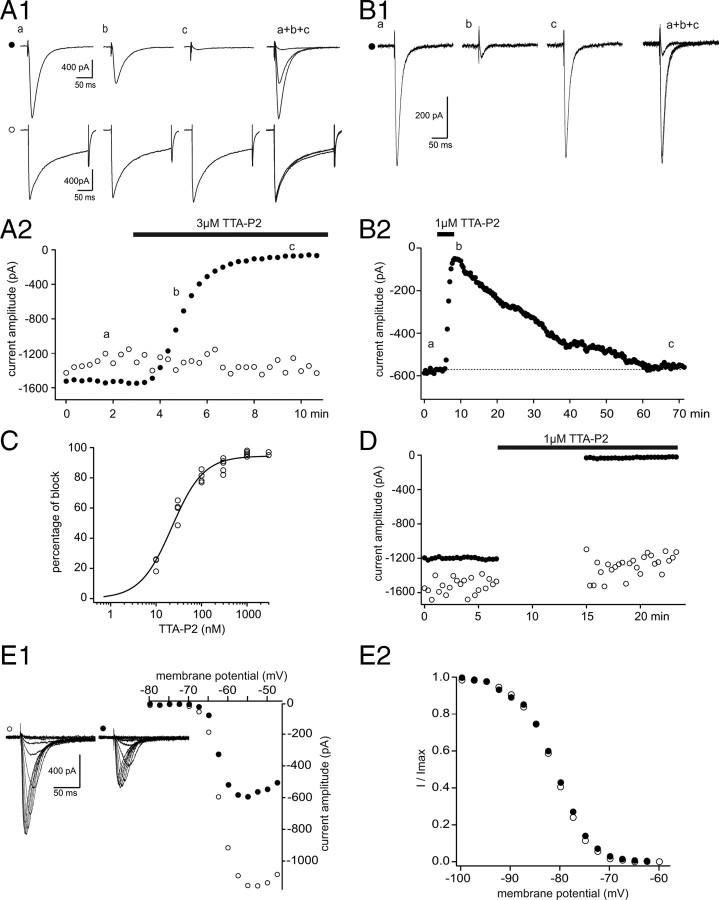
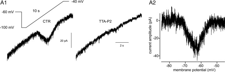
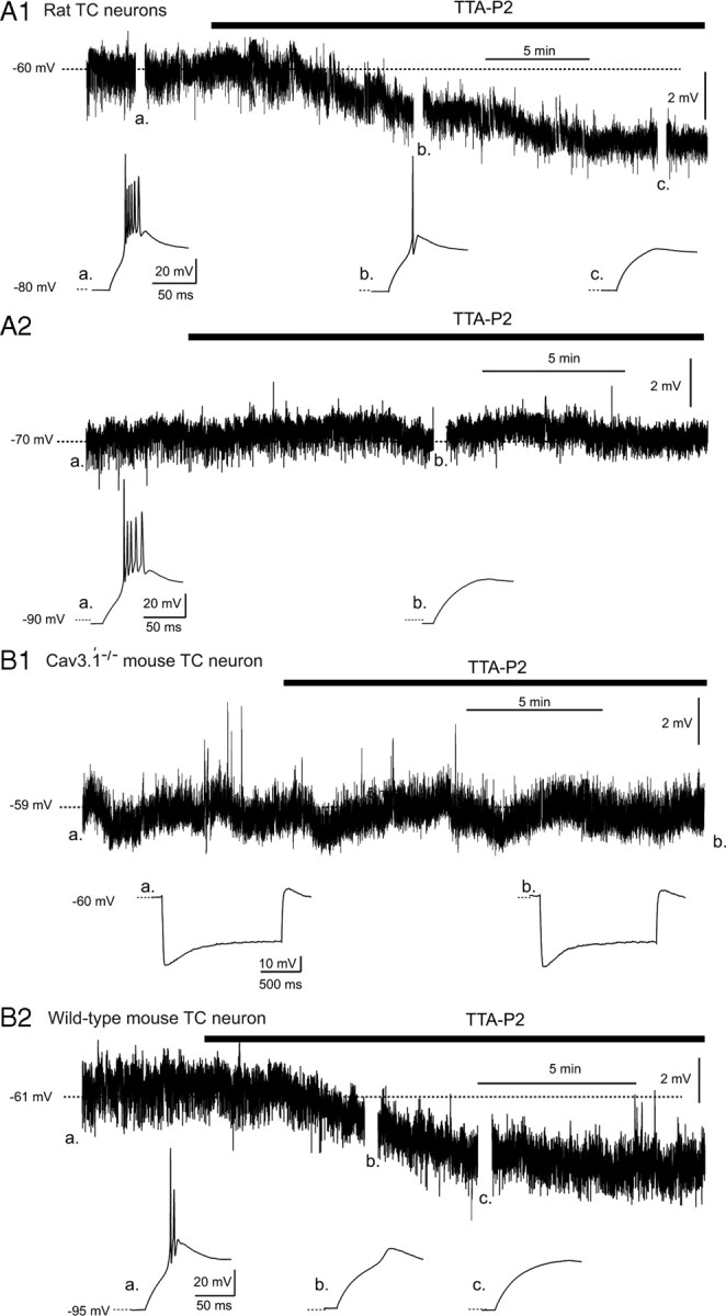
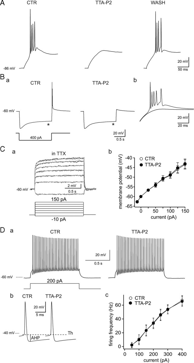
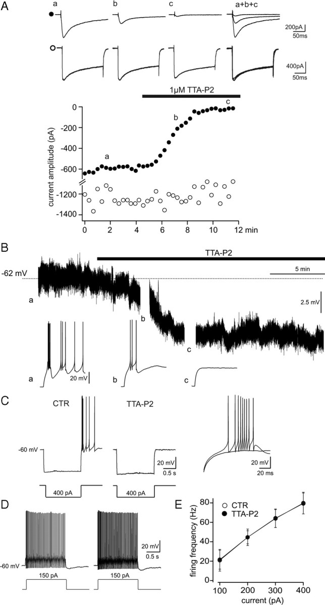
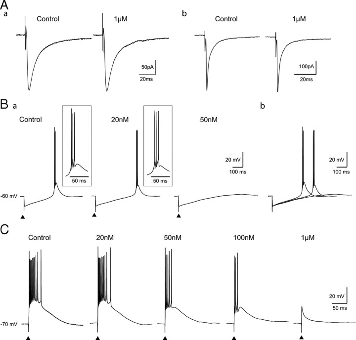
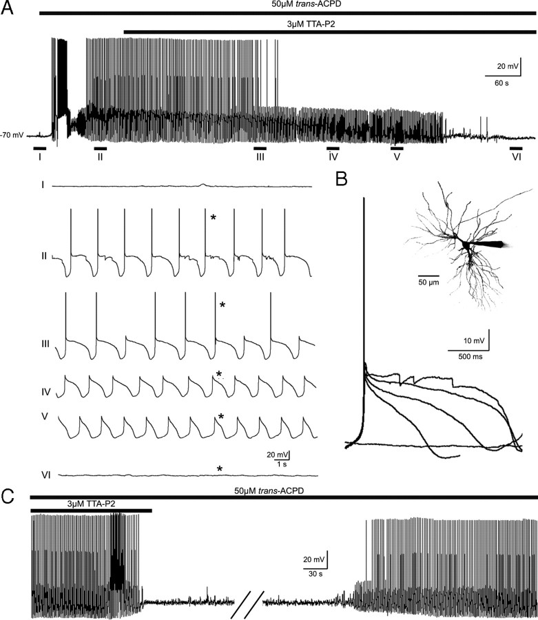
Although it is well established that low-voltage-activated T-type Ca(2+) channels play a key role in many neurophysiological functions and pathological states, the lack of selective and potent antagonists has so far hampered a detailed analysis of the full impact these channels might have on single-cell and neuronal network excitability as well as on Ca(2+) homeostasis. Recently, a novel series of piperidine-based molecules has been shown to selectively block recombinant T-type but not high-voltage-activated (HVA) Ca(2+) channels and to affect a number of physiological and pathological T-type channel-dependent behaviors. Here we directly show that one of these compounds, 3,5-dichloro-N-[1-(2,2-dimethyl-tetrahydro-pyran-4-ylmethyl)-4-fluoro-piperidin-4-ylmethyl]-benzamide (TTA-P2), exerts a specific, potent (IC(50) = 22 nm), and reversible inhibition of T-type Ca(2+) currents of thalamocortical and reticular thalamic neurons, without any action on HVA Ca(2+) currents, Na(+) currents, action potentials, and glutamatergic and GABAergic synaptic currents. Thus, under current-clamp conditions, the low-threshold Ca(2+) potential (LTCP)-dependent high-frequency burst firing of thalamic neurons is abolished by TTA-P2, whereas tonic firing remains unaltered. Using TTA-P2, we provide the first direct demonstration of the presence of a window component of Ca(2+) channels in neurons and its contribution to the resting membrane potential of thalamic neurons and to the Up state of their intrinsically generated slow (<1 Hz) oscillation. Moreover, we demonstrate that activation of only a small fraction of the T-type channel population is required to generate robust LTCPs, suggesting that LTCP-driven bursts of action potentials can be evoked at depolarized potentials where the vast majority of T-type channels are inactivated.


Figures

Figure 1.
Figure 1.
TTA-P2 fully and reversibly blocks T-type Ca2+ current without affecting HVA Ca2+ currents in VB TC neurons. A, IT were evoked every 20 s by step depolarization (200 ms long) to −50 mV after a 1 s hyperpolarizing prepulse to −100 mV. HVA Ca2+ currents were similarly evoked by step depolarization to 10 mV from the −60 mV holding potential. A1, Traces illustrate IT (●, top traces) and HVA Ca2+ currents (○, bottom traces) recorded in the same VB neuron under control condition (a) and in the presence of 3 μm TTA-P2 (b, 2 min application; c, 5 min application). A2, The peak amplitude of IT (●) and HVA Ca2+ currents (○) are plotted against time. TTA-P2 produced a 95% block of IT without any effect on the HVA Ca2+ currents. B1, B2, Same protocols as in A1 and A2 in a different VB TC neuron. A full recovery of the amplitude of IT was obtained after 55 min of wash-out of 1 μm TTA-P2. C, Dose–response curve of the effect of TTA-P2 on the amplitude of IT. Data were fitted with the following equation: y = ymax/(1 + IC50/x)n, where IC50 = 22 nm, ymax = 94.5%, and n = 1.2. D, Same protocols as in A1 and A2. Interruption of the stimulating protocols during the first 8 min of 1 μm TTA-P2 application did not preclude the block of IT. E, Activation and inactivation properties of IT were estimated in the same neuron under control condition and after 20 min of 25 nm TTA-P2 application. The neuron was maintained at −60 mV between protocols. E1, I–V curves were constructed by successive step depolarizations from −80 to −45 mV (2.5 mV increments) preceded by a 1 s hyperpolarizing prepulse to −100 mV. Left traces illustrate currents evoked at the various potentials under control conditions (○) and in the presence of TTA-P2 (●). Despite the strong decrease in current amplitude induced by TTA-P2 application, the apparent voltage dependence of channel activation appears similar in control condition (○) and in the presence of TTA-P2 (●). E2, Normalized steady-state inactivation curves. Inactivation of T-type Ca2+ channels was induced using a 1 s prepulse of increasing potential (from −100 to −60 mV with 2.5 mV increments), and the resulting channel availability was estimated from the normalized current amplitude measured at −50 mV. Note the lack of any significant change in the presence of TTA-P2.
Figure 2.
Figure 2.
Effect of TTA-P2 on ITwindow of VB TC neurons. A1, The window T-type Ca2+ current evoked by a 10-s-long depolarizing voltage ramp from −100 to −40 mV preceded by a 1 s hyperpolarizing prepulse to −100 mV is fully blocked by 1 μm TTA-P2. A2, The voltage dependence of ITwindow is presented. Note that the maximum amplitude of the current occurs around −63 mV.
Figure 3.
Figure 3.
Effect of TTA-P2 on the resting membrane potential of TC neurons. A1, TTA-P2 (1 μm) induces a small hyperpolarization in a Wistar rat TC neuron held at −60 mV (top trace), concomitantly with the block of the LTCP evoked at the end of a hyperpolarizing step (bottom traces, with a–c marking the corresponding breaks in the top trace). A2, TTA-P2 (1 μm) does not hyperpolarize a TC neuron held at −70 mV (top trace) but still blocks the rebound LTCP (bottom traces, with a and b marking the corresponding breaks in the top trace). B1, TTA-P2 (1 μm) has no effect on the membrane potential of a TC neuron from a Cav3.1−/− mouse recorded at −59 mV. Note the absence of a rebound LTCP after the hyperpolarizing steps and the lack of effect of TTA-P2 on the depolarizing sag of the hyperpolarizing response. B2, TTA-P2 (1 μm) induces a small hyperpolarization in a TC neuron from a wild-type mouse held at −61 mV (top trace), concomitantly with the block of the rebound LTCP (bottom traces, with a–c marking the corresponding breaks in the top trace).
Figure 4.
Figure 4.
Effect of TTA-P2 on rat TC neuron excitability. A, Typical example of the reversible block by TTA-P2 (3-min-long application, 1 μm) of the rebound LTCP evoked at the end of a 1.5-s-long hyperpolarizing step in a rat TC neuron. Ba, The block of the rebound LTCP by 1 μm TTA-P2 was not associated with a change in the depolarizing sag (*) of the hyperpolarizing response to an intracellular current step. Bb, The membrane potential responses observed after repolarization in both conditions are superimposed at a larger time scale. Ca, Superimposed voltage traces illustrate the lack of effect of 1 μm TTA-P2 on membrane potential steps induced by injection of depolarizing currents (control, gray traces; TTA-P2, black traces). Cb, An I–V plot for similar experiments as in a from four TC neurons. Da, Typical example of the tonic firing observed in control condition and after 1 μm TTA-P2 application (same cell as in B). A similar number of action potentials (n = 32) is evoked during a 200 pA step depolarization in both control and TTA-P2 conditions. The first action potential recorded in control and in TTA-P2 conditions are enlarged in Db. Note the lack of effect of TTA-P2 on the threshold (Th), amplitude, half-width, and afterhyperpolarization (AHP) of the action potential. The graph in Dc presents the frequency–current plot from six neurons. CTR, Control.
Figure 5.
Figure 5.
Effect of TTA-P2 on NRT neurons. A, Same protocol as in Figure 1A was used to record the IT (●, top traces) and HVA Ca2+ currents (○, bottom traces) in an NRT neuron. TTA-P2 (1 μm) produced a 97% block of IT without a concomitant decrease in the amplitude of the HVA Ca2+ current. B, Continuous voltage record from an NRT neuron showing that 1 μm TTA-P2 induces a small hyperpolarization concomitantly with the block of the rebound LTCP evoked by an hyperpolarizing step (breaks in the trace) before (a) and after a 4 and 8 min wash-in (b, c) of the antagonist. C, D, Same protocols as in Figure 4, B and D, were used to study the effect of 1 μm TTA-P2 on NRT neuron excitability. C, The block of the rebound LTCP by TTA-P2. E, The graph presents the frequency–current plot from nine neurons. CTR, Control.
Figure 6.
Figure 6.
Effect of TTA-P2 on synaptically evoked LTCPs. A, Lack of effect of 1 μm TTA-P2 on evoked IPCSs (a; averages of 8 IPSCs) and EPSCs (b; averages of 7 EPSCs) recorded from two TC neurons. B, In a TC neuron maintained at −60 mV, rebound LTCPs were evoked by stimulations of the NRT at 20 s intervals in control condition and during successive applications of 20 and 50 nm TTA-P2. Ba, A concentration of 20 nm TTA-P2 had no effect on the high-frequency firing, whereas 50 nm TTA-P2 fully blocked the LTCP. However, as clearly seen on the superimposed traces in Bb, 20 nm TTA-P2 slowed the membrane repolarization and delayed the LTCP occurrence by 110 ms. C, In a TC neuron maintained at −70 mV, LTCPs were evoked by repetitive stimulations of the lemniscal pathway every 20 s under control conditions and while successively applying increasing concentrations of TTA-P2 (20, 50, and 100 nm and 1 μm). Note that a reduction in the LTCP-associated firing is observed with TTA-P2 concentrations ≥50 nm, which block >70% of the IT (see Fig. 1C). The full block of the LTCP evoked by the sensory EPSP required a TTA-P2 concentration that abolishes IT (i.e., 1 μm).
Figure 7.
Figure 7.
Block of intrinsic thalamic oscillations by TTA-P2. A, Bath application of trans-ACPD (50 μm) produces a rapid depolarization of a cat MGB TC neuron resulting in continuous tonic firing of action potentials. Hyperpolarization of the cell by steady-state somatic current injection brings about the slow (<1 Hz) sleep oscillation (II). Application of TTA-P2 (3 μm) progressively decreases the duration of the slow oscillation Up state (III and IV) until it eventually resembles the delta oscillation (V), which also is abolished after a few additional minutes of drug exposure (VI). B, Superimposition of the Up states marked by the asterisks (*) in A shows the progressive reduction of their amplitude and duration during the continuous presence of TTA-P2. The inset shows a maximum-intensity z-series reconstruction of the Alexa Fluor 594-filled MGB neuron from which the data illustrated in A and B were collected. C, Reversible block by TTA-P2 of the slow and delta oscillations in a different cat MGB neuron. The break in the trace is 20 min.

Similar articles

Cited by

References

    1. Aizenman CD, Manis PB, Linden DJ. Polarity of long-term synaptic gain change is related to postsynaptic spike firing at a cerebellar inhibitory synapse. Neuron. 1998;21:827–835. - PubMed
    1. Anderson MP, Mochizuki T, Xie J, Fischler W, Manger JP, Talley EM, Scammell TE, Tonegawa S. Thalamic Cav3.1 T-type Ca2+ channel plays a crucial role in stabilizing sleep. Proc Natl Acad Sci U S A. 2005;102:1743–1748. - PMC - PubMed
    1. Bessaih T, Leresche N, Lambert RC. T current potentiation increases the occurrence and temporal fidelity of synaptically evoked burst firing in sensory thalamic neurons. Proc Natl Acad Sci U S A. 2008;105:11376–11381. - PMC - PubMed
    1. Bezprozvanny I, Tsien RW. Voltage-dependent blockade of diverse types of voltage-gated Ca2+ channels expressed in Xenopus oocytes by the Ca2+ channel antagonist mibefradil (Ro 40-5967) Mol Pharmacol. 1995;48:540–549. - PubMed
    1. Bourinet E, Alloui A, Monteil A, Barrere C, Couette B, Poirot O, Pages A, McRory J, Snutch TP, Eschalier A, Nargeot J. Silencing of the Cav3.2 T-type calcium channel gene in sensory neurons demonstrates its major role in nociception. EMBO J. 2005;24:315–324. - PMC - PubMed

Publication types

Substances

LinkOut - more resources