Comparative Study
. 2010 Jan 6;30(1):131-47.
doi: 10.1523/JNEUROSCI.3282-09.2010.

Distinct mechanisms produce functionally complementary actions of neuropeptides that are structurally related but derived from different precursors

Affiliations
Comparative Study

Distinct mechanisms produce functionally complementary actions of neuropeptides that are structurally related but derived from different precursors

Ferdinand S Vilim et al. J Neurosci. .

Abstract

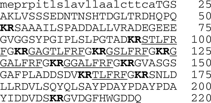
Many bioactive neuropeptides containing RFamide at their C terminus have been described in both invertebrates and vertebrates. To obtain insight into the functional logic of RFamide signaling, we investigate it here in the feeding system of Aplysia. We focus on the expression, localization, and actions of two families of RFamide peptides, the FRFamides and FMRFamide, in the central neuronal circuitry and the peripheral musculature that generate the feeding movements. We describe the cloning of the FRFamide precursor protein and show that the FRFamides and FMRFamide are derived from different precursors. We map the expression of the FRFamide and FMRFamide precursors in the feeding circuitry using in situ hybridization and immunostaining and confirm proteolytic processing of the FRFamide precursor by mass spectrometry. We show that the two precursors are expressed in different populations of sensory neurons in the feeding system. In a representative feeding muscle, we demonstrate the presence of both FRFamides and FMRFamide and their release, probably from the processes of the sensory neurons in the muscle. Both centrally and in the periphery, the FRFamides and FMRFamide act in distinct ways, apparently through distinct mechanisms, and nevertheless, from an overall functional perspective, their actions are complementary. Together, the FRFamides and FMRFamide convert feeding motor programs from ingestive to egestive and depress feeding muscle contractions. We conclude that these structurally related peptides, although derived from different precursors, expressed in different neurons, and acting through different mechanisms, remain related to each other in the functional roles that they play in the system.


Figures

Figure 1.
Figure 1.
Amino acid sequence and processing of the FRFamide precursor protein. A predicted hydrophobic signal peptide (Nielsen et al., 1997) is shown in lowercase letters, and dibasic cleavage sites (Seidah and Chretien, 1999) are shown in bold. The precursor contains six copies of structurally related FRFamide peptides (underlined), as well as multiple linker peptides.
Figure 2.
Figure 2.
Mapping of FRFamide and FMRFamide precursor expression in the central ganglia of Aplysia and in neurons of the buccal ganglion. A, Northern blot analysis of the distribution of FRFamide and FMRFamide precursor mRNA in the different central ganglia of Aplysia (B, buccal; C, cerebral; L, pleural; E, pedal; A, abdominal). The highest levels of expression are in the buccal ganglion for the FRFamide precursor mRNA (A1) and in the pleural ganglia for the FMRFamide precursor mRNA (A2). However, the FMRFamide precursor mRNA is detectable in all of the ganglia, including the buccal ganglion. A3, rRNA stained with methylene blue shows that equal amounts of total RNA were loaded in all lanes. B, C, In situ hybridization showing the distribution of FRFamide and FMRFamide precursor mRNA in neurons of the buccal ganglion. B1, FRFamide in situ hybridization staining of the rostral surface. B2, FRFamide in situ hybridization staining of the caudal surface. C1, FMRFamide in situ hybridization staining of the rostral surface. C2, FMRFamide in situ hybridization staining of the caudal surface. B and C show different ganglia. D, Immunostaining showing the distribution of FRFamide and FMRFamide precursor peptide in neurons and processes of the buccal ganglion. D1, FRFamide immunostaining of the rostral surface. D2, FRFamide immunostaining of the caudal surface. D3, FMRFamide immunostaining of the rostral surface. D4, FMRFamide immunostaining of the caudal surface. The panels in D all show the same ganglion with two different fluorophores, and the immunofluorescence is shown in negative for enhanced visibility. Scale bar, 500 μm. E, Desheathed unstained buccal ganglion showing the location of the S1 and S2 clusters of sensory neurons and the designation of the buccal nerves (CBC, cerebrobuccal connective; RN, radula nerve; EN, esophageal nerve; N1, nerve 1; N2, nerve 2; N3, nerve 3). E1, Rostral surface. E2, Caudal surface.
Figure 3.
Figure 3.
Synthesis of the FRFamide and FMRFamide peptides in buccal sensory neurons. A, B, Radiolabeling and HPLC. Metabolic labeling of the RFamide peptides was accomplished by incubation of buccal ganglia with tritiated phenylalanine. The radiolabeled peptides were extracted from isolated S-clusters and separated by HPLC. The retention times of the radiolabeled peptides (peaks in the count distribution, shown in black) were compared with those of added synthetic RFamides (labeled arrows). A, The first stage of HPLC. B1–B4, The peaks from the first stage were rechromatographed using different columns and counterions that were designed to separate the different RFamides. Radiolabeled peaks corresponding to all five amidated FRFamides A–E can be seen. In B2, two peaks of radioactivity can be seen, one corresponding to FMRFamide (FMRF) and the other to FMRFamide with an oxidized methionine (FM*RF). C, MALDI MS. Representative MALDI mass spectrum (divided into 3 sections) of a single freshly isolated S-cluster neuron exhibits peaks corresponding to all five amidated FRFamides A–E (top section), as well as linker peptides derived from the FRFamide precursor and other known and putative peptides.
Figure 4.
Figure 4.
Representative MALDI mass spectra of cultured S1 and S2 neurons. Some S1 and S2 neurons contained both FMRFamide and the five FRFamides A–E, as well as other peptides (S1 neuron 1 and S2 neuron 1). However, FMRFamide alone was found only in S1 neurons (S1 neuron 2), and the FRFamides alone was found only in S2 neurons (S2 neuron 2).
Figure 5.
Figure 5.
Effects of FRFamide and FMRFamide on feeding motor programs. A1–A4, Feeding motor programs elicited by intracellular stimulation of CBI-2 in the absence (left), superfused presence (middle), and after washout (right) of the indicated concentrations (in molar) of synthetic FRFamide A and FMRFamide. The radula protraction phase of the motor programs was identified by the activity in the extracellular recording from the I2 nerve (I2), the radula retraction phase by the intracellularly recorded depolarization of neuron B4/5 and the activity in the extracellular recording from buccal nerve 2 (BN2), and radula closure by the intracellularly recording firing of the radula closer motor neuron B8 and its reflection in the extracellular recording from the radula nerve (RN) (see Materials and Methods). B1–B4, Analysis of individual experiments corresponding to A1–A4. Plotted is the firing frequency of motor neuron B8 during the retraction phase (y-axis) against its firing frequency during the protraction phase (x-axis), in the absence (circle) and presence (triangle) of the peptide(s) in each experiment. C1–C4, Group data showing the effects on B8 activity during protraction and retraction by the peptides corresponding to examples shown in A1–A4, respectively. Bonferroni's post hoc tests: **p < 0.01; ***p < 0.001. Error bars indicate SEM.
Figure 6.
Figure 6.
MALDI mass spectrum of esophageal nerve extract shows peaks corresponding to FMRFamide as well as the five FRFamides, i.e., FRF A–E. m/z, Mass-to-charge ratio.
Figure 7.
Figure 7.
RFamide release in the ARC muscle. The RFamide content of successive drops of perfusate of the ARC muscle was measured with RIA. A, Calcium dependence of RFamide release. A1, Buccal nerve 3 was extracellularly stimulated at 20 Hz for 3.5 s every 7 s for 5 min, three times as shown by the black bars. For the middle period of stimulation, the calcium in the solution perfusing the muscle was replaced with magnesium. A2, Group data of RFamide measured in the stimulation period before (48.5 ± 6.67), during (3.5 ± 1.53), and after (38.83 ± 5.08) perfusion of calcium-free ASW (means ± SEM; n = 6). B, Block of RFamide release by hexamethonium (Hex). As in A, except that 10−4 m hexamethonium was added to the solution perfusing the muscle for the middle period of stimulation. B2, Group data of RFamide measured in the stimulation period before (57.0 ± 3.65), during (33.2 ± 5.81), and after (48.0 ± 2.21) perfusion of hexamethonium containing ASW (means ± SEM; n = 5). C, Block by hexamethonium of RFamide release elicited by stimulation of the ARC motor neuron B15. As in B except with intracellular stimulation of motor neuron B15 instead of the buccal nerve 3 stimulation. However, the fourth black bar in C1 indicates a period of nerve 3 stimulation added for comparison. C2, Group data of RFamide measured in the stimulation period before (33.25 ± 6.67), during (5.0 ± 1.12), and after (30.75 ± 6.61) perfusion of hexamethonium containing ASW (means ± SEM; n = 4). In A2, B2, and C2, results of statistical analysis of group data via t test is indicated as follows: *p < 0.05; **p < 0.01; ***p < 0.001.
Figure 8.
Figure 8.
RFamides are not present in the ARC motor neurons. ARC muscle immunostaining with the nonspecific rabbit RFamide antibody and rat antibodies to small cardioactive peptide (present in motor neuron B15 processes in the muscle) and myomodulin (present in motor neuron B16 processes). A, Double labeling of the same field for RFamide (A1; rhodamine) and SCP (A2; fluorescein). B, Double labeling of the same field for RFamide (B1; rhodamine) and myomodulin (B2; fluorescein). Note that the RFamide immunostaining pattern does not coincide with that of either ARC motor neuron marker peptide. Negative images are shown for enhanced visibility. Scale bar, 100 μm.
Figure 9.
Figure 9.
FMRFamide is present in the ARC muscle. Purification of FMRFamide from ARC muscle extracts based on bioassay in the ARC neuromuscular preparation of fractions from four sequential stages of HPLC with different conditions. The peak marked with the asterisk in each of A–C was isolated and used in the following stage. A, B, Percentage inhibition (depression) of motor neuron-elicited ARC muscle contractions produced by HPLC fractions eluting at the times (in minutes) shown on the x-axis. C, D, Optical absorbance at 215 nm of the last two HPLC runs. The peak marked with the asterisk in D was subjected to Edman sequencing and had the sequence (with picomoles detected) F(55), M(49), R(17), F(14).
Figure 10.
Figure 10.
Effects of FMRFamide on ARC muscle contractions and EJPs. A, Depression of contractions elicited by alternating bursts of spikes of the two motor neurons B15 and B16 by superfused application of 10−9 m FMRFamide. The panel at right shows washout. B, Dose dependence of the depression of the B16-elicited contractions by progressively increasing concentrations of FMRFamide, indicated in molar. C1, Effect of transient application of 10−8 m FMRFamide on the B16-elicited contractions and the underlying compound EJPs. Representative EJPs before the FMRFamide application (1), at the peak of the depression of contractions (2), and after recovery of the contractions (3) are expanded and superimposed in C2.
Figure 11.
Figure 11.
Effects of the FRFamides on ARC muscle contractions and EJPs. A, Depression of contractions elicited by alternating bursts of spikes of the two motor neurons B15 and B16 by superfused application of increasing concentrations of FRFamide D. B1, Dose–response relations for the depression of the B15- and B16-elicited contraction amplitude by FRFamides C (circles), D (squares), and E (triangles). Each symbol is the mean ± SE of measurements from three to seven (in most cases 6) experiments, normalized to the control contraction amplitude in each experiment. The smooth curves are best fits of the sigmoid function % contraction amplitude = 100/(1 + {[FRF]/KD}a), where [FRF] is the FRFamide concentration, KD is the concentration at half-maximal depression, and a is a constant. B2, Comparison of the KD values ± their SE obtained from the fits in B1. In depressing the contractions elicited by either motor neuron, FRFamide E was significantly less potent than FRFamides C and D (*p < 0.05, one-way ANOVA with post hoc pairwise comparisons using the Holm–Sidak method). C1, Effect of application of 1 μm FRFamide C on the compound EJPs elicited by motor neuron B15. Representative EJPs before (1) and after (2) the FRFamide application are expanded and superimposed (with aligned baselines) in C2.
Figure 12.
Figure 12.
Effects of the FRFamides and FMRFamide on membrane ion current in single voltage-clamped ARC muscle fibers. A, Representative experiment, showing the timing of the slow voltage ramps that were repetitively applied to obtain quasi-steady-state I–V relations before and during superfusion of the peptides (in this experiment, first 10 μm FRF C and then additionally 100 μm FMRFamide). The thick sections of the current and voltage traces were recorded; the thin sections were not recorded and are included to show the relative timing only. The I–V relations plotted in B and C are the currents recorded during selected single ramps during such experiments, plotted against the corresponding voltage. B, FRFamides C–E, applied at 10 μm, all elicited a characteristic outward current that previous work has identified as a modulator-activated K current. C, When compared with FRFamide C in the same fiber, even a 10-fold higher concentration of FMRFamide elicited only a small current. Different fibers were used in A, each of the three panels of B and C; the differences in the amplitudes and apparent I–V characteristics of the elicited currents reflect the interfiber variability rather than any intrinsic differences between the actions of the RFamides.

Similar articles

Cited by

References

    1. Baxter DA, Byrne JH. Feeding behavior of Aplysia: a model system for comparing cellular mechanisms of classical and operant conditioning. Learn Mem. 2006;13:669–680. - PubMed
    1. Bechtold DA, Luckman SM. The role of RFamide peptides in feeding. J Endocrinol. 2007;192:3–15. - PubMed
    1. Brezina V. A simple technique for on-line measurement of contractions of single smooth muscle fibers under current or voltage clamp. Pflugers Arch. 1994;429:126–133. - PubMed
    1. Brezina V, Weiss KR. Analyzing the functional consequences of transmitter complexity. Trends Neurosci. 1997;20:538–543. - PubMed
    1. Brezina V, Evans CG, Weiss KR. Characterization of the membrane ion currents of a model molluscan muscle, the accessory radula closer muscle of Aplysia california. I. Hyperpolarization-activated currents. J Neurophysiol. 1994a;71:2093–2112. - PubMed

Publication types

LinkOut - more resources