Comparative Study
. 2010 Jan 6;30(1):218-29.
doi: 10.1523/JNEUROSCI.3884-09.2010.

Glutamatergic and nonglutamatergic neurons of the ventral tegmental area establish local synaptic contacts with dopaminergic and nondopaminergic neurons

Affiliations
Comparative Study

Glutamatergic and nonglutamatergic neurons of the ventral tegmental area establish local synaptic contacts with dopaminergic and nondopaminergic neurons

Alice Dobi et al. J Neurosci. .

Abstract

The ventral tegmental area (VTA) contributes to reward and motivation signaling. In addition to the well established populations of dopamine (DA) or GABA VTA neurons, glutamatergic neurons were recently discovered in the VTA. These glutamatergic neurons express the vesicular glutamate transporter 2, VGluT2. To investigate whether VTA glutamatergic neurons establish local synapses, we tagged axon terminals from resident VTA neurons by intra-VTA injection of Phaseolus vulgaris leucoagglutinin (PHA-L) or an adeno-associated virus encoding wheat germ agglutinin (WGA) and by immunoelectron microscopy determined the presence of VGluT2 in PHA-L- or WGA-positive terminals. We found that PHA-L- or WGA-positive terminals from tagged VTA cells made asymmetric or symmetric synapses within the VTA. VGluT2 immunoreactivity was detected in the vast majority of PHA-L- or WGA-positive terminals forming asymmetric synapses. These results indicate that both VTA glutamatergic and nonglutamatergic (likely GABAergic) neurons establish local synapses. To examine the possible DAergic nature of postsynaptic targets of VTA glutamatergic neurons, we did triple immunolabeling with antibodies against VGluT2, tyrosine hydroxylase (TH), and PHA-L. From triple-labeled tissue, we found that double-labeled PHA-L (+)/VGluT2 (+) axon terminals formed synaptic contacts on dendrites of both TH-positive and TH-negative cells. Consistent with these anatomical observations, in whole-cell slice recordings of VTA neurons we observed that blocking action potential activity significantly decreased the frequency of synaptic glutamatergic events in DAergic and non-DAergic neurons. These observations indicate that resident VTA glutamatergic neurons are likely to affect both DAergic and non-DAergic neurotransmission arising from the VTA.


Figures

Figure 1.
Figure 1.
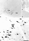
PHA-L injection sites limited to the VTA. A, B, Low (A) and high (B) magnification of a vibratome section containing PHA-L immunoreactivity in the VTA. Rectangle in A delimitates area shown at higher magnification in B. In B, note many PHA-L-immunopositive neurons (arrows). C, Schematic representations of PHA-L-injected sites in the VTA of three different cases (31, 45, and 46). Midbrain sections are ordered rostrocaudally. f, Fasciculus retroflexus; SNr, substantia nigra reticulata. Scale bar: (in B) 550 μm for A; 70 μm for B.
Figure 2.
Figure 2.
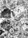
Electron micrographs of PHA-L-labeled axon terminals forming asymmetric or symmetric synapses on dendrites in the VTA. A, Example of an axon terminal (AT1) containing PHA-L immunolabeling [PHAL(+)] and forming an asymmetric synapse (black curved arrow) with a dendrite (De) receiving synapses (white straight arrows) from two unlabeled axon terminals (AT2 and AT3). B, Axon terminal (AT1) containing PHA-L immunolabeling and large dense core vesicles covered by HRP reaction product (arrowhead) forming a symmetric synapse (white curved arrow) with a dendrite that receives an additional symmetric synapse (white straight arrows) from an unlabeled axon terminals (AT2). Scale bar (in B): 0.2 μm for A; 0.26 μm for B.
Figure 3.
Figure 3.
Electron micrographs reveal PHA-L-labeled axon terminals that contain VGluT2 immunolabeling establishing asymmetric synapses on dendrites of local VTA neurons (double immunoelectron microscopy). A, Axon terminal (AT1) containing PHA-L (immunoperoxidase product) and VGluT2 (immunogold-silver particles). PHAL(+)/VGluT2(+) axon terminal makes an asymmetric synapse (black curved arrow) with a dendrite (De). The axon terminal (AT2) containing PHA-L but lacking VGluT2 labeling [PHAL(+)/VGluT2(−)] forms a symmetric synapse (white curved arrow) with a dendrite (De). Note a third axon terminal (AT3) lacking PHA-L but containing VGluT2 [PHAL(−)/VGluT2(+)]. B, A PHA (+)/VGluT2(+) axon terminal (AT1) makes an asymmetric synapse (curved black arrow) with a dendrite (De). A VGluT2-positive terminal (AT2) lacking PHA-L [PHAL(−)/VGluT2(+)] makes an asymmetric synapse (black straight arrow) with a dendrite. An axon terminal (AT3) lacking both PHA-L and VGluT2 [PHAL(−)/VGluT2(−)] makes an asymmetric synapse (black straight arrow) with a dendrite. C, PHA-L(+) and VGluT2(+) axon terminal (AT1) establishes an asymmetric synapse (black curved arrow) with a dendrite (De) receiving a PHA-L and VGluT2-immunonegative [PHAL(−)/VGluT2(−)] axon terminal (AT2) forming a symmetric synapse (white straight arrow). Scale bar (in C): 0.5 μm for A; 0.4 μm for B and C.
Figure 4.
Figure 4.
Electron micrographs of serial sections of a PHA-L/VGluT2 double-labeled axon terminal forming an asymmetric synapse with a dendritic spine of a local VTA neuron. A1, A2, Two consecutive sections showing a double-labeled [PHAL(+)/VGluT2(+)] axon terminal (AT1) that establishes an asymmetric synapse (curved black arrow) with a dendritic spine (S). Note that two axon terminals (AT2 and AT3) lacking PHA-L but containing VGluT2 immunolabeling [PHAL(−)/VGluT2(+)] form asymmetric synapses (black straight arrows) on the same dendrite (De). Scale bar (in A2): A1, A2, 0.5 μm.
Figure 5.
Figure 5.
Electron micrographs showing PHA-L-labeled axon terminals lacking VGluT2 immunolabeling form symmetric synapses with dendrites of local VTA neurons. A, Axon terminal (AT1) containing PHA-L but lacking VGluT2 [PHAL(+)/VGluT2(−)] immunolabeling forms a symmetric synapse (white curved arrow) on a dendrite (De) receiving a symmetric synapse (white straight arrow) from an axon terminal (AT2) lacking both PHA-L and VGluT2 immunoreactivity [PHAL(−)/VGluT2(−)]. Note a third axon terminal (AT3) containing both PHA-L and VGluT2 labeling [PHAL(+)/VGluT2(+)]. B, Axon terminal (AT1) containing PHA-L but lacking VGluT2 [PHAL(+)/VGluT2(−)] immunolabeling forms a symmetric synapse (white curved arrow) on a dendrite (De). Note an axon terminal (AT2) lacking both PHA-L and VGluT2 immunoreactivity [PHAL(−)/VGluT2(−)]. Scale bar (in B): A, B, 0.2 μm.
Figure 6.
Figure 6.
Detection of WGA in VTA neurons. A, A′, WGA-immunoreactive neurons restricted to the VTA. Rectangle in A delimitates low-magnification area shown at higher magnification in A′. B, B′, Ipsilateral striatum to the VTA shown in A and A′. Rectangle in B delimitates nucleus accumbens shown at higher magnification in B′; note lack of WGA-positive fibers. SNr, Substantia nigra reticulata. Scale bar (in A′): 580 μm for A; 75 μm for A′; 2.5 mm for B; 925 μm for B′.
Figure 7.
Figure 7.
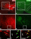
Detection of WGA in VTA neurons that either contain or lack TH immunoreactivity. A, B, TH-immunoreactive neurons (red cells) in the VTA and substantia nigra compacta (SNC). A′, B′, WGA-immunoreactive neurons (green cells) restricted to the VTA. Delimited areas in A′ and B′ are shown at higher magnification in panels C and C′. Transduced cells containing TH immunoreactivity are indicated by a small arrow. Large arrows indicate transfected cells lacking TH immunoreactivity. Scale bar (in C): 566 μm for A and A′; 238 μm for B and B′; 75 μm for C, C′, and merged.
Figure 8.
Figure 8.
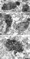
Electron micrographs of WGA-labeled axon terminals forming asymmetric or symmetric synapses on dendrites in the VTA. A, Two axon terminals (AT1 and AT2) containing WGA immunolabeling [WGA(+)], one of which (AT1) forms a symmetric synapse (white curved arrow) with a dendrite (De). B, Two axon terminals (AT1 and AT2) containing VGluT2 immunolabeling [immunogold-silver particles, VGluT2(+)]. While the AT1 also contains WGA immunolabeling [immunoperoxidase product, WGA(+)], the AT2 lacks WGA immunolabeling [WG(−)]. C, An axon terminal (AT1) containing both WGA and VGluT2 immunolabeling [WGA(+)/VGluT2(+)] forms an asymmetric synapse (white curved arrow) with a dendrite (De). Scale bar (in C): 0.4 μm for A; 0.3 μm for B; and 0.2 μm for C.
Figure 9.
Figure 9.
Electron micrographs showing double-labeled PHA-L/VGluT2 axon terminals establishing asymmetric synapses with VTA neurons containing or lacking TH. A1, A2, Two consecutive sections showing that an axon terminal containing PHA-L (peroxidase product) and VGluT2 (immunogold-silver particles) [PHAL(+)/VGluT2(+)] establishes an asymmetric synapse (black curved arrow in A1) with a dendrite (De) containing TH immunolabeling (immunogold-silver particles) [TH(+)]. B1, B2, Two consecutive sections of an axon terminal containing PHA-L (peroxidase product) and VGluT2 (immunogold-silver particles) [PHAL(+)/VGluT2(+)] establishes an asymmetric synapse (black curved arrow in B1) with a dendrite lacking TH immunolabeling [TH(−)]. Scale bar (in B2): 0.2 μm for A1, A2; 0.3 μm for B1, B2.
Figure 10.
Figure 10.
Blocking action potential activity decreases the frequency of excitatory events in VTA whole-cell slice recordings. A, B, Sample traces (calibration in A, 10 pA and 100 ms) and time course (B) of spontaneous glutamatergic events recorded in one neuron under baseline conditions and in the presence of 500 nm TTX in a slice depolarized with 5 mm K+. B, Inset, This recording was made in an Ih(+) neuron (calibration, 200 pA and 50 ms). C, D, For this neuron, the cumulative event plots show that in TTX the amplitudes of spontaneous events slightly decreased (C), while the number of longer interevent intervals greatly increased (D). E, Across cells, 500 nm TTX decreased the amplitude of excitatory events only in 5 mm K+ (n = 10, p < 0.05) but not in control ACSF (n = 6, p > 0.05). F, There was a significant decrease in the frequency of spontaneous excitatory events with 500 nm TTX application both in control ACSF (n = 6) and in 5 mm K+ (n = 10). Application of the AMPAR antagonist DNQX (10 μm) decreased the excitatory event frequency to approximately zero (n = 4). G, In neurons directly identified as TH positive (n = 3) or TH negative (n = 3), in 5 mm K+ there was a trend toward a decrease in excitatory event amplitude with bath application of 500 nm TTX. H, In 5 mm K+, there was a decrease in excitatory event frequency across all neurons tested, and this decrease occurred in both TH-positive and TH-negative neurons. *p < 0.05.

Similar articles

Cited by

References

    1. Bai L, Xu H, Collins JF, Ghishan FK. Molecular and functional analysis of a novel neuronal vesicular glutamate transporter. J Biol Chem. 2001;276:36764–36769. - PubMed
    1. Bayer VE, Pickel VM. Ultrastructural localization of tyrosine hydroxylase in the rat ventral tegmental area: relationship between immunolabeling density and neuronal associations. J Neurosci. 1990;10:2996–3013. - PMC - PubMed
    1. Bayer VE, Pickel VM. GABA-labeled terminals form proportionally more synapses with dopaminergic neurons containing low densities of tyrosine hydroxylase-immunoreactivity in rat ventral tegmental area. Brain Res. 1991;559:44–55. - PubMed
    1. Bellocchio EE, Hu H, Pohorille A, Chan J, Pickel VM, Edwards RH. The localization of the brain-specific inorganic phosphate transporter suggests a specific presynaptic role in glutamatergic transmission. J Neurosci. 1998;18:8648–8659. - PMC - PubMed
    1. Berridge KC, Robinson TE. What is the role of dopamine in reward: hedonic impact, reward learning, or incentive salience? Brain Res Brain Res Rev. 1998;28:309–369. - PubMed

Publication types

LinkOut - more resources