Comparative Study
. 2010 Jan 6;30(1):242-54.
doi: 10.1523/JNEUROSCI.4933-08.2010.

SNAP-25 is a target of protein kinase C phosphorylation critical to NMDA receptor trafficking

Affiliations
Comparative Study

SNAP-25 is a target of protein kinase C phosphorylation critical to NMDA receptor trafficking

C Geoffrey Lau et al. J Neurosci. .

Abstract

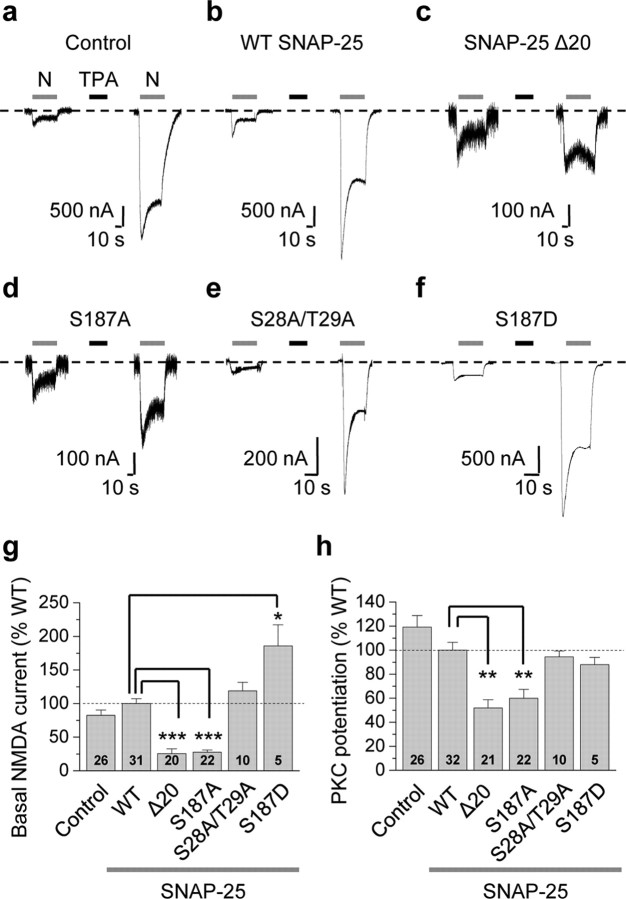
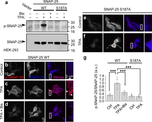
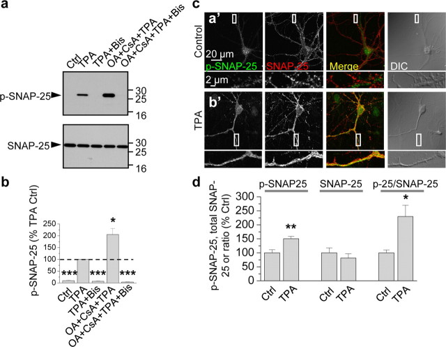
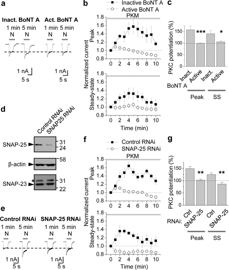
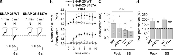
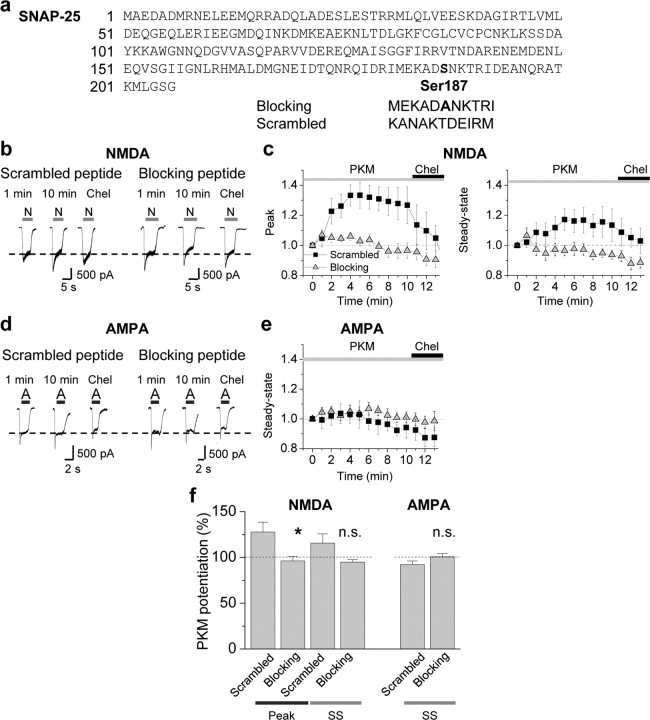
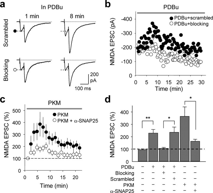
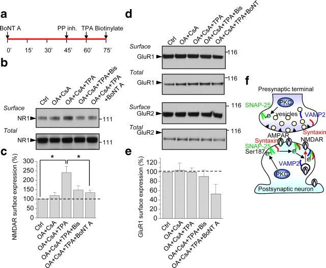
Protein kinase C (PKC) enhances NMDA receptor (NMDAR)-mediated currents and promotes NMDAR delivery to the cell surface via SNARE-dependent exocytosis. Although the mechanisms of PKC potentiation are established, the molecular target of PKC is unclear. Here we show that synaptosomal-associated protein of 25 kDa (SNAP-25), a SNARE protein, is functionally relevant to PKC-dependent NMDAR insertion, and identify serine residue-187 as the molecular target of PKC phosphorylation. Constitutively active PKC delivered via the patch pipette potentiated NMDA (but not AMPA) whole-cell currents in hippocampal neurons. Expression of RNAi targeting SNAP-25 or mutant SNAP-25(S187A) and/or acute disruption of the SNARE complex by treatment with BoNT A, BoNT B or SNAP-25 C-terminal blocking peptide abolished NMDAR potentiation. A SNAP-25 peptide and function-blocking antibody suppressed PKC potentiation of NMDA EPSCs at mossy fiber-CA3 synapses. These findings identify SNAP-25 as the target of PKC phosphorylation critical to PKC-dependent incorporation of synaptic NMDARs and document a postsynaptic action of this major SNARE protein relevant to synaptic plasticity.


Figures

Figure 1.
Figure 1.
Ser187, but not Ser28 and Thr29, of SNAP-25 is critical to PKC potentiation of NMDA currents. a–f, Representative experiments showing PKC potentiation of NMDA-elicited currents recorded from Xenopus laevis oocytes expressing NR1-4b/NR2A and WT or mutant SNAP-25, 24 h after injection of SNAP-25 cRNA. NMDA currents were elicited by bath application of NMDA (N, 300 μm with 10 μm glycine; gray bars above current traces) and recorded by two-electrode voltage clamp at Vh = −60 mV in 1 mm Ca2+ and 0 Mg2+. a, Application of the PKC-activating phorbol ester 12-O-tetradecanoylphorbol 13-acetate (TPA, 100 nm, 10 min, black bar) potentiated NMDA currents by ∼8-fold in control (H2O-injected) oocytes. PKC potentiation is defined as the ratio of steady-state NMDA current after to that before PKC activation. b, Expression of WT SNAP-25 does not detectably alter PKC potentiation of NMDA-elicited currents. c, The dominant-negative mutant of SNAP-25, SNAP25Δ20, markedly reduced the basal NMDA current and PKC potentiation. Note the change in scale. d, A point mutation in which Ser187 was replaced by Ala (S187A) in SNAP-25 reduced both basal NMDA currents and PKC potentiation to an extent indistinguishable from SNAP-25Δ20. e, Replacement of Ser28 and Thr29 by Ala (SNAP-25 S28A/T29A) did not detectably alter basal NMDA currents or PKC potentiation. f, The phosphomimetic mutant, SNAP-25 S187D, markedly enhanced basal NMDA currents without affecting PKC potentiation. g, h, Summary bar graphs showing effects of WT and mutant SNAP-25 on basal NMDA currents (g) and PKC potentiation (h). PKC potentiation in uninjected oocytes and oocytes expressing mutant SNAP-25 constructs was normalized to that in cells expressing WT SNAP-25. Numbers on bars indicate the number of oocytes (n) involving 3–5 independent experiments performed from different batches of oocytes. Data represent means ± SEM. *p < 0.05; **p < 0.01; ***p < 0.001.
Figure 2.
Figure 2.
PKC phosphorylates SNAP-25 in heterologous cells. a, HEK-293 cells overexpressing WT and S187A SNAP-25 were treated with TPA (100 nm, 15 min) in the absence or presence of Bis. Cell lysates were subjected to Western blotting and probed with an antibody that recognizes p-SNAP-25 (top). TPA induced phosphorylation of WT, but not mutant SNAP-25(S187A). Stripping and reprobing with an antibody against total SNAP-25 reveals equal loading of samples and the absence of SNAP-25 in mock-transfected cells (bottom). b–g, HEK-293 cells overexpressing WT SNAP-25 and SNAP-25(S187A) were fixed, permeabilized and colabeled with antibodies to p-SNAP-25 and total SNAP-25, followed by Cy3 (red, p-SNAP-25) and AMCA (blue, SNAP-25)-conjugated secondary antibodies, respectively. b, Under basal conditions, p-SNAP-25 is at background intensity. c, TPA markedly promotes phosphorylation of SNAP-25, particularly evident at the leading edges of the cell. d, Bis (1 μm) abolished TPA-induced phosphorylation of SNAP-25. e, f, TPA does not promote phosphorylation of mutant SNAP-25(S187A). g, Summary bar graph of results shown in b–f. n = 8–12 for each condition; ***p < 0.001.
Figure 3.
Figure 3.
PKC phosphorylates SNAP-25 in neurons. a, Dissociated cortical neurons in culture (DIV 9) were treated with TPA (100 nm, 15 min) in the absence or presence of Bis and the phosphatase inhibitors okadaic acid (OA, 100 nm) and cyclosporine A (CsA, 2 μm), lysed, and protein samples were subjected to Western blots probed with the p-SNAP-25 antibody. TPA induced robust phosphorylation of SNAP-25 at Ser187 (p-SNAP-25, upper blot, second lane), which was enhanced by phosphatase inhibitors (fourth lane) and blocked by the PKC inhibitor Bis (third lane) even in the presence of phosphatase inhibitors (fifth lane). Stripping and reprobing with an antibody to total SNAP-25 revealed equal loading of proteins in all lanes (bottom). b, Summary bar graph showing p-SNAP-25/SNAP-25 normalized to the TPA sample; n = 4 independent experiments; *p < 0.05, ***p < 0.001. c, d, Hippocampal neurons in the absence (control; 0.01% DMSO) or presence of TPA treatment were fixed, permeabilized and colabeled with antibodies for p-SNAP-25 and SNAP-25 and decorated with Alexa Fluor 488- and Cy3-conjugated secondary antibodies, respectively. Representative confocal images showing that neuronal somata and dendrites exhibited low p-SNAP-25 labeling under basal conditions (a′); upon TPA stimulation, p-SNAP-25 immunolabeling markedly increased (b′). Bottom, high-magnification images of regions indicated in boxes above. d, Dendritic regions were selected based on the differential interference contrast (DIC) images, and values of integrated intensity divided by area were plotted. Summary bar graph showing that intensity levels of p-SNAP-25 (left), but not total SNAP-25 (center), increased. Thus, the ratio of p-SNAP-25/SNAP-25 increased (right). *p < 0.05; **p < 0.01.
Figure 4.
Figure 4.
PKC-mediated insertion of NMDARs in neurons requires SNAP-25. a–c, Specific cleavage of SNAP-25 by botulinum toxin A (BoNT A) blocked PKM potentiation of NMDA currents. a, Whole-cell patch-clamp recordings were performed on hippocampal neurons (DIV 10–14) pretreated with inactivated (Inact.; heated at 95°C for 15 min) or active BoNT A (200 ng/ml) for 1 h at 37°C. NMDA-elicited currents were monitored every minute by bath-application of NMDA (N, 100 μm with 10 μm glycine, 5 s). Constitutively active PKC, PKM (2 nm), introduced via the patch pipette, rapidly (within 5 min after break-in) potentiated NMDA currents in cells treated with inactive BoNT A (left). Treatment of neurons with BoNT A abolished PKM potentiation of NMDA currents (right). b, c, Summary time course (b) and bar graph (c) showing that active (○), but not inactive (■), BoNT A abolished PKM potentiation of both peak and steady-state (SS) phases of NMDA currents in hippocampal neurons (n = 9). *p < 0.05; **p < 0.01. d–g, RNAi-mediated knockdown of SNAP-25 blocked PKM potentiation. d, Validation of RNAi-mediated knockdown of endogenous SNAP-25 in N2A cells. N2A cells were transfected with a lentiviral transfer vector driving expression of EGFP and SNAP-25 RNAi cocistronically. Seventy-two hours after transfection, cells were lysed and subjected to Western blotting for SNAP-25. A sequence directed against SNAP-25, but not an unrelated sequence (control), markedly reduced SNAP-25 expression. Stripping and reprobing for β-actin revealed equal loading and that SNAP-23 expression is unaffected. *Nonspecific band. e, Knockdown of SNAP-25 blocks PKM potentiation. Whole-cell recordings were performed on neurons incubated with lentivirus harboring a specific SNAP-25 RNAi sequence for 48–72 h. SNAP-25, but not control, RNAi afforded complete blockade of PKM potentiation. f, g, Summary time course (f) and bar graph (g) showing that SNAP-25 (○), but not control (■), RNAi abolished PKM potentiation of both peak and steady-state (SS) phases of NMDA currents (n = 4). **p < 0.01.
Figure 5.
Figure 5.
Phosphorylation of postsynaptic SNAP-25 at Ser187 is critical to PKC-induced insertion of NMDARs in neurons. a, Representative traces showing PKC potentiation of NMDA responses in neurons expressing SNAP-25/pIRES2-EGFP. Whole-cell patch-clamp recordings were performed on hippocampal neurons (DIV 7–16) expressing SNAP-25 WT or S187A/pIRES2-EGFP, 1 d after transfection. PKM in the patch pipette potentiated NMDA currents expressing SNAP-25 WT (left). Mutant SNAP-25 (S187A) abolished PKC potentiation of NMDA currents (right). b, Summary time course of PKM potentiation in cells expressing WT (■) versus mutant SNAP-25(S187A) (○). PKM potentiated the peak (top) and steady-state (SS, bottom) phases of NMDA-elicited currents (n = 5 each). Current amplitudes were normalized to amplitudes of control responses recorded just after break-in (at 0 min). c, d, Expression of SNAP-25(S187A) did not alter the peak or steady-state phase of basal NMDA currents (c), but abolished PKM-induced potentiation (d). n.s., not significant; *p < 0.05; **p < 0.02.
Figure 6.
Figure 6.
Acute disruption of SNAP-25 interactions inhibits PKC-mediated insertion of NMDARs. a, Peptides corresponding to 11 aa in the distal region of the C terminus of SNAP-25 were synthesized and coapplied with PKM via the patch pipette into neurons. The blocking peptide (MEKADANKTRI) contained the nonphosphorylatable residue Ala in place of Ser at residue 187 flanked on either side by 5 aa. The scrambled peptide (KANAKTDEIRM) had the same amino acid composition as the blocking peptide, but in a randomized order. All peptides were used at 10 μm in the pipette. b–e, PKM potentiated NMDA but not AMPA currents recorded in the same neuron. b, Representative traces showing that in the presence of the scrambled peptide, PKM potentiated NMDA-elicited currents in 5–10 min. The blocking peptide, containing an Ala in the center, completely abolished PKC insertion of NMDARs. c, Summary time course of those experiments described in b. Whereas PKM potentiated NMDAR-mediated currents in the presence of the scrambled (■) peptide, potentiation was abolished in the presence of the blocking peptide (▴). d, Representative traces of AMPA-elicited currents (S-AMPA, 25 μm) recorded in the same cell as in b, following washout of NMDA and glycine, showing that AMPA responses were not potentiated by PKM in presence of either peptides. e, Summary time course of those experiments described in d. f, Summary bar graphs illustrating PKC potentiation of NMDA (left), but not AMPA (right), currents in hippocampal neurons at 9 min. n = 5–7 for each peptide. Error bars represent SEM. *p < 0.05; **p < 0.01; n.s., nonsignificant.
Figure 7.
Figure 7.
SNAP-25 is required for PKC-induced synaptic insertion of NMDARs in hippocampal slices. a–d, Synaptic NMDA currents (NMDA EPSCs) were recorded from CA3 pyramidal neurons in the presence of CNQX, LY303070, and picrotoxin (to block kainate receptors, AMPARs and GABAA receptors), and 1 mm Mg2+ (to reduce Mg2+ block) at Vh = −40 mV. Mossy fibers were activated by paired-pulse electrical stimulation in the dentate gyrus. a, b, Representative traces (a) and time course data (b) showing that application of the PKC-activating phorbol ester PDBu (1 μm, delivered via the recording pipette solution) potentiated NMDA EPSCs in the presence of the scrambled peptide (as in Fig. 6). Loading the postsynaptic cell with the SNAP-25 blocking peptide abolished PKC potentiation. b, Representative time course data showing block of PKC potentiation by blocking peptide (○), but not by scrambled peptide (•). c, An anti-SNAP-25-antibody greatly reduced PKC-induced potentiation of NMDA EPSC. Summary time course showing that the catalytic subunit of PKC, PKM (1 μm, delivered via the patch pipette), potentiated NMDA EPSC; a function-blocking anti-SNAP-25 antibody (α-SNAP-25) inhibited PKM potentiation. Each data point was the average of 6–8 experiments in which responses were binned every minute. d, Summary of data in a–c. All data are expressed as means ± SEMs of percent control response at break in. *p < 0.05; **p < 0.01.
Figure 8.
Figure 8.
PKC activation enhances surface expression of NMDARs via SNAP-25. a, Timeline of experiment. After 45 min incubation with BoNT A (200 ng/ml), cortical neurons (DIV 7–10) were treated with phosphatase inhibitors OA (100 nm) and CsA (2 μm), followed by TPA to activate PKC. Fifteen minutes after TPA incubation, cell surface proteins were biotinylated with EZ-Link Sulfo NHS-SS, cells lysed and surface proteins pulled down with NeutrAvidin beads and subjected to Western blotting for NR1. b, TPA-induced insertion of NMDARs at the cell surface is PKC-mediated and SNARE-dependent. Representative Western blot showing that TPA+OA+CsA enhanced surface expression of NMDARs relative to control (untreated) or OA and CsA alone; the increase in surface expression was blocked by Bis and BoNT A. Treatments did not alter total NR1 levels. c, Summary bar graph showing that BoNT A significantly inhibited the increase in NMDAR surface expression induced by TPA+OA+CsA (n = 4). *p < 0.05. d, e, Representative Western blots probed for GluR1 and GluR2 (d) and summary bar graph (e) showing that TPA, in the presence of OA and CsA, does not enhance surface expression of AMPARs. f, Proposed model showing that activation of PKC phosphorylates SNAP-25 and promotes insertion of new NMDA, but not AMPA, channels at the cell surface, consistent with trafficking of AMPARs and NMDARs in distinct postsynaptic vesicles and delivery to the plasma membrane via distinct pathways of exocytosis.

Similar articles

Cited by

References

    1. Banker G, Goslin K. Developments in neuronal cell culture. Nature. 1988;336:185–186. - PubMed
    1. Ben-Ari Y, Aniksztejn L, Bregestovski P. Protein kinase C modulation of NMDA currents: an important link for LTP induction. Trends Neurosci. 1992;15:333–339. - PubMed
    1. Blanpied TA, Scott DB, Ehlers MD. Dynamics and regulation of clathrin coats at specialized endocytic zones of dendrites and spines. Neuron. 2002;36:435–449. - PubMed
    1. Cheng D, Hoogenraad CC, Rush J, Ramm E, Schlager MA, Duong DM, Xu P, Wijayawardana SR, Hanfelt J, Nakagawa T, Sheng M, Peng J. Relative and absolute quantification of postsynaptic density proteome isolated from rat forebrain and cerebellum. Mol Cell Proteomics. 2006;5:1158–1170. - PubMed
    1. Christensen JK, Paternain AV, Selak S, Ahring PK, Lerma J. A mosaic of functional kainate receptors in hippocampal interneurons. J Neurosci. 2004;24:8986–8993. - PMC - PubMed

Publication types

MeSH terms

Substances