Comparative Study
. 2010 Jan 6;30(1):255-65.
doi: 10.1523/JNEUROSCI.3705-09.2010.

Adult NG2+ cells are permissive to neurite outgrowth and stabilize sensory axons during macrophage-induced axonal dieback after spinal cord injury

Affiliations
Comparative Study

Adult NG2+ cells are permissive to neurite outgrowth and stabilize sensory axons during macrophage-induced axonal dieback after spinal cord injury

Sarah A Busch et al. J Neurosci. .

Abstract

We previously demonstrated that activated ED1+ macrophages induce extensive axonal dieback of dystrophic sensory axons in vivo and in vitro. Interestingly, after spinal cord injury, the regenerating front of axons is typically found in areas rich in ED1+ cells, but devoid of reactive astrocyte processes. These observations suggested that another cell type must be present in these areas to counteract deleterious effects of macrophages. Cells expressing the purportedly inhibitory chondroitin sulfate proteoglycan NG2 proliferate in the lesion and intermingle with macrophages, but their influence on regeneration is highly controversial. Our in vivo analysis of dorsal column crush lesions confirms the close association between NG2+ cells and injured axons. We hypothesized that NG2+ cells were growth promoting and thereby served to increase axonal stability following spinal cord injury. We observed that the interactions between dystrophic adult sensory neurons and primary NG2+ cells derived from the adult spinal cord can indeed stabilize the dystrophic growth cone during macrophage attack. NG2+ cells expressed high levels of laminin and fibronectin, which promote neurite outgrowth on the surface of these cells. Our data also demonstrate that NG2+ cells, but not astrocytes, use matrix metalloproteases to extend across a region of inhibitory proteoglycan, and provide a permissive bridge for adult sensory axons. These data support the hypothesis that NG2+ cells are not inhibitory to regenerating sensory axons and, in fact, they may provide a favorable substrate that can stabilize the regenerating front of dystrophic axons in the inhibitory environment of the glial scar.


Figures

Figure 1.
Figure 1.
Injured sensory axons undergoing axonal dieback are found in the macrophage-filled lesion core. A, Seven days after injury, Texas Red-conjugated dextran 3000 MW (DexTR)-labeled axons (red) are located in areas devoid of GFAP+ astrocyte processes (blue) that have withdrawn from the lesion core. B, Axons are associated with ED1+ cells (green), which have been previously shown to cause axonal dieback following injury. Scale bar: A, B, 50 μm.
Figure 2.
Figure 2.
Dystrophic axons initially associate closely with vimentin+ cells. A–D, Confocal montages of longitudinal sections (10×) of animals receiving a dorsal column crush (DCC) spinal cord injury. The section is oriented so that the caudal end is on the left. A, Two days after injury, GFAP+ astrocytes (blue) and vimentin+ cells (purple) commingle in the lesion center. B, Seven days after injury, there is a distinct alteration in the distribution of vimentin+ and GFAP+ cells, as astrocytes pull away from the lesion center. C, Seven days after injury, ED1+ macrophages (green) have infiltrated the lesion and are found in high numbers in the lesion center, an area also populated by vimentin+ cells (purple). D, ED1+ macrophages are located in the center of the lesion in a pattern opposite that of GFAP+ cells (blue). E, Confocal image (40×) of DexTR-labeled fibers associating with a vimentin+ cell population. F, Colocalization of fibers and vimentin or GFAP. Vimentin and GFAP are significantly different (2-way ANOVA, F(1,170) = 7.33, Tukey's post hoc test, p < 0.01). Overall, 2 d is significantly different from 7 d, and 2 d, 4 d, and 7 d are significantly different from 14 d and 28 d (F(4,170) = 31.87). For vimentin, 2 d is significantly different from 4 d, 7 d, 14 d, and 28 d; 4 d is significantly different from 14 d and 28 d (F(4,170) = 5.28). For GFAP, 2 d, 4 d, and 7 d are significantly different from 14 d and 28 d (F(4,170) = 5.28). **p < 0.001, *p < 0.01. Scale bars: AD, 200 μm. E, 50 μm.
Figure 3.
Figure 3.
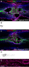
Macrophages typically induce axonal retraction to the caudal end of the lesion which contains GFAP+ and vimentin+ cells by 28 d postlesion. A, Typical outcome of DCC 28 d after injury. Fibers (red) have retracted a considerable distance and rest on GFAP+ (blue) and vimentin+ (purple) cells. Macrophages (green) still remain in the lesion at this time point. B, Inverted grayscale image of superimposed fiber tracings of three sections from the representative animal in A. C, Atypical outcome of DCC 28 d after injury. DexTR fibers associate with vimentin+ cells and remain in the lesion core. D, Inverted grayscale image of superimposed fiber tracings of three sections from the representative animal in C. E, Higher magnification of vimentin (purple) and fibers (red) as seen in C. Scale bars: A–D, 200 μm; E, 50 μm.
Figure 4.
Figure 4.
The vimentin+ cell population expresses the progenitor markers NG2 and nestin. A, Seven days after injury, fibers associate with vimentin+ (purple), nestin+ (green), and NG2+ (blue) cells. B, Confocal image (40×) of NG2- (green) and DexTR- (red) labeled axons, higher magnification of box from C. C, DexTR fibers (red) also associate with an NG2+ (green) cell population. D, DexTR-labeled fibers associate largely with a vimentin+ (green) cell population. E, There is considerable overlap between the NG2+ (green) and vimentin+ (red) populations. F, Nestin+ (green) cells express vimentin (red). Scale bars: A, C–F, 100 μm; B, 50 μm.
Figure 5.
Figure 5.
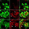
NG2+ cells are permissive to axon outgrowth and express vimentin and nestin. A, Confocal image (40×) showing the association of axons of β-tubulin+ (red) axons with NG2+ (green) cells on a gradient of aggrecan and laminin after 2 d in vitro. Arrowheads indicate NG2+ cells and arrows indicate NG2 negative satellite cells. B, NG2+ cells express vimentin (red). C, NG2+ cells express nestin (red). Scale bars: A–C, 50 μm.
Figure 6.
Figure 6.
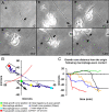
NG2+ cells can stabilize axons during macrophage-mediated axonal dieback in vitro. A, Six representative frames from a time-lapse movie illustrating macrophage/axon interactions on an aggrecan/laminin gradient in the presence of adult mouse spinal cord NG2+ cells. NR8383 macrophages are added to a 2 DIV culture of adult DRG neurons. Times for each frame are given in the lower right of each image, and an asterisk marks a consistent point on the culture dish as a reference for position during frame shifts. An arrow denotes the central domain of the grown cone. Macrophages are added following a 30 min period of observation, and first contact occurs at 103 min. The axon has already undergone a long distance retraction by 110 min and an open arrow indicates the presence of a retraction fiber. The open arrow indicates the presence of a retraction fiber. B, Graph of growth cone position for each frame (30 s) of the time-lapse movie shown in C. The red arc represents the location of the inner rim of the spot. The arrow indicates the initial trajectory of growth. C, Distance from the origin of six dystrophic axons in coculture with NG2+ cells on the aggrecan/laminin spot gradient following contact with macrophages. An arrowhead indicates the position at which the axon has retracted to an NG2 cell. Scale bar: A, 20 μm.
Figure 7.
Figure 7.
NG2+ cells can provide a bridge for regenerating axons. A, NG2+ cells (green) derived from adult mouse spinal cord can cross a proteoglycan gradient, visualized with CS56 (blue), after 5 d in vitro. Adult DRGs, visualized with β-tubulin (red), use adult NG2+ cells to bridge the inhibitory gradient. B, Mature GFAP+ astrocytes (green) do not cross the proteoglycan gradient, and adult DRGs cannot use them as a bridge. Scale bar: A, B, 20 μm.
Figure 8.
Figure 8.
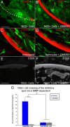
GM6001 prevents NG2+ cells from crossing the inhibitory proteoglycan rim over 5 d in vitro. A, Untreated NG2+ cells (green) 5 DIV cross the inhibitory spot rim (red), denoted by the dotted line. B, GM6001 prevents NG2+ cells (green) from crossing the spot rim, identified by CS56 (red). C, GFAP+ astrocytes (green) do not cross the spot rim (red) over a period of 5 d. D, The lack of astrocytes crossing the rim does not change upon GM6001 treatment. E, Image (16×) of the proteoglycan rim of a spot after 5 d in vitro with NG2+ cells as visualized with CS56. F, Image (16×) of the proteoglycan rim of a spot after 5 d in vitro with NG2+ cells and the broad spectrum MMP inhibitor, GM6001, taken using the same exposure time as E. G, Adult NG2+ cell crossing of the rim is MMP dependent. The Kruskal–Wallis test was used to test overall significance (χ(3)2 = 69.621, p < 0.001). The Mann–Whitney U test was used for pairwise comparisons: #p < 0.02, **p < 0.001. Scale bars: A–F, 100 μm.
Figure 9.
Figure 9.
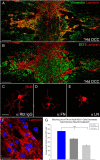
Laminin deposition is mostly associated with vimentin+ cells, and both laminin and fibronectin contribute to axonal outgrowth on NG2+ cells. The orientation of the longitudinal sections shown in A and B is such that caudal is on the left and rostral is on the right. A, Confocal montage (10×) of a lesion 14 d after dorsal column crush injury. Laminin (red) is highly expressed in the lesion core and associated with vimentin+ cells (green). B, Confocal montage (10×) of a lesion 14 d after DCC. ED1+ macrophages (green) and laminin (red). C–E, Adult sensory neurons plated on a confluent culture of adult NG2+ cells for 24 h. C, Adult DRGs visualized with β-tubulin (red) treated with anti-rabbit IgG control antibody for 24 h exhibit extensive process outgrowth. D, Adult DRGs treated with anti-fibronectin (FN) antibody exhibit slightly diminished outgrowth. E, Adult DRGs treated with anti-laminin (LN) antibody exhibit greatly diminished axonal outgrowth. F, Confocal image (40×) of NG2+ cells expressing laminin (red). G, Antibody blocking experiments reveal that the permissive nature of NG2+ cells is mediated by FN and LN. All groups are significantly different from each other (Kruskal–Wallis test, χ(2)2 = 140.790, p < 0.001, followed by Mann–Whitney U test, **p < 0.001). Scale bars: A, B, 100 μm; C–F, 50 μm.
Figure 10.
Figure 10.
Schematic representation of the proximal end of a dorsal column crush lesion 7 d after injury. GFAP+ astrocytes (blue) have pulled away from the lesion core, which is now populated by NG2+ cells (purple) and phagocytic ED1+ macrophages (green). Dorsal root ganglion neurons (red) attempt to regenerate into the lesion core. 1, Typical axon with a dystrophic growth cone that has become susceptible to macrophage attack. 2, Typical axon that has undergone macrophage-mediated retraction back to NG2+ cells and stabilized. 3, Atypical axon that has stabilized further distally within the lesion core upon a contiguous bridge of NG2+ cells. 4, Growth cone of a neuron that has been stimulated or conditioned and has been able to overcome macrophage-induced axonal dieback and extend into the lesion core on NG2+ cells.

Similar articles

Cited by

References

    1. Ben-Hur T, Ben-Yosef Y, Mizrachi-Kol R, Ben-Menachem O, Miller A. Cytokine-mediated modulation of MMPs and TIMPs in multipotential neural precursor cells. J Neuroimmunol. 2006;175:12–18. - PubMed
    1. Bergles DE, Roberts JD, Somogyi P, Jahr CE. Glutamatergic synapses on oligodendrocyte precursor cells in the hippocampus. Nature. 2000;405:187–191. - PubMed
    1. Bodega G, Suárez I, Rubio M, Fernández B. Ependyma: phylogenetic evolution of glial fibrillary acidic protein (GFAP) and vimentin expression in vertebrate spinal cord. Histochemistry. 1994;102:113–122. - PubMed
    1. Bradbury EJ, Moon LD, Popat RJ, King VR, Bennett GS, Patel PN, Fawcett JW, McMahon SB. Chondroitinase ABC promotes functional recovery after spinal cord injury. Nature. 2002;416:636–640. - PubMed
    1. Busch SA, Horn KP, Silver DJ, Silver J. Overcoming macrophage-mediated axonal dieback following CNS injury. J Neurosci. 2009;29:9967–9976. - PMC - PubMed

Publication types

MeSH terms