Comparative Study
. 2010 Jan 6;30(1):304-15.
doi: 10.1523/JNEUROSCI.4256-09.2010.

Reduction of adult hippocampal neurogenesis confers vulnerability in an animal model of cocaine addiction

Affiliations
Comparative Study

Reduction of adult hippocampal neurogenesis confers vulnerability in an animal model of cocaine addiction

Michele A Noonan et al. J Neurosci. .

Abstract

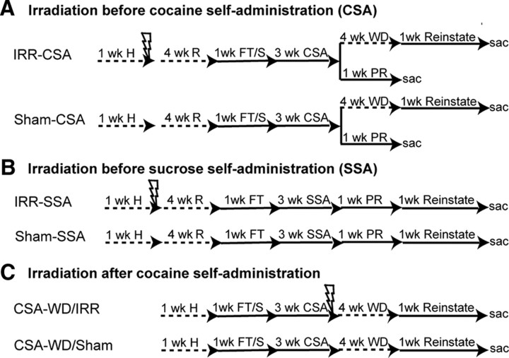
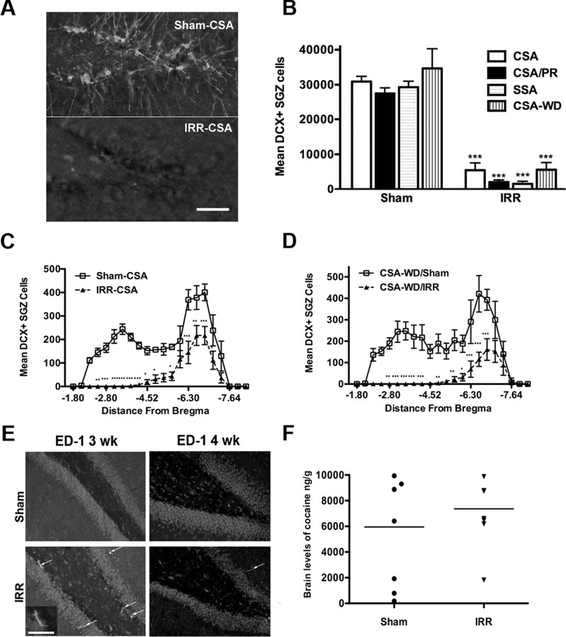
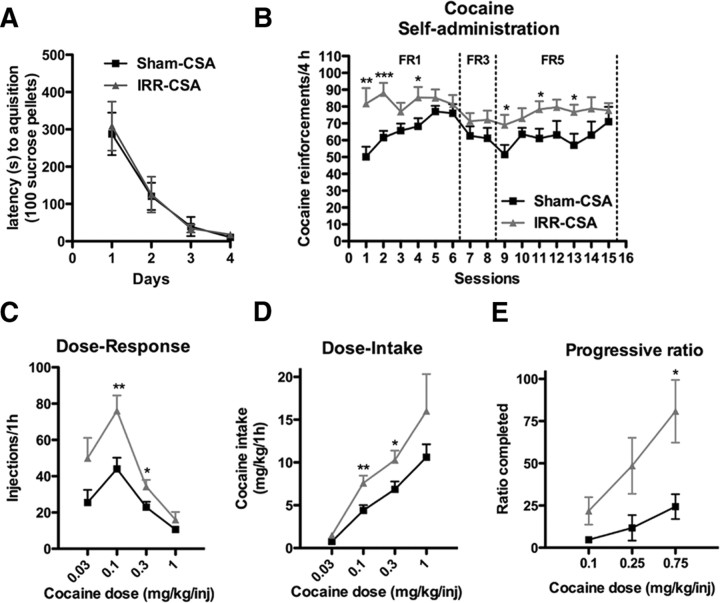
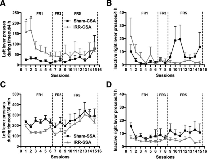
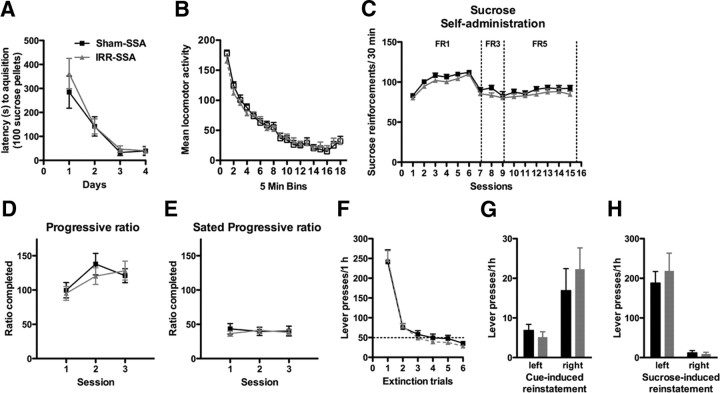
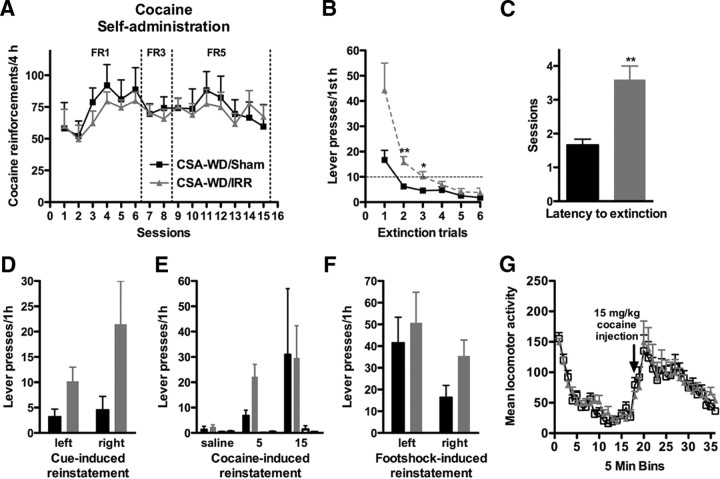
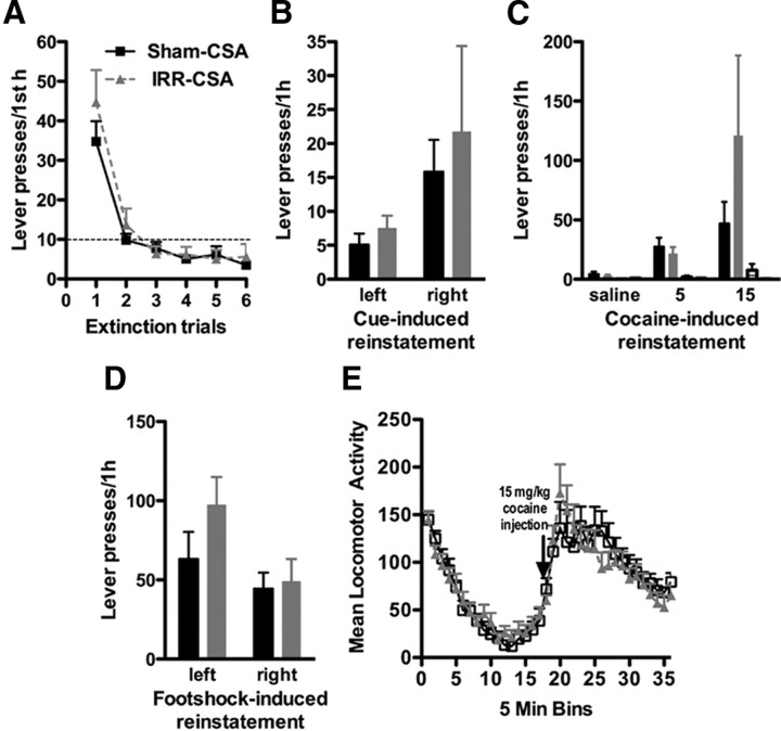
Drugs of abuse dynamically regulate adult neurogenesis, which appears important for some types of learning and memory. Interestingly, a major site of adult neurogenesis, the hippocampus, is important in the formation of drug-context associations and in the mediation of drug-taking and drug-seeking behaviors in animal models of addiction. Correlative evidence suggests an inverse relationship between hippocampal neurogenesis and drug-taking or drug-seeking behaviors, but the lack of a causative link has made the relationship between adult-generated neurons and addiction unclear. We used rat intravenous cocaine self-administration in rodents, a clinically relevant animal model of addiction, to test the hypothesis that suppression of adult hippocampal neurogenesis enhances vulnerability to addiction and relapse. Suppression of adult hippocampal neurogenesis via cranial irradiation before drug-taking significantly increased cocaine self-administration on both fixed-ratio and progressive-ratio schedules, as well as induced a vertical shift in the dose-response curve. This was not a general enhancement of learning, motivation, or locomotion, because sucrose self-administration and locomotor activity were unchanged in irradiated rats. Suppression of adult hippocampal neurogenesis after drug-taking significantly enhanced resistance to extinction of drug-seeking behavior. These studies identify reduced adult hippocampal neurogenesis as a novel risk factor for addiction-related behaviors in an animal model of cocaine addiction. Furthermore, they suggest that therapeutics to specifically increase or stabilize adult hippocampal neurogenesis could aid in preventing initial addiction as well as future relapse.


Figures

Figure 1.
Figure 1.
Cranial irradiation decreases adult neurogenesis. A, IRR-CSA (n = 21) and Sham-CSA (n = 18) rats were habituated to the animal facility for 1 week, anesthetized (50 mg/kg sodium pentobarbital, i.p.), irradiated, and left in the home cage to recover for 4 weeks (with handling every 3 d). Rats completed food training, were implanted with an intravenous catheter, and allowed to recover for several days. Three weeks of CSA followed. Rats were then split into groups for progressive-ratio testing (PR) or dose–response and reinstatement. Progressive-ratio rats had 6 d of testing and then were killed the next day (IRR, n = 7; Sham, n = 5). The remaining rats were given a dose–response test on the 16th day of CSA (IRR, n = 14; Sham, n = 13) and then were placed in the home cage for 4 weeks of WD. Daily reinstatement testing followed, which consisted of an extinction session for at least 1 h, followed by reinstatement testing to several variables as described in Materials and Methods: the context itself, the cue light, saline, intraperitoneal cocaine (5 or 15 mg/kg), and finally footshock stress. A locomotor test (basal and after a 15 mg/kg cocaine injection) was done on the last day. Rats were killed 24 h later. B, The experimental design for IRR-SSA (n = 21) and Sham-SSA (n = 18) rats was similar to A, except that rats were not implanted with an intravenous catheter after food training, and locomotor testing occurred after the last day of food training. Rats completed 3 weeks of SSA, followed by progressive-ratio for sucrose pellets while food restricted for 3 d and while fed ad libitum for 3 d. Rats were then food restricted and restabilized on FR5 timeout 15 s sucrose self-administration for 3 d. Reinstatement testing consisted of extinction of sucrose-seeking, cue-induced, and sucrose-induced reinstatement testing. C, CSA-WD/IRR (n = 15) and CSA-WD/Sham (n = 9) rats were irradiated after 3 weeks of CSA and, after 4 weeks of WD coincident with the recovery period, were then tested for reinstatement. All rats were age matched to the beginning of CSA or SSA. Scale bar, 50 μm. Lightning bolt, 2 d of 10 Gy cranial irradiation, in a 1-cm-diameter circle on the dorsal surface of the head to target the hippocampus as described previously (Snyder et al., 2005). H, Habituation; FT/S, food training and intravenous catheter surgery; r, recovery; sac, sacrifice by intracardial perfusion; Reinstate, reinstatement testing.
Figure 2.
Figure 2.
Cranial irradiation produces a long-term decrease in adult hippocampal neurogenesis. A, Representative images at 200× magnification of DCX+ immature neurons in the dorsal/anterior SGZ near the apex of the ventral and dorsal dentate gyrus limbs in Sham-CSA and IRR-CSA rats 13 weeks after irradiation. DCX+ staining was almost completely absent in dorsal dentate gyrus sections in irradiated rats. Scale bar, 50 μm. B, Quantitative analysis of DCX+ SGZ cell number at various times after cranial irradiation: CSA, 13 weeks; CSA/progressive-ratio (PR), 9 weeks; SSA, 10 weeks; CSA-WD, 5 weeks. Time line of experiments is provided in Figure 1. Regardless of time after irradiation, the number of DCX+ SGZ cells was significantly reduced relative to control rats, with almost complete ablation of DCX+ cells in the dorsal dentate gyrus (A). C, D, Quantitative analysis of DCX+ cell number in relation to distance from bregma in Sham-CSA and IRR-CSA rats (C) and CSA-WD/Sham and CSA-WD/IRR rats (D). Although DCX+ staining was almost completely absent in dorsal dentate gyrus sections (A), this analysis via distance from bregma reveals that DCX+ staining was more evident in ventral dentate gyrus sections. This likely represents the loss of radiation energy as it passed from the dorsal to ventral regions of the brain. E, Representative photomicrographs from ED1/CD-68-stained sections taken from sham and irradiated rats 3 and 4 weeks after irradiation. Arrows indicate ED1/CD-68+ cells in dentate gyrus; inset shows higher magnification of ED1/CD-68+ cell to highlight one of the characteristic ramified shape presented by activated microglia. Consistent with previous publications, this paradigm of irradiation resulted in modest microglial activation at 3 weeks. This activation was transient because only rare positive ED1/CD-68+ cells were observed at 4 weeks after irradiation and none at later time points (data not shown). Scale bar, 20 μm. F, Scatter plot of brain levels of cocaine in rats injected with 20 mg/kg cocaine intraperitoneally. There is no significant difference between the means, and no data points were statistically valid to remove as outliers based on the Grubb's test (Prism; GraphPad Software). Data for B presented as mean ± SEM. *p < 0.5, **p < 0.01, ***p < 0.001.
Figure 3.
Figure 3.
Cranial irradiation before cocaine self-administration increases cocaine reward. A, Irradiated rats did not differ from sham rats in the time it took to obtain 100 sucrose pellets during acquisition of an FR1 schedule during food training. B, Cranial irradiation 4 weeks prior increased the amount of cocaine self-administered at a 0.5 mg/kg infusion dose under both FR1 and FR5 schedules. C, Irradiated rats self-administered more cocaine, causing a vertical shift in the dose–response curve. D, Dose–response data converted into dose-intake curves by multiplying infusions by dose show irradiated rats took more significantly more cocaine. E, Irradiated rats worked harder to get their last infusion of cocaine, suggesting that they find cocaine more rewarding. Data are presented as mean ± SEM. *p < 0.05, **p < 0.01, ***p < 0.001.
Figure 4.
Figure 4.
Cranial irradiation transiently increases impulsive responding during the timeout period of cocaine self-administration but does not alter timeout pressing or inactive right lever pressing during sucrose self-administration. A, B, Left/active lever presses (A) and right/inactive lever presses (B) during the 15 s timeout per daily cocaine self-administration session. Irradiated rats pressed significantly more on the drug-paired lever during the timeout on the first two self-administration sessions but thereafter did not press significantly more than sham rats (A). Irradiated rats did not significantly press more on the inactive lever than sham rats during any session (B). C, D, Left/active lever presses (C) and right/inactive lever presses (D) during the 15 s timeout per daily sucrose self-administration session. Irradiated and sham rats pressed the same amount on the food-paired lever (C) and the inactive lever (D) during the timeout period. Data are presented as mean ± SEM. *p < 0.05.
Figure 5.
Figure 5.
Cranial irradiation before sucrose self-administration does not change natural reward. A, Irradiated rats did not differ from sham rats in the time it took to obtain 100 sucrose pellets during acquisition of an FR1 schedule during food training. B, Basal locomotion was not different between irradiated and sham rats. C, Irradiated rats were not different from sham rats in sucrose pellet self-administration on an FR1, FR3, or FR5 schedule. D, E, Sucrose pellet self-administration on a progressive ratio was not different between irradiated and sham rats when rats were food restricted during testing days (D) or when not food restricted (E). F, Irradiated rats did not differ from sham rats in their ability to extinguish responding on the formerly sucrose-paired lever. G, Irradiated rats did not differ from sham rats in responding to cues formerly paired with sucrose administration. H, Irradiated rats did not differ from sham rats in responding to presentation of sucrose pellets. Data are presented as mean ± SEM.
Figure 6.
Figure 6.
Cranial irradiation after cocaine self-administration enhances resistance to extinction. A, Self-administration of cocaine. Cocaine self-administration in each group before irradiation. Rat were then divided to make two statistically similar groups and assigned to CSA-WD/IRR or CSA-WD/Sham groups. B, C, Context-induced reinstatement. Irradiation after CSA lead to enhanced context-dependent reinstatement. Irradiated rats pressed more on the formerly drug-paired lever (B) and took more sessions to extinguish lever pressing on the formerly drug-paired lever (C) when reexposed daily to the self-administration chamber in the absence of reinforcement after 4 weeks of abstinence. D, Cue-induced reinstatement. Irradiated rats had a nonstatistical trend toward pressing both the formerly drug-paired lever and the inactive lever more than sham-irradiated rats in response to drug cues. E, Drug-induced reinstatement. Response to pressing the left, formerly drug-paired lever (leftmost pair of bars) or right, formerly drug-unpaired lever (rightmost pair of bars) after intraperitoneal injection of saline or 5 or 15 mg/kg cocaine in sham and irradiated rats. Irradiated rats had a nonstatistical trend toward pressing the formerly drug-paired left lever more in response to low-dose (5 mg/kg, i.p.) cocaine exposure. Right lever presses are presented but are under five for both irradiated and sham groups at every dose of cocaine. F, Footshock stress-induced reinstatement. Lever pressing between irradiated and sham-irradiated rats in response to footshock stress. G, Locomotion assessment. Irradiated rats were not impaired in either basal locomotion or cocaine-induced locomotion (15 mg/kg, i.p.). Data are presented as mean ± SEM. *p < 0.05, **p < 0.01.
Figure 7.
Figure 7.
Cranial irradiation before cocaine self-administration does not change relapse to drug-seeking. A, Context-induced reinstatement. Irradiated rats did not press more than sham rats on the formerly drug-paired lever when reexposed to the self-administration chamber after 4 weeks of abstinence. B, Cue-induced reinstatement. Irradiated rats did not differ from sham rats in drug-seeking in response to drug cues. C, Drug-induced reinstatement. Response to pressing the left, formerly drug-paired lever (leftmost pair of bars) or right, formerly drug-unpaired lever (rightmost pair of bars) after intraperitoneal injection of saline or 5 or 15 mg/kg cocaine in sham and irradiated rats. Irradiated rats did not differ from sham rats in drug-seeking in response to low-dose cocaine injections. Right lever presses are presented but are under five for both irradiated and sham groups at each dose of cocaine. D, Footshock stress-induced reinstatement. Lever pressing between irradiated and sham-irradiated rats in response to footshock stress was not significantly different. E, Locomotion assessment. Irradiated rats were not impaired in either basal locomotion or cocaine-induced locomotion (15 mg/kg, i.p.). Data are presented as mean ± SEM.

References

    1. Abrous DN, Adriani W, Montaron MF, Aurousseau C, Rougon G, Le Moal M, Piazza PV. Nicotine self-administration impairs hippocampal plasticity. J Neurosci. 2002;22:3656–3662. - PMC - PubMed
    1. Abrous DN, Koehl M, Le Moal M. Adult neurogenesis: from precursors to network and physiology. Physiol Rev. 2005;85:523–569. - PubMed
    1. Airan RD, Meltzer LA, Roy M, Gong Y, Chen H, Deisseroth K. High-speed imaging reveals neurophysiological links to behavior in an animal model of depression. Science. 2007;317:819–823. - PubMed
    1. Baker DA, Tran-Nguyen TL, Fuchs RA, Neisewander JL. Influence of individual differences and chronic fluoxetine treatment on cocaine-seeking behavior in rats. Psychopharmacology (Berl) 2001;155:18–26. - PubMed
    1. Borders AS, Getchell ML, Etscheidt JT, van Rooijen N, Cohen DA, Getchell TV. Macrophage depletion in the murine olfactory epithelium leads to increased neuronal death and decreased neurogenesis. J Comp Neurol. 2007;501:206–218. - PubMed

Publication types