. 2010 Mar;298(3):C530-41.
doi: 10.1152/ajpcell.00468.2009. Epub 2010 Jan 6.

Mitochondrial matrix K+ flux independent of large-conductance Ca2+-activated K+ channel opening

Affiliations

Mitochondrial matrix K+ flux independent of large-conductance Ca2+-activated K+ channel opening

Mohammed Aldakkak et al. Am J Physiol Cell Physiol. 2010 Mar.

Abstract

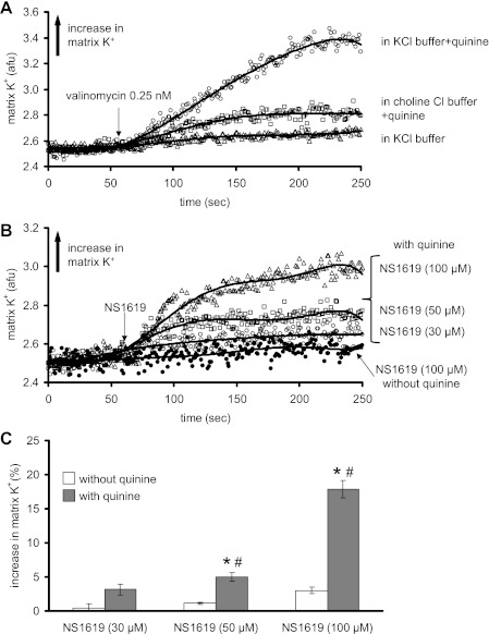
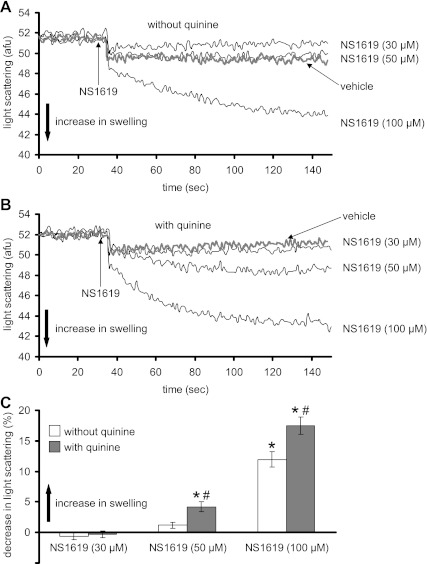
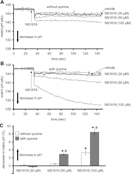
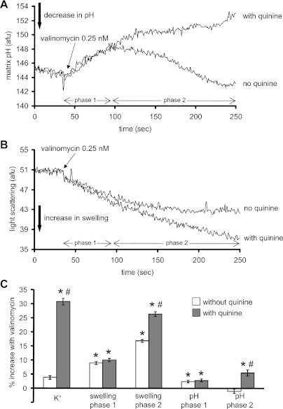
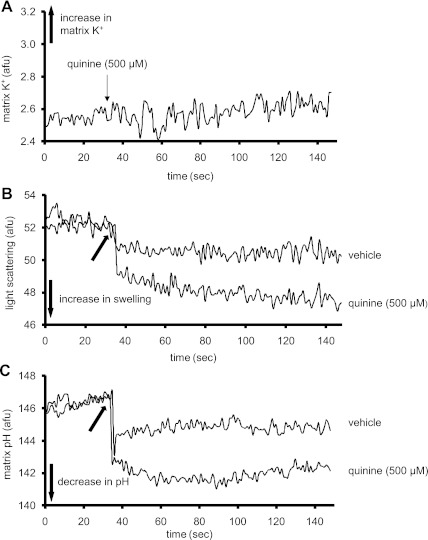
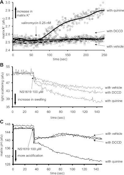
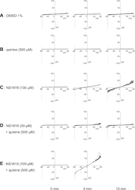
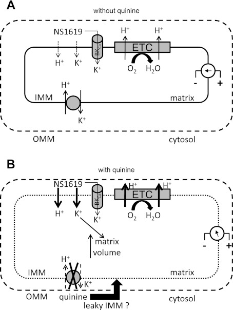
Large-conductance Ca(2+)-activated K(+) channels (BK(Ca)) in the inner mitochondrial membrane may play a role in protecting against cardiac ischemia-reperfusion injury. NS1619 (30 microM), an activator of BK(Ca) channels, was shown to increase respiration and to stimulate reactive oxygen species generation in isolated cardiac mitochondria energized with succinate. Here, we tested effects of NS1619 to alter matrix K(+), H(+), and swelling in mitochondria isolated from guinea pig hearts. We found that 30 microM NS1619 did not change matrix K(+), H(+), and swelling, but that 50 and 100 microM NS1619 caused a concentration-dependent increase in matrix K(+) influx (PBFI fluorescence) only when quinine was present to block K(+)/H(+) exchange (KHE); this was accompanied by increased mitochondrial matrix volume (light scattering). Matrix pH (BCECF fluorescence) was decreased slightly by 50 and 100 microM NS1619 but markedly more so when quinine was present. NS1619 (100 microM) caused a significant leak in lipid bilayers, and this was enhanced in the presence of quinine. The K(+) ionophore valinomycin (0.25 nM), which like NS1619 increased matrix volume and increased K(+) influx in the presence of quinine, caused matrix alkalinization followed by acidification when quinine was absent, and only alkalinization when quinine was present. If K(+) is exchanged instantly by H(+) through activated KHE, then matrix K(+) influx should stimulate H(+) influx through KHE and cause matrix acidification. Our results indicate that KHE is not activated immediately by NS1619-induced K(+) influx, that NS1619 induces matrix K(+) and H(+) influx through a nonspecific transport mechanism, and that enhancement with quinine is not due to the blocking of KHE, but to a nonspecific effect of quinine to enhance current leak by NS1619.


Figures

Fig. 1.
Fig. 1.
Time-dependent changes in mitochondrial matrix K+ (arbitrary fluorescence units, AFU) assessed using the K+ binding fluorescent indicator PBFI-AM. A: K+ influx induced by valinomycin (0.25 nM) in KCl buffer without quinine (▵), in KCl buffer with quinine present at time 0 (○), and in ChCl buffer to substitute for K+ with quinine present at time 0 (□). B: K+ influx induced by different concentrations of NS1619 in KCl buffer with quinine present at time 0 and with 100 μM NS1619 without quinine present. C: summary (n = 12) of the effects of different concentrations of NS1619 on matrix K+ influx with and without quinine present. Note that a concentration-dependent increase in K+ by NS1619 occurred only in the presence of quinine. The effect of the appropriate control (DMSO) on signals was subtracted from the background signal with NS1619 present, which makes control equal to 0. *P < 0.05, NS1619 (30, 50, 100 μM) ± quinine vs. control. #P < 0.05, NS1619 (30, 50, 100 μM) with quinine vs. NS1619 alone.
Fig. 2.
Fig. 2.
Changes in mitochondrial matrix volume (swelling) (AFU) assessed using the 90° light scattering method. A and B: matrix volume changes induced by different concentrations of NS1619 in the absence (A) or presence (B) of quinine at time 0. C: summary (n = 12) of the effects of NS1619 with and without quinine on matrix volume. Note that the concentration-dependent increase in matrix volume by NS1619 was greater in the presence of quinine. The effect of the appropriate control (DMSO) on signal was subtracted from the background signal with NS1619 present, which makes control equal to 0. *P < 0.05, NS1619 (30, 50, 100 μM) ± quinine vs. control. #P < 0.05, NS1619 (30, 50, 100 μM) with quinine vs. NS1619 alone.
Fig. 3.
Fig. 3.
Changes in matrix pH (AFU) assessed using BCECF-AM fluorescence. A and B: matrix pH changes induced by different concentrations of NS1619 in the absence (A) or presence (B) of quinine at time 0. C: summary (n = 12) of the effects of NS1619 with and without quinine on matrix pH. Note that the concentration-dependent increase in matrix acidity by NS1619 was greater in the presence of quinine. The effect of the appropriate control (DMSO) on signal was subtracted from the background signal with NS1619 present, which makes control equal to 0. *P < 0.05, NS1619 (30, 50, 100 μM) ± quinine vs. control. #P < 0.05, NS1619 (30, 50, 100 μM) with quinine vs. NS1619 alone.
Fig. 4.
Fig. 4.
Effects of valinomycin on matrix pH (A) and matrix swelling (B) in the absence or presence of quinine at time 0. C: summary (n = 5) of the effects of valinomycin on matrix K+, pH, and swelling. Note that a valinomycin-induced increase in K+ occurred only in the presence of quinine. Also note the time-dependent differential effects of valinomycin on matrix pH and volume. The effect of the appropriate control (DMSO) on signal was subtracted from the background signal with valinomycin present, which makes control equal to 0. *P < 0.05, valinomycin ± quinine vs. control. #P < 0.05, valinomycin with quinine vs. valinomycin alone.
Fig. 5.
Fig. 5.
Representative traces showing the effects of quinine (500 μM) on matrix K+ (A), matrix swelling (B), and matrix pH (C). Note that quinine caused a very small, gradual increase in matrix K+. Also quinine caused a small increase in matrix volume and a small decrease in matrix pH. These traces were reproducible in other experiments (n = 5). The arrow in B and C at 35 s indicates when vehicle or quinine was administered.
Fig. 6.
Fig. 6.
Effects of valinomycin (0.25 nM) on matrix K+ influx (A), and effects of NS1619 (100 μM) on matrix swelling (B) and matrix pH (C) in the presence of either quinine or N,N′-dicyclohexylcarbodiimide (DCCD) to inhibit K+/H+ exchange (KHE). Note that valinomycin caused a very small increase in matrix K+ influx in the presence of DCCD relative to quinine. Also, NS1619 caused more matrix swelling and more matrix acidification when quinine was used to inhibit KHE relative to DCCD. Also note that changes in matrix K+, pH, and swelling were not different when DCCD was used to block KHE relative to vehicle. These data may indicate another role for quinine in potentiating an ionic leak promoted by either valinomycin or by NS1619 at a higher concentration.
Fig. 7.
Fig. 7.
Representative traces showing the effects of quinine and NS1619 on an artificial lipid bilayer. Currents across the bilayer were recorded during a voltage ramp from −80 to +80 mV from a holding potential of 0 mV. The capacitive current is denoted by the transient inward current at the start of the ramp protocol. Recordings were obtained at 5-min intervals under the following conditions: 1% DMSO (A), 500 μM quinine (B), 100 μM NS1619 (C), 500 μM quinine + 30 μM NS1619 (D), and 500 μM quinine + 100 μM NS1619 (E). These results were reproduced in 3 separate runs.
Fig. 8.
Fig. 8.
Proposed effect of NS1619 on mitochondrial ion fluxes in the absence (A) or presence (B) of quinine to block KHE. A: low concentrations of NS1619 may induce only small K+ and H+ influxes across the inner mitochondrial membrane (IMM), which would accelerate electron flow across the electron transport chain (ETC) but maintain membrane potential gradient (ΔΨm). B: quinine blocks KHE but also induces matrix swelling which may cause the IMM to become more permeable to NS1619-induced ions fluxes (represented by thicker arrows), which will lead to even more swelling. The ΔΨm will become more depolarized and respiration will be accelerated (uncoupling). OMM, outer mitochondrial membrane.

References

    1. Armando-Hardy M, Ellory JC, Ferreira HG, Fleminger S, Lew VL. Inhibition of the calcium-induced increase in the potassium permeability of human red blood cells by quinine. J Physiol 250: 32P–33P, 1975 - PubMed
    1. Azzi A, Casey RP, Nalecz MJ. The effect of N,N′-dicyclohexylcarbodiimide on enzymes of bioenergetic relevance. Biochim Biophys Acta 768: 209–226, 1984 - PubMed
    1. Azzi A, Scarpa A. Inhibition of K+ transport in liver mitochondria. Biochim Biophys Acta 135: 1087–1088, 1967 - PubMed
    1. Azzone GF, Bortolotto F, Zanotti A. Induction of electroneutral exchanges of H+ with K+ in rat liver mitochondria. FEBS Lett 96: 135–140, 1978 - PubMed
    1. Beavis AD, Brannan RD, Garlid KD. Swelling and contraction of the mitochondrial matrix. I. A structural interpretation of the relationship between light scattering and matrix volume. J Biol Chem 260: 13424–13433, 1985 - PubMed

Publication types

MeSH terms