. 2010 Jun;51(6):3226-35.
doi: 10.1167/iovs.09-4833. Epub 2010 Jan 6.

Molecular and biochemical characterization of folate transport proteins in retinal Müller cells

Affiliations

Molecular and biochemical characterization of folate transport proteins in retinal Müller cells

B Renee Bozard et al. Invest Ophthalmol Vis Sci. 2010 Jun.

Abstract

Purpose: To analyze the mechanisms of folate uptake in retinal Müller cells.

Methods: RT-PCR and Western blot analysis were performed in freshly isolated neural retina and RPE/eyecup, primary mouse Müller cells, and rMC-1 cells for the three known folate transport proteins folate receptor alpha (FRalpha), proton-coupled folate transporter (PCFT), and reduced folate carrier (RFC). Laser scanning confocal microscopy (LSCM) and immunoelectron microscopy were used to determine the subcellular location of FRalpha and PCFT in primary Müller cells. The pH dependence of the uptake of [(3)H]-methyltetrahydrofolate ([(3)H]-MTF) was assayed in Müller cells in the presence/absence of thiamine pyrophosphate, an inhibitor of RFC.

Results: FRalpha and PCFT are expressed abundantly in the retina in several cell layers, including the inner nuclear layer; they are present in primary mouse Müller cells and rMC-1 cells. LSCM localized these proteins to the plasma membrane, nuclear membrane, and perinuclear region. Immunoelectron microscopic studies revealed the colocalization of FRalpha and PCFT on the plasma membrane and nuclear membrane and within endosomal structures. Müller cell uptake of [(3)H]-MTF was robust at pH 5.0 to 6.0, consistent with PCFT activity, but also at neutral pH, reflecting RFC function. RFC was expressed in mouse Müller cells that had been allowed to proliferate in culture, but not in freshly isolated primary cells.

Conclusions: FRalpha and PCFT are expressed in retinal Müller cells and colocalize in the endosomal compartment, suggesting that the two proteins may work coordinately to mediate folate uptake. The unexpected finding of RFC expression and activity in cultured Müller cells may reflect the upregulation of this protein under proliferative conditions.


Figures

Figure 1.
Figure 1.
FRα and PCFT expression in mouse retina. (A) Total RNA was isolated from mouse neural retina (NR) and RPE/eyecup (EC). RT-PCR analysis was performed using primers specific for FRα (expected product size, 419 bp) and PCFT (567 bp). FRα and PCFT were expressed in NR and RPE/EC. 18S (315 bp) served as an internal standard. (B) NR, RPE/EC, and intestinal (INT) protein lysates were prepared from mouse and used for Western blot analysis. FRα (∼50 kDa) and PCFT (50–64 kDa) were detected in NR and RPE/EC. PCFT harvested from mouse duodenum migrated at a molecular weight of 55 kDa. β-Actin (∼45 kDa) served as the loading control.
Figure 2.
Figure 2.
Immunodetection of FRα and PCFT in mouse retina. (A, E) Mouse retinal cryosections (harvested from 3-week-old mice) were stained with H&E to demonstrate retinal layers for comparison with cryosections subjected to immunofluorescence detection of FRα (green, B, F) and PCFT (red, C, G). (Yellowish-orange, merged image, D, H) Areas of antibody coimmunofluorescence. (I) Mouse retinal cryosections (harvested from 5-day-old mice) were stained with H&E to demonstrate retinal layers for comparison with cryosections subjected to immunofluorescence detection of FRα (green, J) and PCFT (red, K). (LN) Ganglion cells isolated by immunopanning from neonatal mouse retinas were subjected to immunocytochemistry to detect FRα and PCFT. FRα (green, L) is present in the cell body and processes extending from the cells; PCFT (red, M) is present in the cell body/nucleus of these neurons with minimal detection in processes. The merged image for these two proteins is shown in (N). Scale bars: 20 μm, (AH); 50 μm (IK); 7 μm (LN). GCL, ganglion cell layer; INL, inner nuclear layer; ONL, outer nuclear layer; OLM, outer limiting membrane; RPE, retinal pigment epithelium.
Figure 3.
Figure 3.
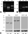
RT-PCR and Western blot analysis of FRα and PCFT in Müller cells. Total RNA was isolated from cells and subjected to RT-PCR using primers specific for mouse FRα and PCFT (419 bp and 567 bp, respectively) in primary Müller cells (A) and for rat FRα and PCFT (638 bp and 646 bp) in rMC-1 cells (B). 18S served as an internal standard. (C) Western blot analysis of FRα and PCFT in primary Müller cells (1°MC) and rMC-1 cells. FRα (∼50 kDa) was detected in both 1°MC and rMC-1 cells, as was PCFT (∼64 kDa). When membranes were incubated with PCFT antibody that had been preabsorbed using a PCFT blocking peptide (BP) and subjected to immunoblotting, no immunoreactive bands were detected. β-Actin (∼45 kDa) served as loading control.
Figure 4.
Figure 4.
Laser-scanning confocal microscopic immunolocalization of FRα and PCFT in primary Müller cells. Müller cells that were freshly isolated from mouse retina were grown on coverslips and subjected to immunofluorescent detection of FRα (red, A) and vimentin (green, B) or PCFT (red, D) and vimentin (green, E). Areas of colocalization of the folate transport proteins with vimentin are shown (merged images, orange-red, C, F). Immunodetection of FRα (green, G), PCFT (red, H), and coimmunolocalization (merged image, orange, I). Detection of the nuclear marker lamin A (green, J), PCFT (red, K), and coimmunolocalization (merged image, orange, L) and the ER marker PDI (green, M), PCFT (red, N), and coimmunolocalization (merged image, orange, O).
Figure 5.
Figure 5.
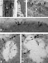
Electron microscopic immunolocalization of FRα and PCFT in Müller cells. Müller cells were fixed, and postembedding electron microscopy immunolocalization was used to detect PCFT (10-nm gold particle, indented arrows) and FRα (18-nm gold particle, flat arrows). (A) Electron microscopic photomicrograph (lower magnification) of a Müller cell. The nucleus (N) is prominent in the cell. e, Endosomes forming along the plasma membrane (pm). (B) PCFT immunolabeling on the plasma membrane. (C) PCFT and FRα immunolabeling along the nuclear membrane. (D) PCFT and FRα immunolabeling along the plasma membrane. (E, F) Immunodetection of PCFT and FRα in endosomes; note several areas in which the two proteins are in proximity.
Figure 6.
Figure 6.
pH profile for 5-MTHF uptake in primary Müller cells. Extravesicular pH was varied from 5 to 8, and uptake was measured for 15 minutes at 37°C. Uptake of [3H]-5-MTHF (40 nM) was measured in the absence (A) and presence (B) of 400 μM TPP in primary mouse Müller cells. Results are mean ± SE of three determinations from at least two experiments.
Figure 7.
Figure 7.
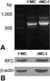
RT-PCR and Western blot analysis of RFC in Müller cells. (A) Total RNA was isolated from cells and subjected to RT-PCR using primers specific for mouse RFC (626 bp) in primary Müller cells and for rat RFC (724 bp) in rMC-1 cells. (B) Western blot analysis of RFC in primary Müller cells (1°MC) and rMC-1 cells. RFC (∼60 kDa) was detected in both 1°MC and rMC-1 cells. β-Actin (∼45 kDa) served as loading control.
Figure 8.
Figure 8.
Immunocytochemical and Western blot analysis of FRα and RFC in freshly isolated retinal Müller cells. (A) Phase-contrast microscopic analysis of primary Müller cells in culture. (B) Lower magnification of the cells shown in (A). Cells were processed for immunofluorescence detection of (C) CRALBP (red), (D) vimentin (green), (E) NF-L (green, negligible in these cells), (F) FRα (green), and (G) RFC (green, negligible in these cells). In all immunofluorescence experiments, DAPI was used to stain the nuclei of the cells (blue). (H) Primary Müller cells, primary ganglion cells, and RPE/eyecup protein lysates were prepared from mouse and used to perform Western blot analysis. RFC (∼65 kDa) was detected in RPE, which served as a positive control. RFC was not expressed in the primary Müller cells (1°MC) or primary ganglion cells (1°GC). β-Actin (∼45 kDa) served as the loading control.

Similar articles

Cited by

References

    1. Knox DL, Chen MF, Guilarte TR, Dang CV, Burnette J. Nutritional amblyopia: folic acid, vitamin B-12, and other vitamins. Retina 1982;2:288–293 - PubMed
    1. Smiddy WE, Green WR. Nutritional amblyopia: a histopathologic study with retrospective clinical correlation. Graefe's Arch Clin Exp Ophthalmol 1987;225:321–324 - PubMed
    1. Golnik KC, Schaible ER. Folate-responsive optic neuropathy. J Neurophthalmol 1994;14:163–169 - PubMed
    1. Miller NR. The optic nerve. Curr Opin Neurol 1996;9:5–15 - PubMed
    1. Sadun A, Rubin R. Residual psychophysical deficits following recovery from the Cuban epidemic of optic neuropathy. In: Lakshminarayan V. ed. Basic and Clinical Applications of Vision Science Dordrecht: Kluwer; 1997:231–234

Publication types

MeSH terms