. 2010 Mar 19;285(12):9018-29.
doi: 10.1074/jbc.M109.065698. Epub 2010 Jan 6.

Flexible recognition of the tRNA G18 methylation target site by TrmH methyltransferase through first binding and induced fit processes

Affiliations

Flexible recognition of the tRNA G18 methylation target site by TrmH methyltransferase through first binding and induced fit processes

Anna Ochi et al. J Biol Chem. .

Abstract

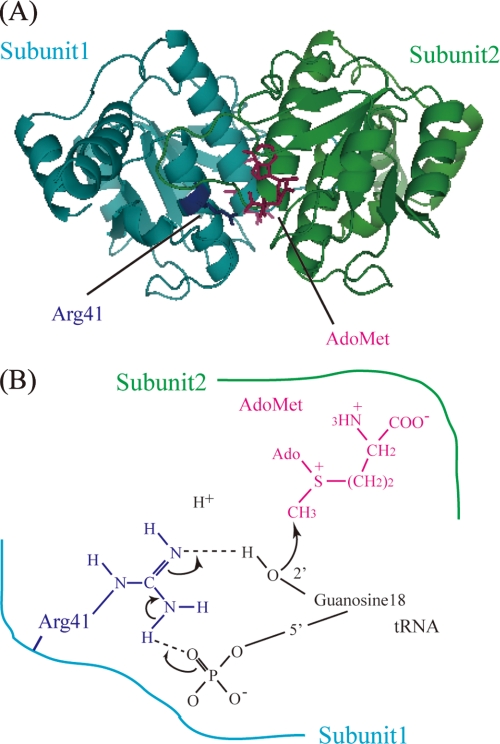
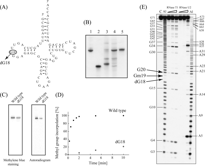
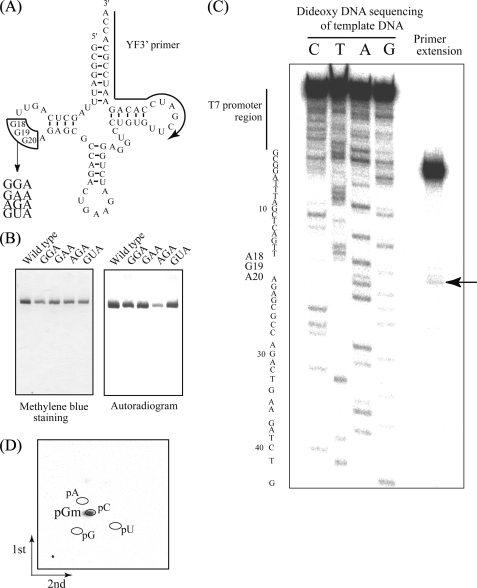
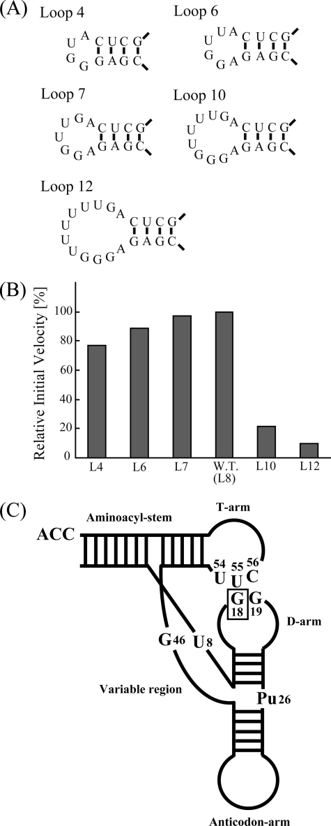
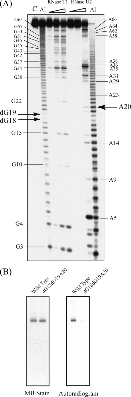
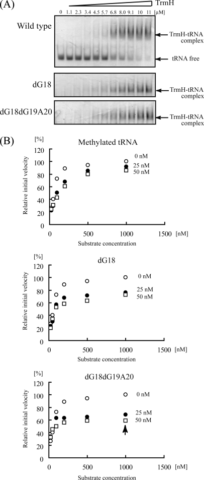
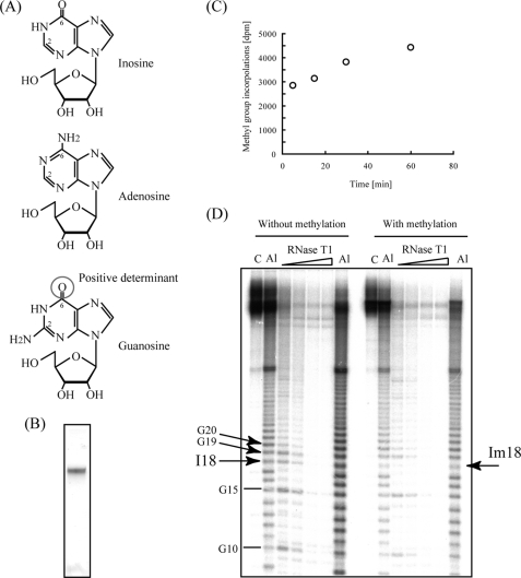
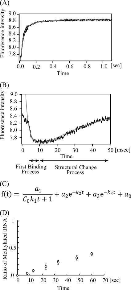
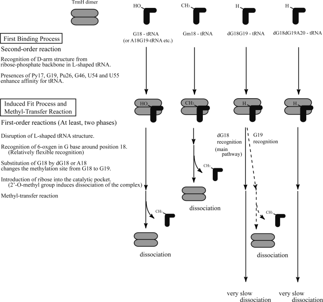
Transfer RNA (Gm18) methyltransferase (TrmH) catalyzes methyl transfer from S-adenosyl-l-methionine to a conserved G18 in tRNA. We investigated the recognition mechanism of Thermus thermophilus TrmH for its guanosine target. Thirteen yeast tRNA(Phe) mutant transcripts were prepared in which the modification site and/or other nucleotides in the D-loop were substituted by dG, inosine, or other nucleotides. We then conducted methyl transfer kinetic studies, gel shift assays, and inhibition experiments using these tRNA variants. Sites of methylation were confirmed with RNA sequencing or primer extension. Although the G18G19 sequence is not essential for methylation by TrmH, disruption of G18G19 severely reduces the efficiency of methyl transfer. There is strict recognition of guanosine by TrmH, in that methylation occurs at the adjacent G19 when the G18 is replaced by dG or adenosine. The fact that TrmH methylates guanosine in D-loops from 4 to 12 nucleotides in length suggests that selection of the position of guanosine within the D-loop is relatively flexible. Our studies also demonstrate that the oxygen 6 atom of the guanine base is a positive determinant for TrmH recognition. The recognition process of TrmH for substrate is inducible and product-inhibited, in that tRNAs containing Gm18 are excluded by TrmH. In contrast, substitution of G18 with dG18 results in the formation of a more stable TrmH-tRNA complex. To address the mechanism, we performed the stopped-flow pre-steady state kinetic analysis. The result clearly showed that the binding of TrmH to tRNA is composed of at least three steps, the first bi-molecular binding and the subsequent two uni-molecular induced-fit processes.


Figures

FIGURE 1.
FIGURE 1.
A, crystal structure of TrmH dimer. Subunit 1 (cyan) acts as the catalytic subunit, although subunit 2 (green) acts as an AdoMet-binding subunit. The catalytic center Arg-41 (dark blue) in subunit 1 and AdoMet (red) in subunit 2 are illustrated using ball-and-stick representations. B, schematic drawing of the proposed catalytic mechanism of TrmH. For binding substrate tRNA, the Arg-41 residue in subunit 1 must come in close proximity to the 5′-phosphate of G18, whereupon the negative charge of the phosphate attracts the guanidino group of Arg-41. Activated Arg-41 displaces or excludes the 2′-OH proton of G18 ribose, and methyl transfer then occurs by nucleophilic attack of the deprotonated oxygen atom of the ribose against the methyl group in AdoMet. The detached proton is depicted in the figure.
FIGURE 2.
FIGURE 2.
Methylation of a yeast tRNAPhe-dG18 variant. A, cloverleaf structure of wild-type yeast tRNAPhe, with the substitution site G18 enclosed by a circle. B, preparation of yeast tRNAPhe-dG18. RNA fragments were analyzed by electrophoresis in 10% polyacrylamide gels (7 m urea) and then stained with methylene blue. Lane 1, wild-type tRNAPhe (control); lane 2, 5′-half-fragment containing dG18; lane 3, 3′-half-fragment; lane 4, ligation sample of 5′- and 3′-half-fragments; lane 5, purified tRNAPhe-dG18. C, methylation of tRNA-dG18 monitored by 10% PAGE (7 m urea). TrmH was pre-incubated with [14C]AdoMet and then with either wild-type tRNAPhe (left lanes) or tRNAPhe-dG18 (right lanes) prior to analysis by 10% PAGE (7 m urea). The gel was stained with methylene blue (left panel), and an autoradiographic image of the same gel was then captured using a Fuji BAS2000 imaging analyzer (right panel). D, time course experiment of tRNA-dG18 methylation (performed under the same conditions as shown in C). Filled and open circles indicate rates of methylation of wild-type tRNAPhe and tRNA-dG18, respectively. E, identification of the tRNA-dG18 methylation site. Analyses using RNase T1 partial digestion and alkaline-limited hydrolysis reactions are described under “Experimental Procedures.” Lane C, untreated control RNA; lane Al (left), alkaline hydrolysis (95 °C, 2 min); lane RNase T1 included 0.03, 0.1, or 0.3 units of enzyme; lanes RNase U2 included 0.01, 0.03, or 0.1 units of enzyme; lane Al (right), alkaline hydrolysis (95 °C, 4 min). Positions of G and A nucleotides are marked on both sides of the gel, and those of dG18, Gm19, and G20 are indicated with arrows.
FIGURE 3.
FIGURE 3.
A, substitution sites (G18G19G20) and replaced sequences depicted on a tRNAPhe clover-leaf schematic. The substitution site (G18G19G20) is enclosed by lines, and the region complementary to the primer used for extension (C) is marked with an arrow. B, methylation of tRNAPhe variants. Methyl group acceptance activities of tRNAPhe variants GGA, GAA, AGA, and GUA were analyzed using the same method as Fig. 2C. C, primer extension to locate sites of modification. Following methylation of tRNAPhe AGA using TrmH and AdoMet, primer extension was performed as described under “Experimental Procedures.” To generate the DNA sequence ladder on the left, dideoxy DNA sequencing was performed using the same 5′-32P-labeled primer. The terminated band (marked with an arrow) shows 2′-O-methylation at G19. D, two-dimensional thin layer chromatography analysis of the modified nucleotide. The tRNA AGA variant was methylated using TrmH and [14C]AdoMet, digested with nuclease P1, and then separated on two-dimensional thin layer chromatography plates prior to autoradiography.
FIGURE 4.
FIGURE 4.
Methylation of tRNA D-loop size variants. A, sequence and structure of the D-loop regions (4, 6, 7, 10 and 12 nucleotides in length) in five tRNA variants. B, relative initial velocities for methylation of tRNA D-loop size variants. All tRNAs were methylated, although variants containing 10 and 12 nucleotide D-loops were modified at a significantly slower velocity relative to the wild-type (W.T.) yeast tRNAPhe. C, model of TrmH recognition elements within structured tRNA. In an earlier study (7), we presented a model in which TrmH recognizes the D-arm structure and conserved nucleotides (U8, Pu26, G46, U54, U55, and C56) in tRNA that affect methylation efficiency. The enzyme recognizes the modification site G18 by its position relative to the ribose phosphate backbones in the three-dimensional core of tRNA.
FIGURE 5.
FIGURE 5.
tRNA-dG18dG19A20 variant was not methylated by TrmH. A, RNA sequencing of tRNA-dG18dG19A20 variant using the Donis-Keller method. Lane C, untreated control RNA; lane Al (left), alkaline hydrolysis (95 °C, 2 min); lanes RNase T1 included 0.03, 0.1, or 0.3 units of enzyme; lanes RNase U2 included 0.01, 0.03, or 0.1 units of enzyme; Lane Al (right), alkaline hydrolysis (95 °C, 4 min). Positions of G and A nucleotides are marked on both sides of the gel, and those of dG18, Gm19, and G20 are indicated with arrows. B, elimination of the methyl group acceptance activity by substitution of G18G19G20 by dG18dG19A20. Methylation of tRNA-dG18 monitored by 10% PAGE (7 m urea). TrmH was pre-incubated with [14C]AdoMet, and then with either wild-type tRNAPhe (left lanes) or tRNAPhe-dG18dG19A20 (right lanes) prior to analysis by 10% PAGE (7 m urea). The gel was stained with methylene blue (MB, left panel), and an autoradiographic image of the same gel was then captured using a Fuji BAS2000 imaging analyzer (right panel).
FIGURE 6.
FIGURE 6.
Gel shift assay and inhibition experiments with tRNA-dG18 and -dG18dG19A20 variants. A, steady-state binding of TrmH to wild-type tRNAPhe (upper panel), tRNA-dG18 (middle panel), and tRNA-dG18dG19A20 (lower panel) variants. Transcripts incubated with increasing concentrations of TrmH protein in a binding reaction were then resolved by 6% native PAGE. Gels were stained sequentially with Coomassie Brilliant Blue and methylene blue for visualization of protein and RNA, respectively. B, inhibition of methyl transfer to wild-type tRNA in the presence of methylated tRNAs containing Gm18 (upper panel), tRNA-dG18 (middle panel), or tRNA-dG18dG19A20 (lower panel). The concentration of TrmH was fixed at 80 nm, whereas the concentrations of the tRNA variant heading each graph are 0 nm (open circles), 25 nm (closed circles), and 50 nm (open squares).
FIGURE 7.
FIGURE 7.
Catalytic pocket of TrmH. Structure of a TrmH dimer represented by a space-filling model (left). Conserved amino acid residues in the catalytic pocket include Arg-41 (dark blue), Ser-150 (yellow), and AdoMet (red) in the catalytic pocket (right).
FIGURE 8.
FIGURE 8.
Substitution of G18 by inosine. A, structural representations of inosine (upper panel), adenosine (middle panel), and guanosine (lower panel). The oxygen 6 in guanosine is enclosed by a circle. B, purified tRNA-I18 analyzed by 10% PAGE (7 m urea) and stained with methylene blue. C, time-dependent increase in steady-state levels of methyl group incorporation into tRNA-I18. The time course experiment showed that more than 70% of the variant was methylated within 5 min. D, determination of the modification site of the tRNA-I18 variant using the Donis-Keller method. Unmodified RNA was cleaved by RNase T1 at I18 (left), whereas alkali and RNase T1 ladder rungs corresponding to cleavage at I18 disappeared following methylation by TrmH (right). Amounts of RNase T1 were 0.03, 0.05, 0.07, and 0.1 units per one sample (from left to right). Positions of nucleotides I18, G19, G20, and Im18 are marked with arrows.
FIGURE 9.
FIGURE 9.
Pre-steady state kinetic study by stopped-flow fluorescence measurements. Changes of fluorescence intensity derived from tryptophan residues in TrmH with tRNA binding were monitored (A). This figure shows the average of 20 scans of 7.7 μm TrmH and tRNA complex formation. The first 50 ms is highlighted (B). The dead time of the measurement was 3 ms. The data could be fitted by an equation (C) using IGOR Pro 6.10 (WaveMetrics, Inc.). The abbreviations used are as follows: a0, the fluorescence intensity when t = ∞; a1a3, each amplitude; C0, the initial concentration of TrmH and tRNA (m); k1, second-order rate constant (m−1s−1); k2k3, each first-order rate constant (s−1); t, time (s). The theoretical fitting curves are indicated in gray (A and B). The calculated parameters are given in Table 2. D, ratio of the methylated tRNA was monitored by filter assay with [3H]AdoMet.
FIGURE 10.
FIGURE 10.
Model for TrmH recognition of wild-type and variant tRNA molecules. An outline of the initial binding process is reiterated from a previous study (7). The second process in the model, which we refer to as :induced fit,“ involves at least three steps as follows: disruption of L-shaped tRNA structure, recognition of oxygen 6 in the G base, and introduction of ribose into the TrmH catalytic pocket. Although recognition of the oxygen 6 in guanine is essential, the position of the target guanine within D-loop is somewhat flexible. tRNA methylated at G18 (Gm18) is excluded from TrmH binding in the induced fit process. Mechanistic details of the proposed model are discussed in the text.

Similar articles

Cited by

References

    1. Rozenski J., Crain P. F., McCloskey J. A. (1999) Nucleic Acids Res. 27, 196–197 - PMC - PubMed
    1. Dunin-Horkawicz S., Czerwoniec A., Gajda M. J., Feder M., Grosjean H., Bujnicki J. M. (2006) Nucleic Acids Res. 34, D145–D149 - PMC - PubMed
    1. Garcia G., R., Goodenough-Lashhua D. M. (1998) in Modification and Editing of RNA (Grosjean H., Benne R. eds) pp. 555–560, American Society for Microbiology, Washington, D. C.
    1. Robertus J. D., Ladner J. E., Finch J. T., Rhodes D., Brown R. S., Clark B. F., Klug A. (1974) Nature 250, 546–551 - PubMed
    1. Kim S. H., Sussman J. L., Suddath F. L., Quigley G. J., McPherson A., Wang A. H., Seeman N. C., Rich A. (1974) Proc. Natl. Acad. Sci. U.S.A. 71, 4970–4974 - PMC - PubMed

Publication types

LinkOut - more resources