. 2010 Mar 5;285(10):6867-78.
doi: 10.1074/jbc.M109.073064. Epub 2010 Jan 6.

A tyrosine-based motif localizes a Drosophila vesicular transporter to synaptic vesicles in vivo

Affiliations

A tyrosine-based motif localizes a Drosophila vesicular transporter to synaptic vesicles in vivo

Anna Grygoruk et al. J Biol Chem. .

Abstract

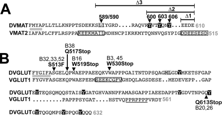
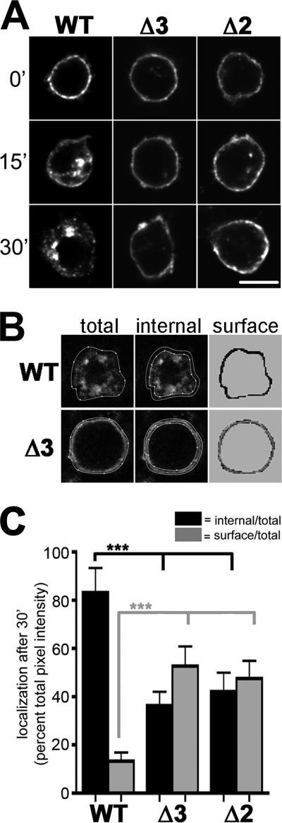
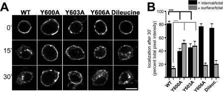
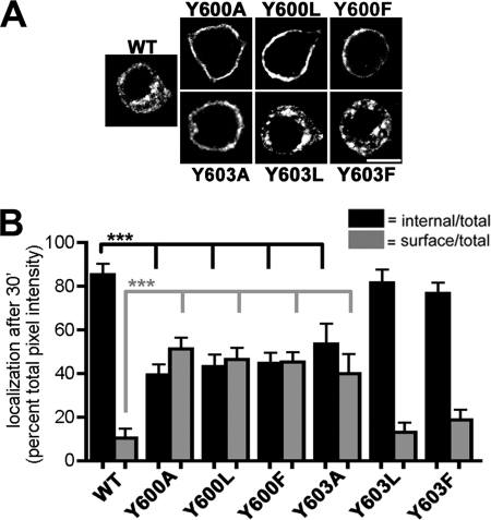
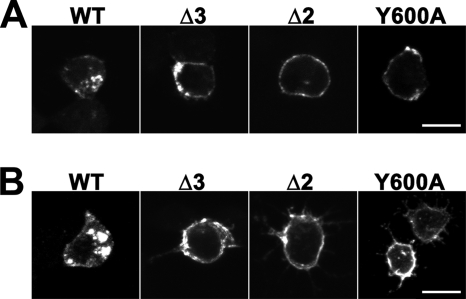
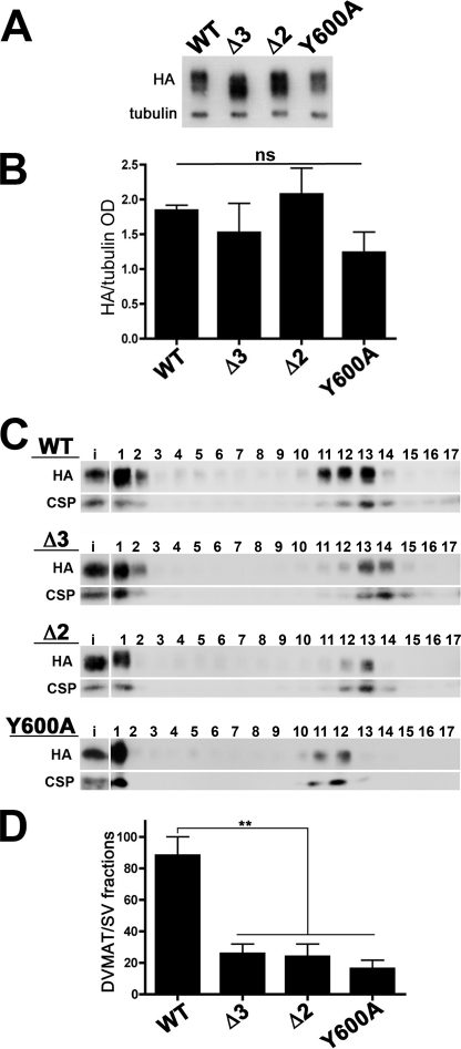
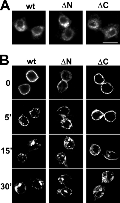
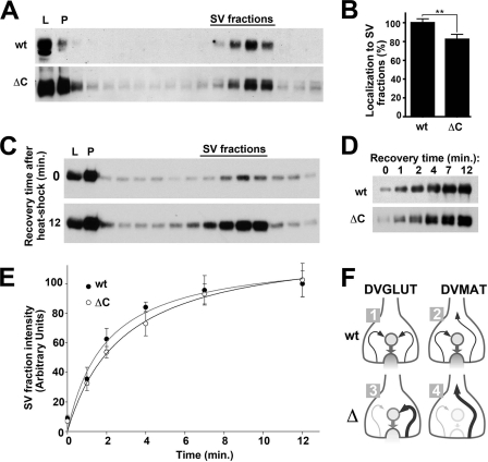
Vesicular neurotransmitter transporters must localize to synaptic vesicles (SVs) to allow regulated neurotransmitter release at the synapse. However, the signals required to localize vesicular proteins to SVs in vivo remain unclear. To address this question we have tested the effects of mutating proposed trafficking domains in Drosophila orthologs of the vesicular monoamine and glutamate transporters, DVMAT-A and DVGLUT. We show that a tyrosine-based motif (YXXY) is important both for DVMAT-A internalization from the cell surface in vitro, and localization to SVs in vivo. In contrast, DVGLUT deletion mutants that lack a putative C-terminal trafficking domain show more modest defects in both internalization in vitro and trafficking to SVs in vivo. Our data show for the first time that mutation of a specific trafficking motif can disrupt localization to SVs in vivo and suggest possible differences in the sorting of VMATs versus VGLUTs to SVs at the synapse.


Figures

FIGURE 1.
FIGURE 1.
DVMAT-A and DVGLUT C-terminal mutants. The distal end of the last transmembrane domain and terminal residues are respectively indicated with gray bars and gray numbers. A, the cytoplasmic C termini of rat VMAT2 and DVMAT-A. Boxed residues in VMAT2 indicate trafficking motifs, including a dileucine motif with upstream acidic residues (KEEKMAIL) and downstream acidic patch (DDEESESD). For DVMAT-A, we indicate the extent of three deletions (Δ1–3) and the position of point mutants, including tyrosines 600, 603, and 606 (white text, black background) and a potential dileucine motif at 589/590 (black text, gray background). B, the C terminus of DVGLUT and rat VGLUT1 are shown. Boxed residues in VGLUT1 (SEEKCGFV) indicate a proposed dileucine-like motif present in VGLUT1, -2, and -3 (19). A second signal (underlined PPRPPPP) is present in VGLUT1 but not VGLUT2 or -3. For DVGLUT, the site of mutations identified in the suppressor screen are indicated (see also Table 1). Tyrosines (white text, black background) and a pair of hydrophobic residues in DVGLUT (gray background) are highlighted as potential endocytosis motifs.
FIGURE 2.
FIGURE 2.
Endocytosis of DVMAT-A deletion mutants in cultured Drosophila S2 cells. A, S2 cells transiently transfected with dVMAT-A wt, Δ3, or Δ2 constructs were incubated in HA antibody for 1 h on ice. Cells were then either fixed immediately on ice (0′), or incubated for 15 min (15′) or 30 min (30′) at 23 °C to allow endocytosis. Wild-type DVMAT-A (wt) was largely internalized following 30-min incubation, whereas both the Δ3 and Δ2 mutants remained on the cell surface. Scale bar, 5 μm. B, to quantitate internalization, cells were manually divided into total and internal regions. These are shown enclosed by dotted white lines in representative examples of wt and Δ3 cells (left and middle panels as indicated). The surface/perimembranous region (right hand panels), was obtained by digitally masking (shown in gray) both the area outside of the cell (outside the “total” boundary) and the area within the “internal” boundary. Pixels within the remaining ring were then quantified as “surface.” To normalize for cell-to-cell variation in total cellular expression, the pixel intensity in the surface and internal regions was expressed as a ratio of either internal/total or surface/total intensity respectively (see “Experimental Procedures” for equations). Note that images used for quantitation in B were obtained at lower magnification than those shown in A and therefore appear more pixilated. C, quantitation of pixel intensity at the 30-min time point for internalized (black columns) and cell surface immunoreactivity (gray columns), both normalized for total immunoreactivity per cell. For DVMAT-A wt, 82.2 ± 4.7% was internalized (mean ± S.D., ≥77 cells from ≥3 separate experiments for each genotype). In contrast, 36.3 ± 5.9% of DVMAT-A Δ3 and 42.1 ± 7.8% of DVMAT-A Δ2 internalized. One-way ANOVA, p < 0.0001, with Bonferroni post test, p < 0.001 (black asterisks) between internal wt and both Δ2 and Δ3; one-way ANOVA, p < 0.0001, with Bonferroni post test, p < 0.001 (gray asterisks) between cell surface wt and both Δ2 and Δ3.
FIGURE 3.
FIGURE 3.
Endocytosis of DVMAT-A point mutants in S2 cells. A, endocytosis assays were performed as in Fig. 2. DVMAT-A wt, Y606A, and dileucine mutant (LI589/590AA) constructs were largely internalized following 30-min incubation, whereas both the Y600A and Y603A constructs remained mostly on the cell surface. Scale bar, 5 μm. B, quantitation of pixel intensity at the 30-min time point as in Fig. 2 shows that, for DVMAT-A wt, 81 ± 4% internalized (black columns, mean ± S.D., ≥35 cells, from ≥3 separate experiments for each genotype). Similarly, 76.6 ± 4.7% of DVMAT-A Y606A and 74.3 ± 7% of the dileucine mutant internalized. In contrast, only 39.1 ± 4.8% of DVMAT-A Y600A and 44.7 ± 6.4% of DVMAT-A Y603A internalized. One-way ANOVA, p < 0.0001; Bonferroni post test, p < 0.001 (black asterisks) between internal wt and internal Y600A and Y603A. Bonferroni post test, p < 0.001 (gray asterisks), between cell surface wt and cell surface Y600A and Y603A.
FIGURE 4.
FIGURE 4.
Endocytosis of DVMAT-A bulky group point mutants in S2 cells. A, endocytosis assays were performed as in Fig. 2. DVMAT-A wt, Y603L, and Y603F constructs largely internalized following 30-min incubation, whereas DVMAT-A Y600A, Y600L, Y600F, and Y603A constructs remained mostly on the cell surface. Scale bar, 5 μm. B, quantitation of pixel intensity at the 30-min time point as in Fig. 2 shows that 85.3 ± 5% of DVMAT-A wt internalized (black columns; mean ± S.D., ≥31 cells from ≥3 separate experiments for each genotype). In contrast, only 39.4 ± 4.8% of DVMAT-A Y600A internalized. Similarly, DVMAT-A Y600L and Y600F showed 43.2 ± 5.5% and 44.7 ± 4.9% internalization, respectively. Similar to wt, but in contrast to Y603A, which showed 53.5 ± 9.3% internalization, 81.5 ± 6.1% of Y603L and 76.7 ± 4.9% of Y603F internalized from the cell surface. One-way ANOVA, p < 0.0001; Bonferroni post test, p < 0.001 (black asterisks), between internal wt and internal Y600A, Y603A, Y600F, and Y600L. Bonferroni post test, p < 0.001 (gray asterisks) between cell surface wt and cell surface Y600A, Y603A, Y600L, and Y600F.
FIGURE 5.
FIGURE 5.
Endocytosis of wt and mutant DVMAT-A constructs in Drosophila DmBG2C6 cells. Endocytosis assays using Drosophila DmBG2C6 cells were performed as for S2 cells. DVMAT-A wt was largely internalized following 30-min incubation, whereas DVMAT-A Δ3, Δ2, and Y600A constructs remained mostly on the cell surface as shown in individual optical slices (A) and projections (B) of confocal images. Scale bars, 5 μm.
FIGURE 6.
FIGURE 6.
Glycerol velocity gradients of fly heads expressing DVMAT-A. A, comparison of fly lines expressing DVMAT-A wt, Y600A, Δ3, and Δ2 pan-neuronally using an elav-GAL4 driver (see “Experimental Procedures” for details of genotypes). Homogenates from each genotype were probed on Western blots using mAbs to HA.11 (top panel) and the Drosophila tubulin protein (bottom panel). B, three Western blots per genotype (panel A and data not shown, mean ± S.E.) were quantified and normalized to the tubulin loading controls. Expression of wt and mutant DVMAT-As is not statistically different (“ns”). C, the postnuclear homogenates from fly heads expressing DVMAT-A wt, Y600A, Δ3, or Δ2 were separated by glycerol velocity gradient centrifugation, and fractions were probed by Western blots (fraction #1 is the bottom of the gradient; fraction #17 is the top of the gradient). The mAb to the HA tag shows the position of DVMAT-A (top panel of immunoblots in C). DCSP (bottom panel of immunoblots in C) serves as a marker for SV fractions. A 1:10 dilution of the homogenate loaded onto the gradient (input or “i” here and in the text) was probed in parallel. The amounts of HA-tagged DVMAT-A and DCSP were expressed as a percentage of total input loaded onto the gradient (see “Experimental Procedures” for equations). Representative blots show that DVMAT-A wt sedimented in SV fractions to a greater extent than Δ3, Δ2 or Y600A. D, quantitation of immunoblots (n = 3 for each genotype, mean ± S.E.) shows that DVMAT-A wt was mostly found in synaptic vesicle fractions, and that the localization of DVMAT-A Y600A, Δ3, and Δ2 to synaptic vesicle fractions was reduced relative to wt. One-way ANOVA, p = 0.0009. **, Bonferroni post test, p < 0.01, for wt control versus each of the mutants.
FIGURE 7.
FIGURE 7.
Endocytosis of DVGLUT in S2 cells. S2 cells expressing DVGLUT wt and deletions of the N (ΔN) and C(ΔC) terminus. A, the steady-state expression of each construct was similar. B, internalization assays performed as described for DVMAT-A show that ΔN did not appear to differ from wt, whereas ΔC may show a slight reduction in internalization relative to wt at 5 and 15 min, but not at 30 min.
FIGURE 8.
FIGURE 8.
Localization of DVGLUT to SVs in vivo. A, steady-state localization. Homogenates from flies expressing the DVGLUT C-terminal deletion mutant were subjected to glycerol velocity gradient fractionation as described for DVMAT-A in Fig. 6. Fractions were probed on Western blots using primary antibodies to endogenous DVGLUT (wt, top) and to the GFP tag in the ΔC mutant (ΔC, bottom). B, quantitation of three independent experiments shows that the mutant localized to SVs ∼80% of wild-type levels. C, return of DVGLUT wt to SVs after endocytosis. Flies shifted to the non-permissive temperature for shits1 were assayed immediately (top panel, 0 min), or after 12-min recovery at the permissive temperature (bottom panel, 12 min). D, flies expressing DVGLUT-ΔC were assayed as in C, and the relative amount of protein localizing to SV fractions assayed as in A and C at the indicated time points. E, quantitation of D and two additional experiments show that DVGLUT wt and ΔC appeared to return to SVs at similar rates. F, model of possible differences between VMAT and VGLUT recycling at the synapse. Our data and previous studies suggest that most if not all trafficking at the synapse returns wt VGLUTs to SVs (F, 1). In contrast, we propose that a portion of VMAT may sort into a separate pathway (F, 2). In consequence, mutation of one or more internalization motifs in VMATs can shunt the protein into this pathway and away from SVs (F, 4). In contrast, because of its access to multiple pathways to SVs, disruption of any single pathway does not prevent the VGLUTs from sorting primarily to SVs (F, 3).

Similar articles

Cited by

References

    1. Régnier-Vigouroux A., Tooze S. A., Huttner W. B. (1991) EMBO J. 10, 3589–3601 - PMC - PubMed
    1. Hannah M. J., Schmidt A. A., Huttner W. B. (1999) Annu. Rev. Cell Dev. Biol. 15, 733–798 - PubMed
    1. Jahn R., Rizzoli S. O. (2007) Traffic 8, 1121–1122 - PubMed
    1. Barbosa J., Jr., Ferreira L. T., Martins-Silva C., Santos M. S., Torres G. E., Caron M. G., Gomez M. V., Ferguson S. S., Prado M. A., Prado V. F. (2002) J. Neurochem. 82, 1221–1228 - PubMed
    1. Estes P. S., Roos J., van der Bliek A., Kelly R. B., Krishnan K. S., Ramaswami M. (1996) J. Neurosci. 16, 5443–5456 - PMC - PubMed

Publication types

MeSH terms

Substances

LinkOut - more resources