. 2010 Mar 5;285(10):7312-23.
doi: 10.1074/jbc.M109.091546. Epub 2010 Jan 6.

Unique extracellular matrix heparan sulfate from the bivalve Nodipecten nodosus (Linnaeus, 1758) safely inhibits arterial thrombosis after photochemically induced endothelial lesion

Affiliations

Unique extracellular matrix heparan sulfate from the bivalve Nodipecten nodosus (Linnaeus, 1758) safely inhibits arterial thrombosis after photochemically induced endothelial lesion

Angélica M Gomes et al. J Biol Chem. .

Abstract

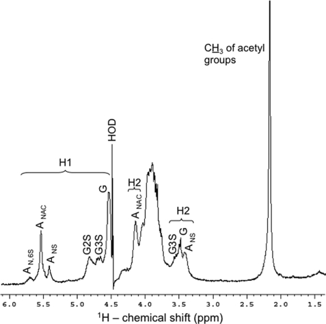
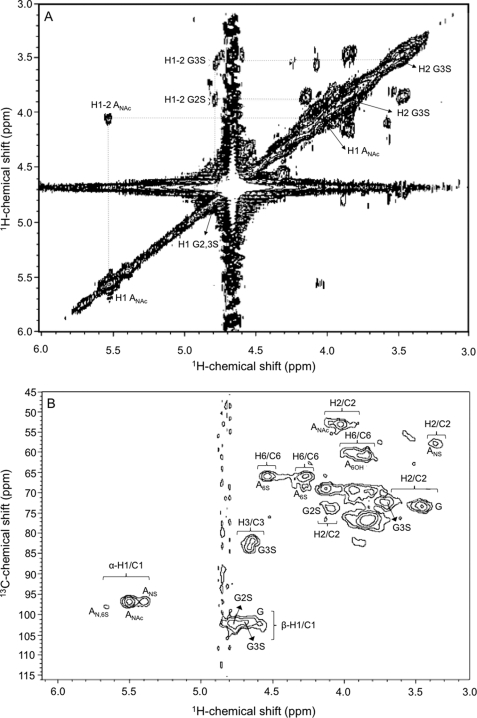
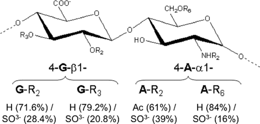
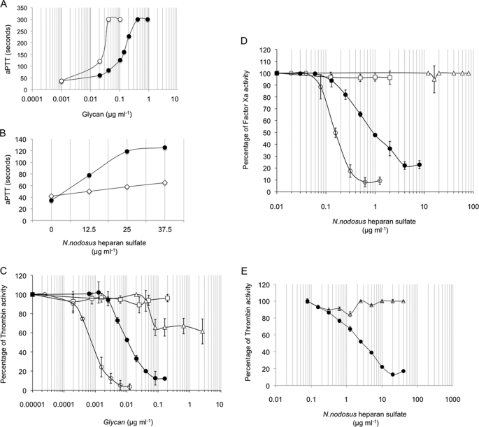
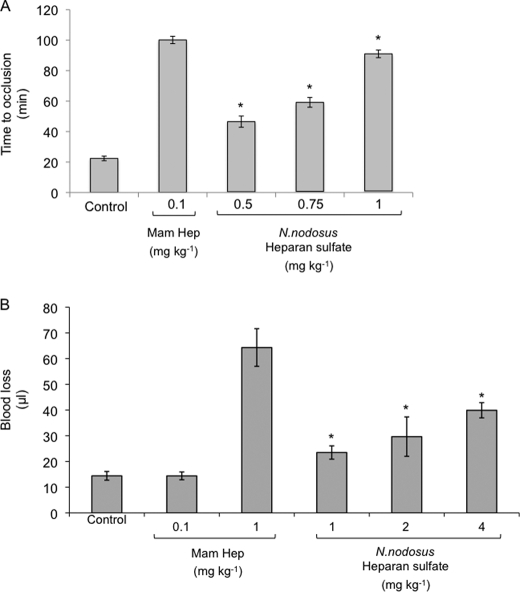
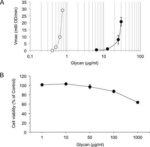
Heparin-like glycans with diverse disaccharide composition and high anticoagulant activity have been described in several families of marine mollusks. The present work focused on the structural characterization of a new heparan sulfate (HS)-like polymer isolated from the mollusk Nodipecten nodosus (Linnaeus, 1758) and on its anticoagulant and antithrombotic properties. Total glycans were extracted from the mollusk and fractionated by ethanol precipitation. The main component (>90%) was identified as HS-like glycosaminoglycan, representing approximately 4.6 mg g(-1) of dry tissue. The mollusk HS resists degradation with heparinase I but is cleaved by nitrous acid. Analysis of the mollusk glycan by one-dimensional (1)H, two-dimensional correlated spectroscopy, and heteronuclear single quantum coherence nuclear magnetic resonance revealed characteristic signals of glucuronic acid and glucosamine residues. Signals corresponding to anomeric protons of nonsulfated, 3- or 2-sulfated glucuronic acid as well as N-sulfated and/or 6-sulfated glucosamine were also observed. The mollusk HS has an anticoagulant activity of 36 IU mg(-1), 5-fold lower than porcine heparin (180 IU mg(-1)), as measured by the activated partial thromboplastin time assay. It also inhibits factor Xa (IC(50) = 0.835 microg ml(-1)) and thrombin (IC(50) = 9.3 microg ml(-1)) in the presence of antithrombin. In vivo assays demonstrated that at the dose of 1 mg kg(-1), the mollusk HS inhibited thrombus growth in photochemically injured arteries. No bleeding effect, factor XIIa-mediated kallikrein activity, or toxic effect on fibroblast cells was induced by the invertebrate HS at the antithrombotic dose.


Figures

FIGURE 1.
FIGURE 1.
Histological analysis of sections from N. nodosus stained with alcian blue. A, schematic drawing of N. nodosus showing the internal organs. The sections from the gills (A–D) and mantle (E and F) were stained with alcian blue before (B and E) or after incubation with chondroitinase AC (C) or nitrous acid treatment (D and F). The arrows indicate alcian blue staining of the basement membrane. Arrowheads indicate alcian blue staining of the basement membrane after chondroitinase AC treatment. The dashed arrows indicate alcian blue staining of the basement membrane after nitrous acid treatment. The asterisks indicate alcian blue staining at the extracellular matrix. The black stars indicate alcian blue staining at the extracellular matrix after chondroitinase AC treatment. The white stars in black circles indicate alcian blue staining at the extracellular matrix after nitrous acid treatment. Bars, 200 μm.
FIGURE 2.
FIGURE 2.
Distribution and initial characterization of glycosaminoglycans in N. nodosus. A, glycosaminoglycans were detected in the organs by the amount (mg g−1) of uronic acid, determined by the carbazole reaction (23). B, agarose gel electrophoresis of the glycans isolated from adductor muscle and viscera (pool of different organs: gills, mantle, digestive tract, and heart). C, agarose gel electrophoresis of the total glycans from the organs before (−) or after (+) incubation with chondroitinase AC (Chase AC), chondroitinase ABC (Chase ABC), keratanase, and nitrous acid. DS, dermatan sulfate.
FIGURE 3.
FIGURE 3.
Fractionation and electrophoretic analysis of the N. nodosus glycans. A, Total glycans isolated from the viscera of N. nodosus were precipitated with increasing concentrations of ethanol, 35.6% (P-1), 70% (P-2), and 95% (P-3) and analyzed by agarose gel electrophoresis. B, the purified HS obtained in the 70% precipitate (P-2) was analyzed by agarose gel electrophoresis before (−) or after (+) deaminative cleavage with nitrous acid or incubation with heparanase I. C, the purified mollusk HS was analyzed by polyacrylamide gel electrophoresis along molecular weight standard glycans: dextran 500 (S1) (average molecular weight, 500,000); chondroitin 6-sulfate from shark cartilage (S3) (average molecular weight, 54,000); chondroitin 4-sulfate from whale cartilage (S2) (average molecular weight, 36,000); and dextran 8 (S4) (average molecular weight, 8,000).
FIGURE 4.
FIGURE 4.
1H NMR spectrum of the N. nodosus HS. Approximately 5 mg of the purified sample were dissolved in 0.5 ml of D2O, and the one-dimensional NMR spectrum was recorded at 50 °C. The residual water signal was suppressed by presaturation. Chemical shifts are relative to external trimethylsilylpropionic acid at 0 ppm. The H1 signals correspond to the both α- and β-anomeric protons at δH 5.8–5.3 and δH 4.9–4.5 ppm, respectively. The H2 signals denote protons at the 2-position in the sugar rings. AN,6S, N,6-di-sulfated-d-glucosaminyl; ANAC, N-acetyl-d-glucosaminyl; ANS, N-sulfated-d-glucosaminyl; G2S, 2-sulfated-glucuronate; G3S, 3-sulfated glucuronate; G, glucuronate. The percentages (relative integral values of all α-glucosamine residues) of the AN,6s, ANAC, and ANS signals are 16:61:23, respectively. The percentages (also based on relative integral values of 1H-anomeric) of the different types of glucuronates are 50.7:28.42:20.81, respectively for G, G2S, and G3S.
FIGURE 5.
FIGURE 5.
1H/1H COSY (A) and 1H/13C HSQC (B) NMR spectra of the N. nodosus HS. Approximately 5 mg of the purified sample were dissolved in 0.5 ml of D2O, and the two-dimensional NMR spectra were recorded at 50 °C. The residual water signal was suppressed by presaturation. Chemical shifts are relative to external trimethylsilylpropionic acid at 0 ppm for 1H and relative to methanol for 0 ppm of 13C. The H1 signals correspond to the both α- and β-anomeric protons at δH 5.8–5.3 and δH 4.9–4.5 ppm, respectively. The numbers at the right side of H (A) or H/C (B) indicate the positions of the nuclei in the sugar rings. AN,6S, N,6-di-sulfated-d-glucosaminyl; ANAC, N-acetyl-d-glucosaminyl; ANS, N-sulfated-d-glucosaminyl; G2S, 2-sulfated-glucuronate; G3S, 3-sulfated glucuronate; G, glucuronate.
FIGURE 6.
FIGURE 6.
Suggested structure of the major disaccharide unit of the N. nodosus HS. The percentages of the radicals are based in integral values of the NMR signals (Fig. 3 and 4). The residues denoted with G and A are glucoronate and d-glucosamine, respectively. Rn represents the radicals, where their respective chemical groups are down listed, and where the respective subscript numbers correspond to the positions in the sugar ring.
FIGURE 7.
FIGURE 7.
Anticoagulant properties of N. nodosus HS. Measurement of the aPTT was carried according to the manufacture's specifications (Biolab-Merieux AS, Rio de Janeiro, Brazil). Normal human (A) was incubated with mammalian heparin (○) or purified N. nodosus HS (●) (0.001–100 μg ml−1) at 37 °C for 1 min. Then 100 μl of APTT reagent (Celite; Biolab) were added and incubated at 37 °C. After 2 min of incubation, 100 μl of 0.25 m CaCl2 were added to the mixtures, and the clotting time was recorded in a coagulometer (Amelung KC4A). B, normal (●) or antithrombin/heparin cofactor II-free plasma (◇) was incubated with purified N. nodosus HS (0–37.5 μg ml−1), and the clotting time recorded in a coagulometer (Amelung KC4A), as described above. C–E, inhibition of thrombin (C) or factor Xa (D) activity by AT or HCII (E) in the presence of intact (○) or nitrous acid-treated (□) mammalian or intact (●) or nitrous acid-treated (▵) N. nodosus HS. AT (50 nm) or HCII (68 nm) was incubated with thrombin (15 nm) or factor Xa (20 nm) in the presence of various concentrations of glycans. After 60 s, the remaining thrombin or factor Xa activity was determined with a chromogenic substrate (ΔA405/min). The results are expressed as percentages of thrombin activity.
FIGURE 8.
FIGURE 8.
In vivo antithrombotic and bleeding effect of N. nodosus HS. A, antithrombotic effect. Mammalian or N. nodosus HS was administered intravenously at the doses indicated. Then 15 min later, vascular injury was initiated by injection of rose bengal, and the time to thrombotic occlusion was determined. The error bars represent the means ± S.D. Five animals were used per dose. *, p ≤ 0.002 versus control (no glycan). B, bleeding effect. Mammalian or N. nodosus HS were infused into rats at the doses indicated and allowed to circulate for 5 min. The rat tail was cut 3 mm from the tip and immersed in 40 ml of distilled water at room temperature. Blood loss was determined 60 min later by measuring the hemoglobin in the water. The error bars represent the means ± S.D. Five animals were used per dose. *, p ≤ 0.002 versus control (no glycan).
FIGURE 9.
FIGURE 9.
Effect of glycans on factor XII activation and cell viability. A, factor XII activation. Normal human plasma was incubated with increasing concentrations of oversulfated chondroitin (○) or purified N. nodosus HS (●). After 60 s of incubation at 37 °C, 0.3 mm of chromogenic substrate for plasma kallikrein was added. The increase in absorbance at 405 nm was expressed by milli optical density min-1 (means ± S.E., n = 3). B, cell viability. The cells were treated with the indicated concentration of purified N. nodosus HS for 24 h. The percentage of viable cells was determined using 3-(4,5-dimethylthiazol-2-yl)-2,5-diphenyltetrazolium bromide assay. The data representative of three independent experiments are expressed as the means ± S.E.

Similar articles

Cited by

References

    1. Hirsh J., Anand S. S., Halperin J. L., Fuster V. (2001) Circulation 103, 2994–3018 - PubMed
    1. Mackman N. (2008) Nature 451, 914–918 - PMC - PubMed
    1. Lindahl U., Bäckström G., Thunberg L., Leder I. G. (1980) Proc. Natl. Acad. Sci. U.S.A. 77, 6551–6555 - PMC - PubMed
    1. Casu B., Oreste P., Torri G., Zoppetti G., Choay J., Lormeau J. C., Petitou M., Sinäy P. (1981) Biochem. J. 197, 599–609 - PMC - PubMed
    1. Choay J., Lormeau J. C., Petitou M., Sinaÿ P., Fareed J. (1981) Ann. N.Y. Acad. Sci. 370, 644–649 - PubMed

Publication types

MeSH terms