DIP2A functions as a FSTL1 receptor
- PMID: 20054002
- PMCID: PMC2844162
- DOI: 10.1074/jbc.M109.069468
DIP2A functions as a FSTL1 receptor
Abstract
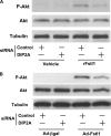
FSTL1 is an extracellular glycoprotein whose functional significance in physiological and pathological processes is incompletely understood. Recently, we have shown that FSTL1 acts as a muscle-derived secreted factor that is up-regulated by Akt activation and ischemic stress and that FSTL1 exerts favorable actions on the heart and vasculature. Here, we sought to identify the receptor that mediates the cellular actions of FSTL1. We identified DIP2A as a novel FSTL1-binding partner from the membrane fraction of endothelial cells. Co-immunoprecipitation assays revealed a direct physical interaction between FSTL1 and DIP2A. DIP2A was present on the cell surface of endothelial cells, and knockdown of DIP2A by small interfering RNA reduced the binding of FSTL1 to cells. In cultured endothelial cells, knockdown of DIP2A by small interfering RNA diminished FSTL1-stimulated survival, migration, and differentiation into network structures and inhibited FSTL1-induced Akt phosphorylation. In cultured cardiac myocytes, ablation of DIP2A reduced the protective actions of FSTL1 on hypoxia/reoxygenation-induced apoptosis and suppressed FSTL1-induced Akt phosphorylation. These data indicate that DIP2A functions as a novel receptor that mediates the cardiovascular protective effects of FSTL1.
Figures


Similar articles
-
Follistatin-like 1 attenuates apoptosis via disco-interacting protein 2 homolog A/Akt pathway after middle cerebral artery occlusion in rats.Stroke. 2014 Oct;45(10):3048-3054. doi: 10.1161/STROKEAHA.114.006092. Epub 2014 Aug 19. Stroke. 2014. PMID: 25139876 Free PMC article.
-
Blocking the FSTL1-DIP2A Axis Improves Anti-tumor Immunity.Cell Rep. 2018 Aug 14;24(7):1790-1801. doi: 10.1016/j.celrep.2018.07.043. Cell Rep. 2018. PMID: 30110636
-
Dynamic resistance exercise increases skeletal muscle-derived FSTL1 inducing cardiac angiogenesis via DIP2A-Smad2/3 in rats following myocardial infarction.J Sport Health Sci. 2021 Sep;10(5):594-603. doi: 10.1016/j.jshs.2020.11.010. Epub 2020 Nov 24. J Sport Health Sci. 2021. PMID: 33246164 Free PMC article.
-
Disco interacting protein 2 homolog A (DIP2A): A key component in the regulation of brain disorders.Biomed Pharmacother. 2023 Dec;168:115771. doi: 10.1016/j.biopha.2023.115771. Epub 2023 Oct 26. Biomed Pharmacother. 2023. PMID: 37897975 Review.
-
Follistatin-like 1 in development and human diseases.Cell Mol Life Sci. 2018 Jul;75(13):2339-2354. doi: 10.1007/s00018-018-2805-0. Epub 2018 Mar 29. Cell Mol Life Sci. 2018. PMID: 29594389 Free PMC article. Review.
Cited by
-
ERG transcriptional networks in primary acute leukemia cells implicate a role for ERG in deregulated kinase signaling.PLoS One. 2013;8(1):e52872. doi: 10.1371/journal.pone.0052872. Epub 2013 Jan 3. PLoS One. 2013. PMID: 23300998 Free PMC article.
-
Systems genetics uncover new loci containing functional gene candidates in Mycobacterium tuberculosis-infected Diversity Outbred mice.bioRxiv [Preprint]. 2023 Dec 22:2023.12.21.572738. doi: 10.1101/2023.12.21.572738. bioRxiv. 2023. Update in: PLoS Pathog. 2024 Jun 11;20(6):e1011915. doi: 10.1371/journal.ppat.1011915. PMID: 38187647 Free PMC article. Updated. Preprint.
-
DIP-2 suppresses ectopic neurite sprouting and axonal regeneration in mature neurons.J Cell Biol. 2019 Jan 7;218(1):125-133. doi: 10.1083/jcb.201804207. Epub 2018 Nov 5. J Cell Biol. 2019. PMID: 30396999 Free PMC article.
-
Acute and Chronic Increases of Circulating FSTL1 Normalize Energy Substrate Metabolism in Pacing-Induced Heart Failure.Circ Heart Fail. 2018 Jan;11(1):e004486. doi: 10.1161/CIRCHEARTFAILURE.117.004486. Circ Heart Fail. 2018. PMID: 29317401 Free PMC article.
-
Effects of FSTL1 on cell proliferation in breast cancer cell line MDA‑MB‑231 and its brain metastatic variant MDA‑MB‑231‑BR.Oncol Rep. 2017 Nov;38(5):3001-3010. doi: 10.3892/or.2017.6004. Epub 2017 Sep 26. Oncol Rep. 2017. PMID: 29048681 Free PMC article.
References
-
- Shibanuma M., Mashimo J., Mita A., Kuroki T., Nose K. (1993) Eur. J. Biochem. 217, 13–19 - PubMed
-
- Sumitomo K., Kurisaki A., Yamakawa N., Tsuchida K., Shimizu E., Sone S., Sugino H. (2000) Cancer Lett. 155, 37–46 - PubMed
-
- Chan Q. K., Ngan H. Y., Ip P. P., Liu V. W., Xue W. C., Cheung A. N. (2009) Carcinogenesis 30, 114–121 - PubMed
-
- Kawabata D., Tanaka M., Fujii T., Umehara H., Fujita Y., Yoshifuji H., Mimori T., Ozaki S. (2004) Arthritis Rheum. 50, 660–668 - PubMed
-
- Le Luduec J. B., Condamine T., Louvet C., Thebault P., Heslan J. M., Heslan M., Chiffoleau E., Cuturi M. C. (2008) Am. J. Transplant. 8, 2297–2306 - PubMed
Publication types
MeSH terms
Substances
Grants and funding
LinkOut - more resources
Full Text Sources
Other Literature Sources
Molecular Biology Databases
Miscellaneous

