

doi: 10.1107/S1744309109043814.
Epub 2009 Nov 27.
Structure of SurE protein from Aquifex aeolicus VF5 at 1.5 A resolution
Affiliations
- PMID: 20054112
- PMCID: PMC2802864
- DOI: 10.1107/S1744309109043814
Item in Clipboard
Structure of SurE protein from Aquifex aeolicus VF5 at 1.5 A resolution
Acta Crystallogr Sect F Struct Biol Cryst Commun.
.
Abstract
SurE is a stationary-phase survival protein found in bacteria, eukaryotes and archaea that exhibits a divalent-metal-ion-dependent phosphatase activity and acts as a nucleotidase and polyphosphate phosphohydrolase. The structure of the SurE protein from the hyperthermophile Aquifex aeolicus has been solved at 1.5 A resolution using molecular replacement with one dimer in the asymmetric unit and refined to an R factor of 15.6%. The crystal packing reveals that two dimers assemble to form a tetramer, although gel-filtration chromatography showed the presence of only a dimer in solution. The phosphatase active-site pocket was occupied by sulfate ions from the crystallization medium.
Figures

Crystals of A. aeolicus VF5 SurE.
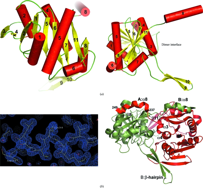
(a) Two orthogonal orientations of the AaSurE subunit showing the secondary structure and overall fold. The core globular domain consists of nine β-strands (yellow) and six helices (red cylinders), with helix α7 linking the C-terminal β-hairpin to the main body of the molecule. The dimer interface is located on the subunit edge where the β-hairpin (strands β9 and β10) and helix α8 project away from the main body of the molecule. (b) The AaSurE dimer (right) as found in the asymmetric unit, indicating the secondary-structure elements involved in forming salt bridges and/or hydrogen bonds between the two subunits A (red) and B (green). The domain-swapped α8 helices are well ordered. The quality of the electron-density map (left) is shown for A α8 at the 1σ contour level. The overall fold of AaSurE compared with the SurE dimers from other organisms give root-mean-square deviations for Cα atoms of 2.9 Å (PDB code 1j9j , the molecular-replacement search model) and 2.6 Å for 1ilv (both from Thermotoga maritima), 2.1 Å for 1l5x from P. aerophilum and 6.7 Å for 2e6e from Thermus thermophilus. This figure was drawn using Coot (Emsley & Cowtan, 2004 ▶) and PyMOL (DeLano, 2008 ▶).

(a) A tetramer of AaSurE, generated by applying symmetry operations to the dimer, is shown in two orthogonal views. The main contacts forming the tetramer interfaces occur between the β-hairpin strands of adjacent A (red) subunits and adjacent B (green) subunits. The metal-binding site that gives the enzyme its phosphatase activity is shown for each subunit, with a water molecule or Na ion (magenta sphere) occupying the position usually taken by a divalent metal ion and with a sulfate ion (red and yellow spheres) filling the active-site pocket. (b) Close-up of the active site of subunit A (red sticks) compared with the equivalent sites in SurE from Thermotoga maritima with Mg2+ (PDB entry 1j9j , green) or water (1ilv , magenta) at the metal-binding site, from Thermus thermophilus with water and a sulfate ion in the active site (2e69 , cyan) or an empty active site (2e6e , yellow) and from P. aerophilum (1l5x , blue) with water at the active site. The numbering of the active-site ligands follows the sequence of AaSurE.
Similar articles
-
Structure of D-lactate dehydrogenase from Aquifex aeolicus complexed with NAD(+) and lactic acid (or pyruvate).Acta Crystallogr Sect F Struct Biol Cryst Commun. 2009 Dec 1;65(Pt 12):1209-13. doi: 10.1107/S1744309109044935. Epub 2009 Nov 27. Acta Crystallogr Sect F Struct Biol Cryst Commun. 2009. PMID: 20054113 Free PMC article.
-
Structural and functional studies on a mesophilic stationary phase survival protein (Sur E) from Salmonella typhimurium.FEBS J. 2008 Dec;275(23):5855-64. doi: 10.1111/j.1742-4658.2008.06715.x. FEBS J. 2008. PMID: 19021761
-
Structure and function of an archaeal homolog of survival protein E (SurEalpha): an acid phosphatase with purine nucleotide specificity.J Mol Biol. 2003 Mar 7;326(5):1559-75. doi: 10.1016/s0022-2836(03)00056-1. J Mol Biol. 2003. PMID: 12595266
-
The structure of an archaeal ribose-5-phosphate isomerase from Methanocaldococcus jannaschii (MJ1603).Acta Crystallogr Sect F Struct Biol Cryst Commun. 2009 Dec 1;65(Pt 12):1214-7. doi: 10.1107/S1744309109044923. Epub 2009 Nov 27. Acta Crystallogr Sect F Struct Biol Cryst Commun. 2009. PMID: 20054114 Free PMC article.
-
Surface-Induced Dissociation: An Effective Method for Characterization of Protein Quaternary Structure.Anal Chem. 2019 Jan 2;91(1):190-209. doi: 10.1021/acs.analchem.8b05071. Epub 2018 Dec 18. Anal Chem. 2019. PMID: 30412666 Free PMC article. Review.
Cited by
-
Improved Assessment of Globularity of Protein Structures and the Ellipsoid Profile of the Biological Assemblies from the PDB.Biomolecules. 2023 Feb 17;13(2):385. doi: 10.3390/biom13020385. Biomolecules. 2023. PMID: 36830752 Free PMC article.
-
Structural and functional insights into the stationary-phase survival protein SurE, an important virulence factor of Brucella abortus.Acta Crystallogr F Struct Biol Commun. 2016 May;72(Pt 5):386-96. doi: 10.1107/S2053230X16005999. Epub 2016 Apr 22. Acta Crystallogr F Struct Biol Commun. 2016. PMID: 27139831 Free PMC article.
-
Dramatic structural changes resulting from the loss of a crucial hydrogen bond in the hinge region involved in C-terminal helix swapping in SurE: a survival protein from Salmonella typhimurium.PLoS One. 2013;8(2):e55978. doi: 10.1371/journal.pone.0055978. Epub 2013 Feb 7. PLoS One. 2013. PMID: 23409101 Free PMC article.
-
Crystallization and preliminary X-ray analysis of stationary phase survival protein E (SurE) from Xylella fastidiosa in two crystal forms.Acta Crystallogr Sect F Struct Biol Cryst Commun. 2012 Apr 1;68(Pt 4):464-7. doi: 10.1107/S1744309112007129. Epub 2012 Mar 28. Acta Crystallogr Sect F Struct Biol Cryst Commun. 2012. PMID: 22505421 Free PMC article.
References
-
- Abola, A., Bernstein, F. C., Bryant, S. H., Koetzle, T. F. & Weng, J. (1987). Crystallographic Databases – Information Content, Software Systems, Scientific Applications, edited by F. H. Allen, G. Bergerhoff & R. Sievers, pp. 107–132. Bonn/Cambridge/Chester: Data Commission of the International Union of Crystallography.
-
- DeLano, W. L. (2008). PyMOL Molecular Viewer. DeLano Scientific, Palo Alto, California, USA. http://www.pymol.org.
-
- Ellis, M. J., Antonyuk, S. & Hasnain, S. S. (2002). Acta Cryst. D58, 456–458. - PubMed
-
- Emsley, P. & Cowtan, K. (2004). Acta Cryst. D60, 2126–2132. - PubMed
-
- Huber, R. & Stetter, K. O. (2001). Methods Enzymol.330, 11–24. - PubMed
Publication types
MeSH terms
Substances
Associated data
- Actions
- Actions
LinkOut - more resources
Full Text Sources
