Review
. 2010;25(1):41-54.
doi: 10.1159/000272062. Epub 2009 Dec 22.

P63 (CKAP4) as an SP-A receptor: implications for surfactant turnover

Affiliations
Review

P63 (CKAP4) as an SP-A receptor: implications for surfactant turnover

Sandra R Bates. Cell Physiol Biochem. 2010.

Abstract

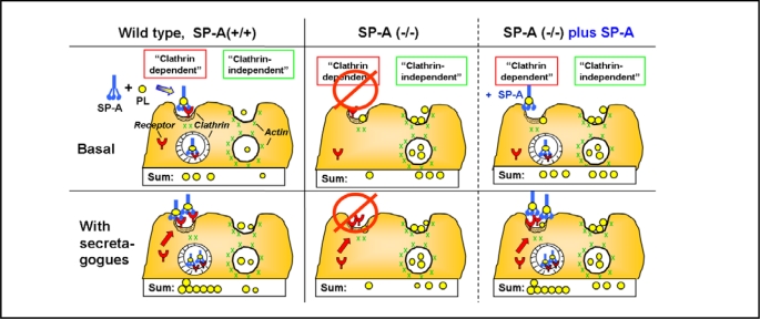
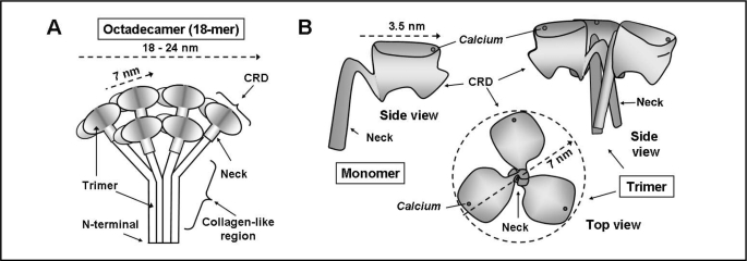
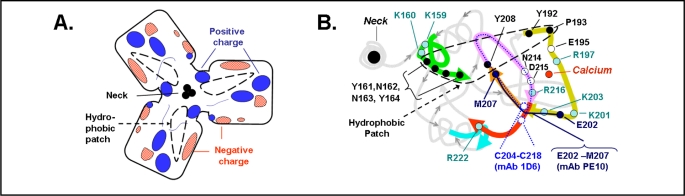
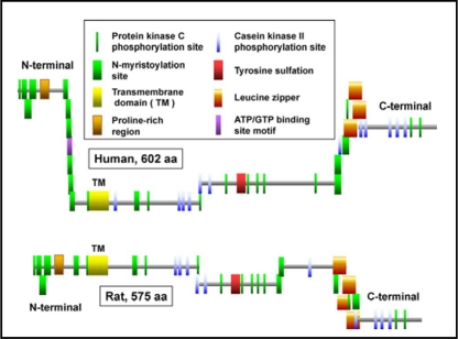
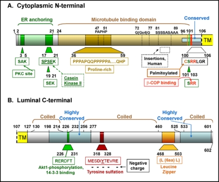
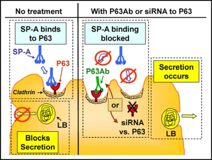
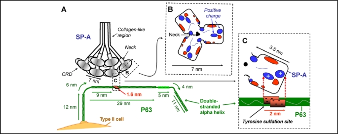
Surfactant protein-A (SP-A) plays an important role in the clearance of surfactant from the lung alveolar space and in the regulation of surfactant secretion and uptake by type II pneumocytes in culture. Two pathways are important for the endocytosis of surfactant by type II cells and the intact lung, a receptor-mediated clathrin-dependent pathway and a non-clathrin, actin-mediated pathway. The critical role of the clathrin/receptor-mediated pathway in normal mice is supported by the finding that SP-A gene-targeted mice use the actin-dependent pathway to maintain normal clearance of surfactant. Addition of SP-A to the surfactant of the SP-A null mice "rescued" the phenotype, further emphasizing the essential role of the SP-A/receptor-mediated process in surfactant turnover. This review presents an overview of the structure of SP-A and its function in surfactant turnover. The evidence that the interaction of SP-A with type II cells is a receptor-mediated process is presented. A newly identified receptor for SP-A, P63/CKAP4, is described in detail, with elucidation of the specific structural features of this 63 kDa, nonglycosylated, highly coiled, transmembrane protein. The compelling evidence that P63 functions as a receptor for SP-A on type II cells is summarized. Regulation of P63 receptor density on the surface of pneumocytes may be a novel approach for the regulation of surfactant homeostasis by the lung.


Figures

Fig. 1
Fig. 1
Pathways for lipid clearance by wild type (SP-A +/+) and SP-A gene-targeted (SP-A −/−) mice lungs. Data are adapted from reference [13]. 3H-dipalmitoyl-phosphatidylcholine labeled phospholipid (PL) liposomes (circles) were instilled into the trachea of mice and the lungs placed in an isolated perfused lung system under basal (upper panels) or secretagogue-stimulated (lower panels) conditions. Uptake of liposomes sensitive to the clathrin-inhibitor, amantadine, is “Clathrin-dependent.” Uptake of liposomes that is inhibited by cytochalasin D, an actin blocker, but not affected by amantadine is “Clathrin-independent.” (Left) SP-A, present in the surfactant of wild-type mice, associates with the instilled liposomes, while (Middle) SP-A is absent in the SP-A null mice. (Right) SP-A (−/−) plus SP-A: liposomes containing SP-A were instilled into the lungs of SP-A (−/−) mice. The addition of SP-A to the liposomes served to “rescue” the phenotype of the SP-A (−/−) mice by restoring the clathrin-mediated, secretagogue-sensitive process used by the wild type mice which had SP-A present.
Fig. 2
Fig. 2
Diagrammatic representation of the structure of SP-A. A. The octadecameric structure of the complete SP-A molecule based on Voss et al [14] and Palanyar et al. [17]. Three monomers form a collagen-like region, a neck region and a globular carbohydrate recognition domain (CRD). Six trimers join to form the “bunch of tulips” or “broccoli” shape. B. Representation of the carbohydrate-recognition domain (CRD) and neck region of SP-A based on the crystal structure from Head, et al [18]. The side view of one monomer and the side and top view of a trimer are shown. The side view of the trimer demonstrates the “T”-like shape and the top view the “boat propeller” configuration. The position of the calcium ion shown in each view is approximated from the crystal structure.
Fig. 3
Fig. 3
Details of the region on the top surface face of the carbohydrate recognition domain of SP-A. A. Diagram of the electrostatic surface charges of an SP-A trimer with calcium bound, based on the data of Head, et al [18]. The area resembles a flattened boat propeller with the location of three neck regions in the center as also depicted in Fig. 2B. The hydrophobic “patch” described by Head, et al [18] is outlined with a dashed black line. B. A string diagram representation of the surface of a monomer of SP-A taken from the RCSR Protein Data Rank Protein Workshop 3.4, protein code 1R13, using the Molecular Biology Toolkit [65] from the data of Head, et al. [18]. The string of amino acids located on the surface is shown in thick lines while the rest of the SP-A structure is in light gray. Hydrophobic amino acids in the area of the hydrophobic patch (dashed black line) are shown in black. Positively charged amino acids postulated to interact with a negatively charged receptor are filled circles. Amino acids recognized by mAb 1D6 are shown in with a dotted line and amino acids recognized by mAb PE10 are shown in with a solid line [19, 20, 21, 22]. Neck region is on the left.
Fig. 4
Fig. 4
Rule-based predicted features of P63 comparing the features of the human and mouse protein [66]. The figures are based on the diagrams of P63 from the ScanProsite web page www.expasy.ch/tools/scanprosite whose figures are under copyright and are the property of the Swiss Institute of Bioinformatics. The proteins are aligned by the transmembrane domain. The location of the transmembrane domains and the predicted area of tyrosine sulfation have been added.
Fig. 5
Fig. 5
Structural characteristics of the human P63 protein. A. The N-terminal cytoplasmic portion from amino acids 1-106. B. The C-terminal luminal portion from amino acid 107-602. The enzymes or proteins are listed and the amino acid sequence sites predicted to be involved with them are shown. The amino acid sequences that are highly conserved between human, mouse, and rat are indicated. The data are summarized from the amino acid composition, the ScanProsite predicted features shown in Fig. 4, and the data of Hauri's group [43, 47, 48, 49]. TM, transmembrane domain.
Fig. 6
Fig. 6
Effect of P63 on SP-A regulation of surfactant secretion from type II cells. (Left) Without treatment, SP-A binds to P63 and blocks surfactant release from lamellar bodies. (Right) Preventing SP-A/P63 interactions using anti P63 antibody (Ab) or reduction of P63 protein through siRNA directed against P63 allows surfactant secretion to proceed. LB, lamellar body.
Fig. 7
Fig. 7
Hypothesized interaction of SP-A and P63. Diagrams are drawn approximately to scale. A. The positively charged areas on the surface of the carbohydrate recognition domain (CRD) of SP-A is hypothesized to interact with the double stranded P63 protein at the negatively charged region of P63 predicted to contain a sulfated tyrosine. The length of the P63 luminal C-terminal was drawn as 69 nm, the predicted size of a double stranded helix of 602 amino acids [49]. Structural features of P63 are based on the amino acid composition, the ScanProsite predicted features, and the data of Hauri's group [43, 47, 48, 49] as shown in Fig. 4 and 5. P63 is drawn as a curved rod as seen in EM photomicrographs [49]. R. View of the top of the 7nm long SP-A trimer CRD region, with the location of the electrostatic surface charges shown after calcium binding [18]. Positive charged regions enlarged in diagram C are indicated. C. View of one SP-A molecule in the trimeric CRD of SP-A, estimated to be approx. 3.5 nm in length. In the P63 molecule, the area from 318 to 328 containing 5 negatively charged amino acids is estimated to be approx. 1.6 nm long forming a patch of 2 nm due to the double strands of the coiled coil of P63. The positively charged area on SP-A could interact with the negatively charged tyrosine sulfation site on P63.

Similar articles

Cited by

References

    1. Fisher AB. Lung Surfactant Clearance and Cellular Processing. In: Rooney SA, Landes RG, editors. Lung surfactant: cellular and molecular processing Medical Intelligence Unit 5. Austin, TX: Landes Bioscience; 1998. pp. 165–189.
    1. Khubchandani KR, Snyder JM. Surfactant protein A (SP-A): the alveolus and beyond. Faseb J. 2001;15:59–69. - PubMed
    1. Kuroki Y, Mason RJ, Voelker DR. Chemical modification of surfactant protein A alters high affinity binding to rat alveolar type II cells and regulation of phospholipid secretion. J Biol Chem. 1988;263:17596–17602. - PubMed
    1. Wright, Borchelt JD, Hawgood S. Lung surfactant apoprotein SP-A (26-36 kDa) binds with high affinity to isolated alveolar type II cells. Proc Natl Acad Sci U S A. 1989;86:5410–5414. - PMC - PubMed
    1. Pastva AM, Wright, Williams KL. Immunomodulatory roles of surfactant proteins A and D: implications in lung disease. Proc Am Thorac Soc. 2007;4:252–257. - PMC - PubMed

Publication types

MeSH terms

Substances