. 2010 Feb 28;42(2):122-31.
doi: 10.3858/emm.2010.42.2.014.

Intramarrow injection of beta-catenin-activated, but not naive mesenchymal stromal cells stimulates self-renewal of hematopoietic stem cells in bone marrow

Affiliations

Intramarrow injection of beta-catenin-activated, but not naive mesenchymal stromal cells stimulates self-renewal of hematopoietic stem cells in bone marrow

Ji Yeon Ahn et al. Exp Mol Med. .

Abstract

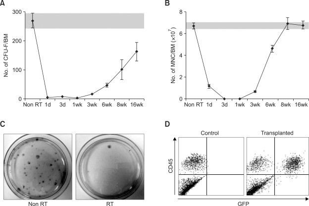
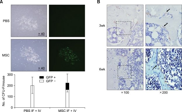
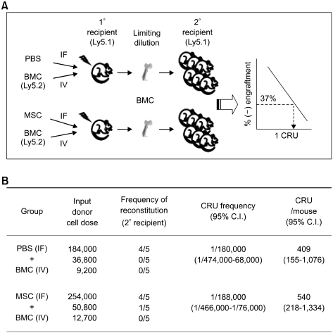
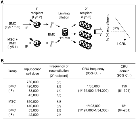
Bone marrow mesenchymal stromal cells (MSCs) have been implicated in the microenvironmental support of hematopoietic stem cells (HSCs) and often co-transplanted with HSCs to facilitate recovery of ablated bone marrows. However, the precise effect of transplanted MSCs on HSC regeneration remains unclear because the kinetics of HSC self-renewal in vivo after co-transplantation has not been monitored. In this study, we examined the effects of intrafemoral injection of MSCs on HSC self-renewal in rigorous competitive repopulating unit (CRU) assays using congenic transplantation models in which stromal progenitors (CFU-F) were ablated by irradiation. Interestingly, naïve MSCs injected into femur contributed to the reconstitution of a stromal niche in the ablated bone marrows, but did not exert a stimulatory effect on the in-vivo self-renewal of co-transplanted HSCs regardless of the transplantation methods. In contrast, HSC self-renewal was four-fold higher in bone marrows intrafemorally injected with beta-catenin-activated MSCs. These results reveal that naive MSCs lack a stimulatory effect on HSC self-renewal in-vivo and that stroma must be activated during recoveries of bone marrows. Stromal targeting of wnt/beta-catenin signals may be a strategy to activate such a stem cell niche for efficient regeneration of bone marrow HSCs.


Figures

Figure 1
Figure 1
Recoveries of CFU-F in the irradiated bone marrows. Mice were total body irradiated (900 rad) and bone marrow cells (1 × 10⁵/mice) from transgenic mice expressing GFP were intravenously injected. Bone marrows (2 femurs and 2 tibias) of recipient mice were then harvested at various time points after irradiation and aliquots of the bone marrow cells were plated for 14 days to form CFU-F. The numbers of CFU-Fs formed in the plates were measured by crystal violet staining. Shown are the mean ± SEM of CFU-F numbers (A) and total mononuclear cell (MNC) numbers (B) contained in two femurs and tibia of mice at each time point of recovery (3 independent experiments). (C) Representative image of crystal violet staining for CFU-F obtained after plating 1 × 10⁷ bone marrow cells from non-radiated and irradiated (24 h before) mice. (D) Lack of stromal contribution from intravenously transplanted bone marrows cells. CFU-Fs generated in (A) were trypsinized and passage cultured to enrich non-hematopoietic (CD45-negative) stromal cells and examined for stromal cells of donor origin (CD45-neg, GFP +). Shown are the representative flowcytometry profiles for subculture of CFU-Fs obtained from non-transplanted mice (control) and mice transplanted with GFP-transgenic mice bone marrow cells (transplanted).
Figure 2
Figure 2
MSCs intrafemorally (IF) injected into bone marrows contribute to stromal regeneration in-vivo. (A) Contribution of injected MSCs to CFU-F pool of recipients. Mice were intrafemorally injected with PBS buffer or MSCs transduced with retroviral vector expressing GFP (MPG) (1 × 10⁵ cells/ mice) and IV injected with helper cells (1 × 10⁵ bone marrow cells/ mice). Recipient bone marrow cells were harvested 14 weeks after transplantation, plated and examined for GFP-positive CFU-F colonies by fluorescent microscope. Shown are representative photographs of GFP-positive colonies under light microscopy and fluorescence microscopy (upper) and the number of GFP-positive and GFP-negative CFU-F colonies (lower) (n = 3). (B) Bone marrows of mice injected with GFP-positive MSCs were immunostained with antibody against GFP at 3 and 6 weeks after transplantation. Shown are the representative images of trabecular bone marrows, at the indicated magnifications (dotted area), as visualized by DAB (brown). Arrows indicate positive staining for GFP.
Figure 3
Figure 3
Effects of naïve MSCs on in vivo self-renewal of co-transplanted HSCs. Lethally irradiated mice (Ly5.1) were intravenously transplanted with congeneic (Ly5.2) donor bone marrow cells (BMCs) along with intrafemoral (IF) injection of MSCs or PBS buffer. Sixteen weeks after transplantation, the number of regenerated CRUs in the recipient mice was determined by limiting dilution transplantations of primary recipient mice marrow into secondary recipients. (A) Schematic illustration of the experimental design is shown. (B) The results of the CRU assays are shown. CRU frequencies and 95% confidence interval (C.I.) of the frequencies were obtained by applying Poisson statistics to the percent of negatively engrafted mice at different cell doses. The number of CRUs regenerated in mice was calculated based on the assumption that the cells in two femurs and tibias represent 25% of the total bone marrow as described (Boggs, 1984).
Figure 4
Figure 4
Direct administration of HSCs and naïve MSCs into femur as a mixture do not have stimulatory effects on the in-vivo self-renewal of HSCs. Effects of naïve MSCs on HSCs were examined by directly transplanting HSCs and MSCs as a mixture into femur. The mixture of BMC (Ly5.1) plus MSCs or BMC alone (F1 hybrid Ly5.1/5.2) were transplanted by intrafemoral (IF) injection into recipient mice. Eight weeks after transplantation, BMCs were harvested from the injected femurs of mice from each group (n = 3 for each group), mixed together in a 1:1 ratio and subjected to limiting-dilution transplantations into secondary recipients. Engraftment of each donor origin cells in the same secondary recipient mice at 13 weeks after transplantation was discerned by surface marker (Ly5.1 or Ly5.1/5.2 hybrid). The experimental design (A) and the results of the CRU analysis for the injected femurs (2 experiments) are shown (B). CRU frequencies with 95% C.I. and CRU numbers were calculated as described in Methods.
Figure 5
Figure 5
Effects of β-catenin stabilized MSCs on the in vivo self-renewal of co-transplanted HSCs. Recipient mice were intrafemorally (IF) co-injected with a mixture of BMCs (Ly5.1) and MSCs transduced with MPG control vector (MPG/MSC), or with a mixture of BMCs (F1 hybrid Ly5.1/5.2) and MSCs transduced with stable form β-catenin (β-catenin/MSCs). Eight weeks after transplantation, the total number of MNCs in the injected bone marrows and lineage differentiation of the donor-derived cells were examined. BMCs harvested from the intrafemoral-injected femurs were pooled (n = 6 for each group) and pool of each treatment group were mixed in a 1:1 ratio and subjected to a competitive limiting-dilution transplantation into secondary recipients. Engraftment of each donor origin cells in the secondary recipient mice at 12 weeks after transplantation was discerned by surface marker (Ly5.1 or Ly5.1/5.2 hybrid). (A) Schematic illustration of the experimental design is shown. (B, C) The total number of MNCs in the injected bone marrows of the primary recipient mice (B) and the lineages of the engrafted cells (C) are shown with error bars representing SEM (n = 3). (D) The results of the CRU analysis for the injected femurs are shown (2 experiments). CRU frequencies with 95% C.I. and total CRU numbers were calculated as described in Methods.

Similar articles

Cited by

References

    1. Adams GB, Scadden DT. A niche opportunity for stem cell therapeutics. Gene Ther. 2008;15:96–99. - PubMed
    1. Ball LM, Bernardo ME, Roelofs H, Lankester A, Cometa A, Egeler RM, Locatelli F, Fibbe WE. Cotransplantation of ex vivo expanded mesenchymal stem cells accelerates lymphocyte recovery and may reduce the risk of graft failure in haploidentical hematopoietic stem-cell transplantation. Blood. 2007;110:2764–2767. - PubMed
    1. Bartsch K, Al-Ali H, Reinhardt A, Franke C, Hudecek M, Kamprad M, Tschiedel S, Cross M, Niederwieser D, Gentilini C. Mesenchymal stem cells remain host-derived independent of the source of the stem-cell graft and conditioning regimen used. Transplantation. 2009;87:217–221. - PubMed
    1. Bhatia M, Bonnet D, Kapp U, Wang JC, Murdoch B, Dick JE. Quantitative analysis reveals expansion of human hematopoietic repopulating cells after short-term ex vivo culture. J Exp Med. 1997;186:619–624. - PMC - PubMed
    1. Boggs DR. The total marrow mass of the mouse: a simplified method of measurement. Am J Hematol. 1984;16:277–286. - PubMed

Publication types

MeSH terms

LinkOut - more resources