

Opposing microRNA families regulate self-renewal in mouse embryonic stem cells
- PMID: 20054295
- PMCID: PMC2894702
- DOI: 10.1038/nature08725
Opposing microRNA families regulate self-renewal in mouse embryonic stem cells
Erratum in
- Nature. 2010 Mar 4;464(7285):126
Abstract
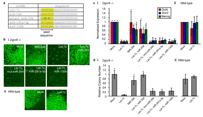
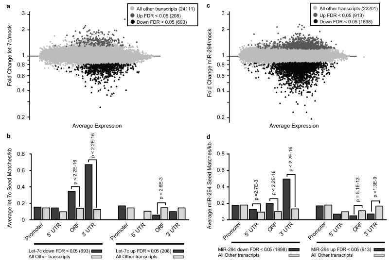
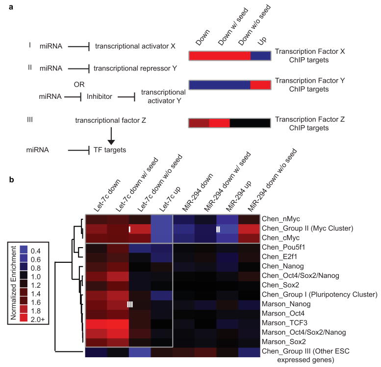
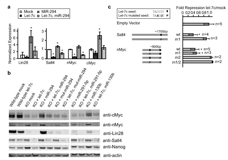
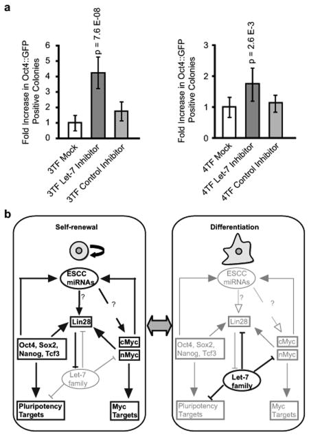
When embryonic stem cells (ESCs) differentiate, they must both silence the ESC self-renewal program and activate new tissue-specific programs. In the absence of DGCR8 (Dgcr8(-/-)), a protein required for microRNA (miRNA) biogenesis, mouse ESCs are unable to silence self-renewal. Here we show that the introduction of let-7 miRNAs-a family of miRNAs highly expressed in somatic cells-can suppress self-renewal in Dgcr8(-/-) but not wild-type ESCs. Introduction of ESC cell cycle regulating (ESCC) miRNAs into the Dgcr8(-/-) ESCs blocks the capacity of let-7 to suppress self-renewal. Profiling and bioinformatic analyses show that let-7 inhibits whereas ESCC miRNAs indirectly activate numerous self-renewal genes. Furthermore, inhibition of the let-7 family promotes de-differentiation of somatic cells to induced pluripotent stem cells. Together, these findings show how the ESCC and let-7 miRNAs act through common pathways to alternatively stabilize the self-renewing versus differentiated cell fates.
Figures
Comment in
-
Stem cells: Big roles for small RNAs.Nature. 2010 Feb 4;463(7281):616. doi: 10.1038/463616a. Nature. 2010. PMID: 20130638 No abstract available.
Similar articles
-
Reprogramming of 3' untranslated regions of mRNAs by alternative polyadenylation in generation of pluripotent stem cells from different cell types.PLoS One. 2009 Dec 23;4(12):e8419. doi: 10.1371/journal.pone.0008419. PLoS One. 2009. PMID: 20037631 Free PMC article.
-
Induced multipotency in adult keratinocytes through down-regulation of ΔNp63 or DGCR8.Proc Natl Acad Sci U S A. 2014 Feb 4;111(5):E572-81. doi: 10.1073/pnas.1319743111. Epub 2014 Jan 21. Proc Natl Acad Sci U S A. 2014. PMID: 24449888 Free PMC article.
-
Suppression of epithelial-mesenchymal transition and apoptotic pathways by miR-294/302 family synergistically blocks let-7-induced silencing of self-renewal in embryonic stem cells.Cell Death Differ. 2015 Jul;22(7):1158-69. doi: 10.1038/cdd.2014.205. Epub 2014 Dec 12. Cell Death Differ. 2015. PMID: 25501598 Free PMC article.
-
Embryonic stem cell microRNAs: defining factors in induced pluripotent (iPS) and cancer (CSC) stem cells?Curr Stem Cell Res Ther. 2009 Sep;4(3):168-77. doi: 10.2174/157488809789057400. Curr Stem Cell Res Ther. 2009. PMID: 19492978 Review.
-
microRNAs: important regulators of stem cells.Stem Cell Res Ther. 2017 May 11;8(1):110. doi: 10.1186/s13287-017-0551-0. Stem Cell Res Ther. 2017. PMID: 28494789 Free PMC article. Review.
Cited by
-
miR-302 Is Required for Timing of Neural Differentiation, Neural Tube Closure, and Embryonic Viability.Cell Rep. 2015 Aug 4;12(5):760-73. doi: 10.1016/j.celrep.2015.06.074. Epub 2015 Jul 23. Cell Rep. 2015. PMID: 26212322 Free PMC article.
-
Regulatory non-coding RNAs in pluripotent stem cells.Int J Mol Sci. 2013 Jul 11;14(7):14346-73. doi: 10.3390/ijms140714346. Int J Mol Sci. 2013. PMID: 23852015 Free PMC article. Review.
-
MiR-371-373 cluster acts as a tumor-suppressor-miR and promotes cell cycle arrest in unrestricted somatic stem cells.Tumour Biol. 2015 Sep;36(10):7765-74. doi: 10.1007/s13277-015-3519-7. Epub 2015 May 5. Tumour Biol. 2015. PMID: 25941115
-
Regulators of pluripotency and their implications in regenerative medicine.Stem Cells Cloning. 2015 Apr 21;8:67-80. doi: 10.2147/SCCAA.S80157. eCollection 2015. Stem Cells Cloning. 2015. PMID: 25960670 Free PMC article. Review.
-
Epigenetics: judge, jury and executioner of stem cell fate.Epigenetics. 2012 Aug;7(8):823-40. doi: 10.4161/epi.21141. Epub 2012 Jul 18. Epigenetics. 2012. PMID: 22805743 Free PMC article. Review.
References
-
- Hornstein E, Shomron N. Canalization of development by microRNAs. Nat Genet. 2006;38:S20–24. - PubMed
Publication types
MeSH terms
Substances
Associated data
- Actions
Grants and funding
LinkOut - more resources
Full Text Sources
Other Literature Sources
Molecular Biology Databases
Research Materials
