

Case Reports
doi: 10.1038/leu.2009.275.
Epub 2010 Jan 7.
Failure is not fatal: long-term remission in refractory acute myeloid leukemia (AML) after graft failure of cord blood stem cells
- PMID: 20054352
- PMCID: PMC10405622
- DOI: 10.1038/leu.2009.275
Item in Clipboard
Case Reports
Failure is not fatal: long-term remission in refractory acute myeloid leukemia (AML) after graft failure of cord blood stem cells
Leukemia.
2010 Mar.
No abstract available
Conflict of interest statement
Disclaimer: The authors state that they have no conflict of interest related to this article
Figures
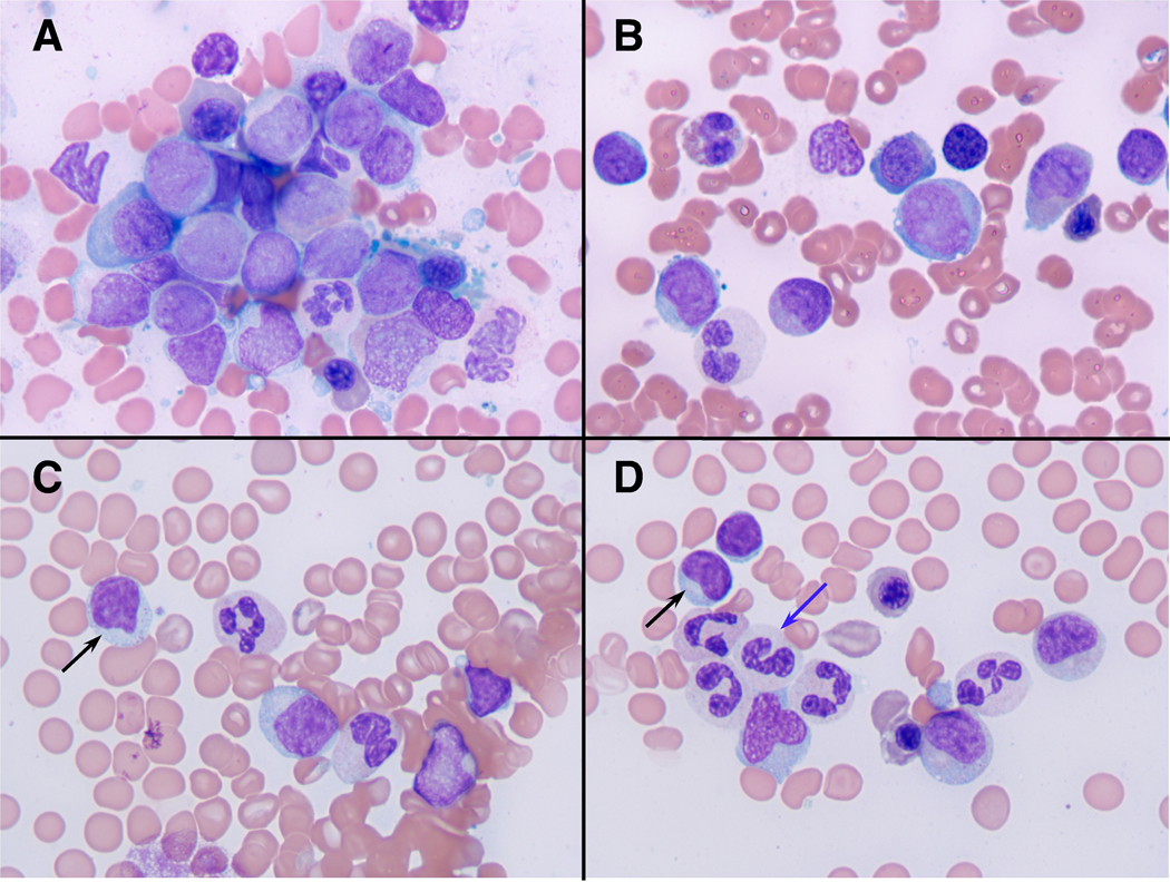
Bone marrow morphology before (A, B) and after cord blood stem cell transplantation (C, D). Fig. 1 A and B depict bone marrow aspirate smears at the initial presentation with myeloblasts in a background of dysplastic erythroid and myeloid elements (Wright-Giemsa stains, photographed at 1000x). Fig. 1 C and D display photomicrographs of bone marrow aspirate smears after CB-SCT that are consistent with a complete remission and that depict scattered large granular lymphocytes (black arrows) in a background of mildly dysplastic myeloid cells, some with hypogranular cytoplasm (blue arrow).
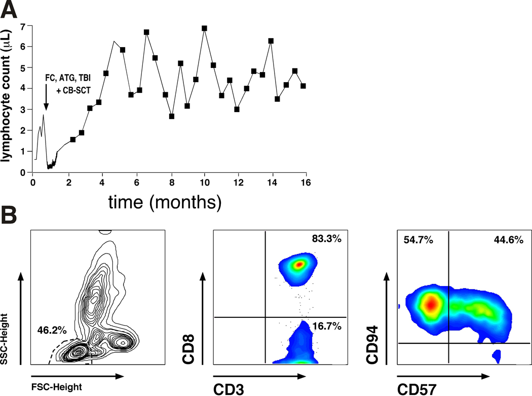
Expansion of autologous, CD8+ T cells after CB-SCT. Fig. 2A depicts the kinetics of the lymphocytosis after non-myeloablative conditioning with FC, ATG and TBI, and subsequent CB-SCT (indicated by the arrow). Fig. 2B illustrates the immunophenotype of the expanded T cells. Displayed are the forward (FSC) and sideward scatter (SSC) characteristics with gating on the lymphocyte population (left hand box), and staining with anti-CD3 and anti-CD8 mAbs, revealing that 83.3% of the lymphocytes were CD8 positive (center box). Staining with anti-CD57 and anti-CD94 mAbs (right hand box) also revealed that these CD8+ T cells displayed a phenotype consistent with large granular lymphocytes (LGL), which is in keeping with the morphology of these activated lymphocytes (Fig. 1C, D).
Similar articles
-
Potential role of adoptively transferred allogeneic WT1-specific CD4+ and CD8+ T lymphocytes for the sustained remission of refractory AML.Bone Marrow Transplant. 2010 Mar;45(3):597-9. doi: 10.1038/bmt.2009.191. Epub 2009 Aug 17. Bone Marrow Transplant. 2010. PMID: 19684628 No abstract available.
-
Comparison of outcomes after unrelated cord blood and unmanipulated haploidentical stem cell transplantation in adults with acute leukemia.Leukemia. 2015 Sep;29(9):1891-900. doi: 10.1038/leu.2015.98. Epub 2015 Apr 17. Leukemia. 2015. PMID: 25882700
-
CD4(+)and CD8(+)T-cell reactions against leukemia-associated- or minor-histocompatibility-antigens in AML-patients after allogeneic SCT.Immunobiology. 2014 Apr;219(4):247-60. doi: 10.1016/j.imbio.2013.10.008. Epub 2013 Oct 27. Immunobiology. 2014. PMID: 24315637
-
Haploidentical hematopoietic cell transplantation for adult acute myeloid leukemia: a position statement from the Acute Leukemia Working Party of the European Society for Blood and Marrow Transplantation.Haematologica. 2017 Nov;102(11):1810-1822. doi: 10.3324/haematol.2017.176107. Epub 2017 Sep 7. Haematologica. 2017. PMID: 28883081 Free PMC article. Review.
-
Alternative donors hematopoietic stem cells transplantation for adults with acute myeloid leukemia: Umbilical cord blood or haploidentical donors?Best Pract Res Clin Haematol. 2010 Jun;23(2):207-16. doi: 10.1016/j.beha.2010.06.002. Best Pract Res Clin Haematol. 2010. PMID: 20837332 Review.
References
-
- Daccache A, Kizhakekuttu T, Siebert J, Veeder M. Hematologic and cytogenetic spontaneous remission in acute monocytic leukemia (FAB M5b) with trisomy 8. J Clin Oncol 2007. Jan 20; 25(3): 344–346. - PubMed
-
- Delmer A, Heron E, Marie JP, Zittoun R. Spontaneous remission in acute myeloid leukaemia. Br J Haematol 1994. Aug; 87(4): 880–882. - PubMed
-
- Mitterbauer M, Fritzer-Szekeres M, Mitterbauer G, Simonitsch I, Knobl P, Rintelen C, et al. Spontaneous remission of acute myeloid leukemia after infection and blood transfusion associated with hypergammaglobulinaemia. Ann Hematol 1996. Oct; 73(4): 189–193. - PubMed
-
- Narayanan MN, Lewis MJ. Spontaneous complete remission of acute myeloid leukaemia with interstitial deletion of chromosome 5. Clinical and laboratory haematology 1991; 13(4): 391–395. - PubMed
-
- Muller CI, Trepel M, Kunzmann R, Lais A, Engelhardt R, Lubbert M. Hematologic and molecular spontaneous remission following sepsis in acute monoblastic leukemia with translocation (9;11): a case report and review of the literature. European journal of haematology 2004. Jul; 73(1): 62–66. - PubMed
Publication types
MeSH terms
Grants and funding
LinkOut - more resources
Full Text Sources
Medical
