. 2009 Dec;74(6):1459-70.
doi: 10.1111/j.1365-2958.2009.06947.x.

The Francisella tularensis pathogenicity island encodes a secretion system that is required for phagosome escape and virulence

Affiliations

The Francisella tularensis pathogenicity island encodes a secretion system that is required for phagosome escape and virulence

Jeffrey R Barker et al. Mol Microbiol. 2009 Dec.

Abstract

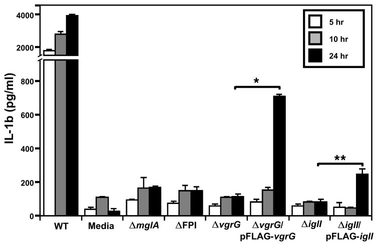
Francisella tularensis causes the human disease tularemia. F. tularensis is able to survive and replicate within macrophages, a trait that has been correlated with its high virulence, but it is unclear the exact mechanism(s) this organism uses to escape killing within this hostile environment. F. tularensis virulence is dependent upon the Francisella pathogenicity island (FPI), a cluster of genes that we show here shares homology with type VI secretion gene clusters in Vibrio cholerae and Pseudomonas aeruginosa. We demonstrate that two FPI proteins, VgrG and IglI, are secreted into the cytosol of infected macrophages. VgrG and IglI are required for F. tularensis phagosomal escape, intramacrophage growth, inflammasome activation and virulence in mice. Interestingly, VgrG secretion does not require the other FPI genes. However, VgrG and other FPI genes, including PdpB (an IcmF homologue), are required for the secretion of IglI into the macrophage cytosol, suggesting that VgrG and other FPI factors are components of a secretion system. This is the first report of F. tularensis FPI virulence proteins required for intramacrophage growth that are translocated into the macrophage.


Figures

Figure 1
Figure 1. Comparison of F. tularensis FPI with T6SS gene clusters in Pseudomonas aeruginosa and Vibrio cholerae
A. Genes exhibiting homology between all three gene clusters are depicted by open arrows and similar gene designations. Shown are the FPI gene annotations for Ftt FPI-I and FPI-II, which correspond to FTN1309-FTN1325 of Ftn. Alignments are provided in Fig. S1. B. Schematic alignment of F. tularensis VgrG with V. cholerae VgrG-1, VgrG-2, and VgrG-3. See text for more details.
Figure 2
Figure 2. Secretion of F. tularensis FPI proteins
A. Secretion of FPI protein-CyaA fusions in infected macrophages. The macrophage cells were infected with Ftn strains expressing protein-CyaA fusions, and intramacrophage cAMP levels measured (Materials & Methods). J774 macrophages were infected with Ftn strains U112 (WT), KKF219 (ΔFPI), KKF101 (ΔicmF), or KKF102 (ΔvgrG), carrying plasmids pKEK894 (“-“; does not express CyaA fusion), pKEK1072 (signal sequenceless BlaB-CyaA), pKEK939 (PepO-CyaA), pKEK1012 (VgrG-CyaA), pKEK1016 (Hcp-CyaA), or pKEK1051 (IglI-CyaA). Assay was performed in triplicate (*P < 0.05, **P < 0.001, P < 0.001 as determined by Student’s two-tailed t-test). B. Secretion of FPI proteins into culture supernatants. Culture supernatants (supe) and corresponding whole cell lysates (WC) of Ftn strains U112 (“WT”), KKF102 (ΔvgrG), or KKF219 (ΔFPI) carrying plasmids pKEK1191 (ΔNBlaB-FLAG), pKEK1157 (FLAG-VgrG), or pKEK1175 (FLAG-IglI), were examined by Western immunoblot utilizing mouse monoclonal antibody against FLAG. Lysates and supernatants were matched to equivalent protein concentrations.
Figure 3
Figure 3. Intramacrophage growth and Phagosome Escape of F. tularensis FPI mutant strains
A. Ftn strains U112 (wt), KKF102 (ΔvgrG), and KKF108 (ΔiglI) either without plasmid, or carrying plasmid pKEK1157 (pFLAG-VgrG) or pKEK1175 (pFLAG-IglI) were inoculated at a MOI of ~10:1 into J774 cells, and intracellular bacteria were enumerated at 1, 24, and 48 h. The assay was performed in triplicate. B. Ftn strains U112 (wt), KKF102 (ΔvgrG) either without plasmid or carrying plasmid pKEK1157 (pFLAG-VgrG), KKF24 (ΔiglC), and KKF219 (ΔFPI) were inoculated at a MOI of 10:1 into mouse BMM, and % cytoplasmic bacteria determined as described (Materials & Methods). C. Ftn strains U112 (wt), and KKF108 (ΔiglI) either without plasmid or carrying plasmid pKEK1175 (pFLAG-IglI)) were inoculated at a MOI of 10:1 into mouse BMM, and % cytoplasmic bacteria determined as described (Materials & Methods) D. Representative immunofluorescent images of phagosomal integrity assay used to determine % cytoplasmic bacteria in BMM infected with Ftn strains (listed on right) at 4 h post infection, showing cytoplasmic Ftn following treatment with digitonin (“F. nov dig”; green), all intracellular Ftn (both cytoplasmic and phagosomal) following treatment with saponin (“F. nov. sap”; red), calnexin staining for ER (blue), and the merge of all three.
Figure 4
Figure 4. Detection of VgrG and IglI within infected macrophages by immunofluorescence
The J774 macrophage cell line was infected with Ftn strains labeled with anti-Ftn LPS (green) expressing FLAG-tagged proteins, fixed at 40 min post-infection, and processed for immunofluorescence using anti-FLAG-Cy3 antibody (red) and DAPI (blue). A. Ftn strain U112 (WT) expressing ΔNBlaB-FLAG, B. Ftn strain KKF102 (ΔvgrG) expressing FLAG-VgrG, C. Ftn strain KKF219 (ΔFPI) expressing FLAG-VgrG, D. Ftn strain KKF108 (ΔiglI) expressing FLAG-IglI, E. Ftn strain KKF102 (ΔvgrG) expressing FLAG-IglI, and F. Ftn strain KKF219 (ΔFPI) expressing FLAG-IglI. For A–F, shown is larger image including macrophage nucleus, arrow depicts intracellular bacterium, while insets show magnification of bacterium with individual red and green channels, and merge. (Scale bars: 10 μM; Insets, 2 μM)
Figure 5
Figure 5. IL-1β signalling in macrophages infected with F. tularensis FPI mutants
Ftn strains U112 (wt), KKF34 (mglA), KKF219 (ΔFPI), KKF102 (ΔvgrG) either with or without plasmid pKEK1157 (pFLAG-VgrG), and KKF108 (ΔiglI) with or without plasmid pKEK1175 (pFLAG-IglI) were used to infect mouse BMM at an MOI of 100. Supernatants were collected at selected time points and Il-1β levels were measured by ELISA. Assay was performed in triplicate (*P < 0.0001, **P < 0.01 as determined by Student’s two-tailed t-test).

References

    1. Bonemann G, Pietrosiuk A, Diemand A, Zentgraf H, Mogk A. Remodelling of VipA/VipB tubules by ClpV-mediated threading is crucial for type VI protein secretion. EMBO J. 2009;28:315–325. - PMC - PubMed
    1. Bosio CM, Bielefeldt-Ohmann H, Belisle JT. Active suppression of the pulmonary immune response by Francisella tularensis Schu4. J Immunol. 2007;178:4538–4547. - PubMed
    1. Bosio CM, Elkins KL. Susceptibility to secondary Francisella tularensis live vaccine strain infection in B-cell-deficient mice is associated with neutrophilia but not with defects in specific T-cell-mediated immunity. Infect Immun. 2001;69:194–203. - PMC - PubMed
    1. Broms JE, Lavander M, Sjostedt A. A conserved alpha-helix essential for a type VI secretion-like system of Francisella tularensis. J Bacteriol. 2009;191:2431–2446. - PMC - PubMed
    1. Brotcke A, Weiss DS, Kim CC, Chain P, Malfatti S, Garcia E, Monack DM. Identification of MglA-regulated genes reveals novel virulence factors in Francisella tularensis. Infect Immun. 2006;74:6642–6655. - PMC - PubMed

MeSH terms