Review
. 2010:50:295-322.
doi: 10.1146/annurev.pharmtox.011008.145533.

Metabotropic glutamate receptors: physiology, pharmacology, and disease

Affiliations
Review

Metabotropic glutamate receptors: physiology, pharmacology, and disease

Colleen M Niswender et al. Annu Rev Pharmacol Toxicol. 2010.

Abstract

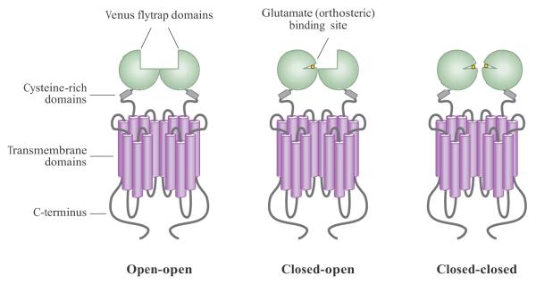
The metabotropic glutamate receptors (mGluRs) are family C G-protein-coupled receptors that participate in the modulation of synaptic transmission and neuronal excitability throughout the central nervous system. The mGluRs bind glutamate within a large extracellular domain and transmit signals through the receptor protein to intracellular signaling partners. A great deal of progress has been made in determining the mechanisms by which mGluRs are activated, proteins with which they interact, and orthosteric and allosteric ligands that can modulate receptor activity. The widespread expression of mGluRs makes these receptors particularly attractive drug targets, and recent studies continue to validate the therapeutic utility of mGluR ligands in neurological and psychiatric disorders such as Alzheimer's disease, Parkinson's disease, anxiety, depression, and schizophrenia.


Figures

Figure 1
Figure 1
Schematic diagram of the mGluR dimer in different activity states. mGluR dimers contain two large extracellular domains called the Venus flytrap domains (VFDs), which bind glutamate and other orthosteric ligands. The cysteine-rich domain links the VFDs to seven transmembrane-spanning domains; the C-terminus faces intracellularly and is often subject to alternative splicing to generate different C-terminal protein tails. The open-open state (left) is the inactive state and can be stabilized by antagonists. Either one or two VFDs can then bind glutamate, resulting in active receptor conformations.
Figure 2
Figure 2
Simple schematic representation of the positive allosteric regulator (PAM) and negative allosteric regulator (NAM) activity using an in vitro functional assay. (a) Monitoring of a functional assay such as calcium mobilization shows that increasing PAM concentrations progressively shift the glutamate concentration response for an mGluR to the left. Depending on the assay used, PAMs can also cause an increase in the maximal agonist response. (b) Increasing NAM concentrations progressively shift the magnitude of the concentration response curve and produce little change in potency, indicating a noncompetitive form of antagonism.
Figure 3
Figure 3
Protein-protein interactions mediating the activity of mGluR7. It is proposed that in the resting state, mGluR7 binds PICK1, MacMARCKs (MacM), and Gβγ subunits; this binding prevents Gβγ subunits from inhibiting voltage-sensitive calcium channels (VSCCs; blue). Rises in calcium induce displacement of MacMARCKs and Gβγ subunits by calcium-calmodulin (Ca2+-CaM), promoting Gβγ subunit inhibition of VSCCs. Adapted from (52, 54).
Figure 4
Figure 4
Schematic representation of mGluRs at the synapse. In general, group I mGluRs (green) are localized postsynaptically, and group II (blue) and III (red) receptors are localized in presynaptic locations, although exceptions occur. In presynaptic locations, mGluRs 2, 3, 4, and 8 are generally found in extrasynaptic locations, and mGluR7 is highly localized to the active zone (136). Group II and III receptors inhibit release of glutamate (left, yellow circles) or GABA (right, red circles), whereas group I receptors promote release when present. At the postsynaptic terminal, the glutamate gated ion channels N-methyl-D-asparate (NMDA), α-amino-3-hydroxyl-5-methyl-4-isoxazole-propionate (AMPA) and kainate respond to glutamate with increases in intracellular sodium or calcium, promoting cell excitability. Group I mGluRs signal via Gq proteins to increase intracellular calcium; additionally, mGluR5 and NMDA receptors are closely linked signaling partners reciprocally regulated by phosphorylation (black circle) (71). Postsynaptic mGluR2/3 and GABAB1/2 receptors couple to cAMP inhibition. GABAA chloride channels (pink) modulate intracellular chloride. Expression of mGluR3 and mGluR5 on glia is now emerging as another key site for mGluR regulation of synaptic activity, although the signaling pathways and consequences of receptor activation on these cells are not presently well understood.

Similar articles

Cited by

References

    1. Pin JP, Galvez T, Prezeau L. Evolution, structure, and activation mechanism of family 3/C G-protein-coupled receptors. Pharmacol. Ther. 2003;98(3):325–54. - PubMed
    1. Tsuchiya D, et al. Structural views of the ligand-binding cores of a metabotropic glutamate receptor complexed with an antagonist and both glutamate and Gd3+ Proc. Natl. Acad. Sci. USA. 2002;99(5):2660–65. - PMC - PubMed
    1. Kunishima N, Shimada Y, Tsuji Y, Sato T, Yamamoto M, et al. Structural basis of glutamate recognition by a dimeric metabotropic glutamate receptor. Nature. 2000;407(6807):971–77. - PubMed
    1. Muto T, Tsuchiya D, Morikawa A, Jingami H. Structures of the extracellular regions of the group II/III metabotropic glutamate receptors. Proc. Natl. Acad. Sci. USA. 2007;104(10):3759–64. - PMC - PubMed
    1. Jingami H, Nakanishi S, Morikawa K. Structure of the metabotropic glutamate receptor. Curr. Opin. Neurobiol. 2003;13(3):271–78. - PubMed

Publication types

MeSH terms

Substances

LinkOut - more resources