

Exogenous thymosin beta4 prevents apoptosis in human intervertebral annulus cells in vitro
- PMID: 20055734
- PMCID: PMC2886502
- DOI: 10.3109/10520290903116884
Exogenous thymosin beta4 prevents apoptosis in human intervertebral annulus cells in vitro
Abstract
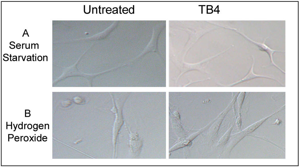
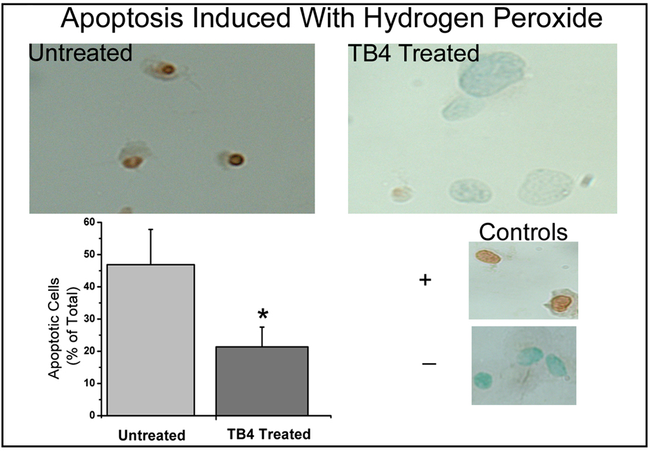
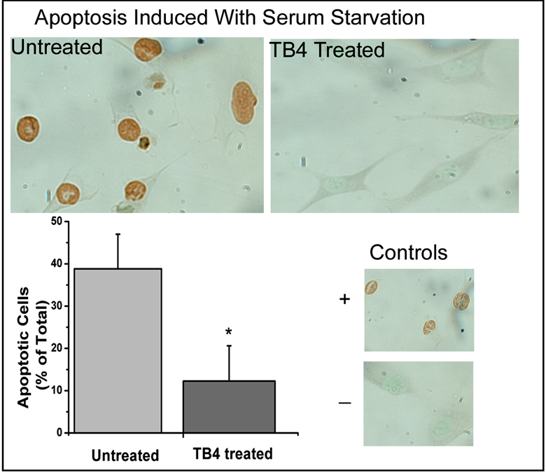
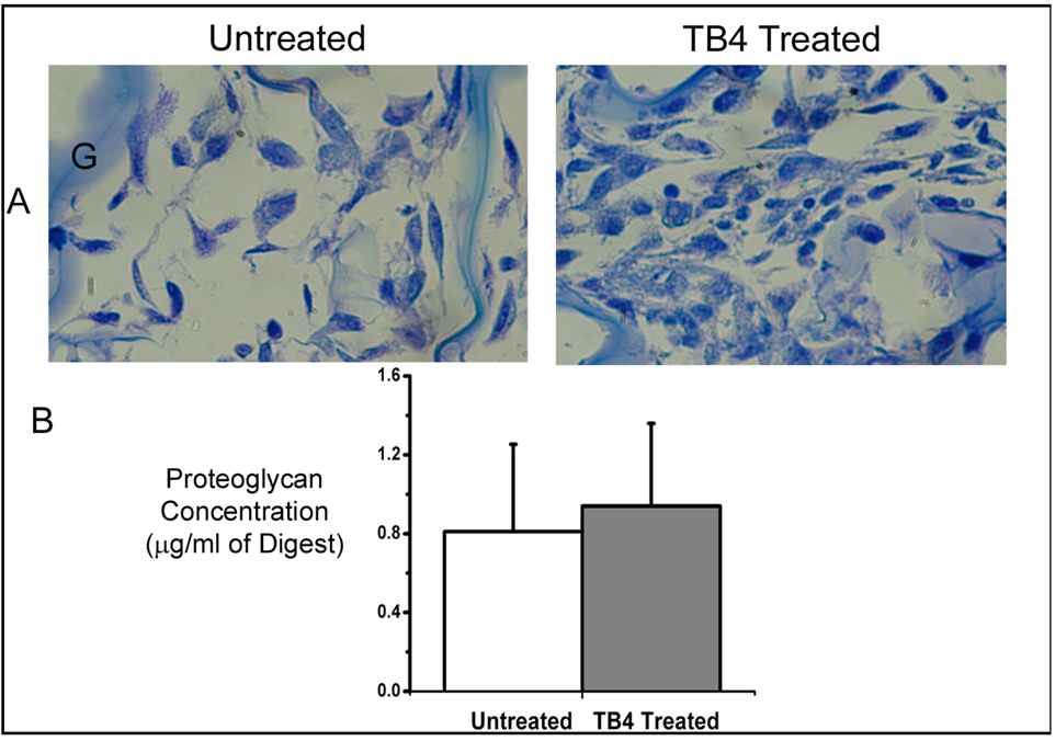
Loss of cells in the human disc due to programmed cell death (apoptosis) is a major factor in the aging and degenerating human intervertebral disc. Our objective here was to determine if thymosin beta(4) (TB4), a small, multifunctional 5 kDa protein with diverse activities, might block apoptosis in human annulus cells cultured in monolayer or three-dimensional (3D) culture. Apoptosis was induced in vitro using hydrogen peroxide or serum starvation. Annulus cells were processed for identification of apoptotic cells using the TUNEL method. The percentage of apoptotic cells was determined by cell counts. Annulus cells also were treated with TB4 for determination of proliferation, and proteoglycan production was assessed using cell titer and 1,2 dimethylmethylamine (DMB) assays and histological staining. A significant reduction in disc cell apoptosis occurred after TB4 treatment. The percentage of cells undergoing apoptosis decreased significantly in TB4 treated cells in both apoptosis induction designs. TB4 exposure did not alter proteoglycan production as assessed by either DMB measurement or histological staining. Our results indicate the need for further studies of the anti-apoptotic effect of TB4 and suggest that TB4 may have therapeutic application in future biological therapies for disc degeneration.
Figures
References
-
- Bock-Marquette I, Saxena A, White MD, Dimaio JM, Srivastava D. Thymosin beta4 activates integrin-linked kinase and promotes cardiac cell migration, survival and cardiac repair. Nature. 2004;432:466–472. - PubMed
-
- Bonnet D, Lemoine FM, Frobert Y, Bonnet ML, Baillou C, Najman A, Guigon M. Thymosin beta4, inhibitor for normal hematopoietic progenitor cells. Exp. Hematol. 1996;24:776–782. - PubMed
-
- Bubb MR. Thymosin beta 4 interactions. Vitam. Horm. 2003;66:297–316. - PubMed
-
- Choi SY, Kim DK, Eun B, Kim K, Sun W, Kim H. Anti-apoptotic function of thymosin-beta in developing chick spinal motor neurons. Biochem. Biophys. Res. Commun. 2006;346:872–878. - PubMed
-
- Choi SY, Noh MR, Kim DK, Sun W, Kim H. Neuroprotective function of thymosin-beta and its derivative peptides on the programmed cell death of chick and rat neurons. Biochem. Biophys. Res. Commun. 2007;362:587–593. - PubMed
Publication types
MeSH terms
Substances
Grants and funding
LinkOut - more resources
Full Text Sources