Decay accelerating factor is essential for successful corneal engraftment
- PMID: 20055803
- PMCID: PMC3520429
- DOI: 10.1111/j.1600-6143.2009.02961.x
Decay accelerating factor is essential for successful corneal engraftment
Abstract
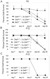
In contrast to immune restrictions that pertain for solid organ transplants, the tolerogenic milieu of the eye permits successful corneal transplantation without systemic immunosuppression, even across a fully MHC disparate barrier. Here we show that recipient and donor expression of decay accelerating factor (DAF or CD55), a cell surface C3/C5 convertase regulator recently shown to modulate T-cell responses, is essential to sustain successful corneal engraftment. Whereas wild-type (WT) corneas transplanted into multiple minor histocompatibility antigen (mH), or HY disparate WT recipients were accepted, DAF's absence on either the donor cornea or in the recipient bed induced rapid rejection. Donor or recipient DAF deficiency led to expansion of donor-reactive IFN-gamma producing CD4(+) and CD8(+) T cells, as well as inhibited antigen-induced IL-10 and TGF-beta, together demonstrating that DAF deficiency precludes immune tolerance. In addition to demonstrating a requisite role for DAF in conferring ocular immune privilege, these results raise the possibility that augmenting DAF levels on donor corneal endothelium and/or the recipient bed could have therapeutic value for transplants that clinically are at high risk for rejection.
Figures


Similar articles
-
IFN-γ blocks CD4+CD25+ Tregs and abolishes immune privilege of minor histocompatibility mismatched corneal allografts.Am J Transplant. 2013 Dec;13(12):3076-84. doi: 10.1111/ajt.12466. Epub 2013 Oct 1. Am J Transplant. 2013. PMID: 24119152 Free PMC article.
-
Donor deficiency of decay-accelerating factor accelerates murine T cell-mediated cardiac allograft rejection.J Immunol. 2008 Oct 1;181(7):4580-9. doi: 10.4049/jimmunol.181.7.4580. J Immunol. 2008. PMID: 18802060 Free PMC article.
-
Role of CD4+ T cells in immunobiology of orthotopic corneal transplants in mice.Invest Ophthalmol Vis Sci. 1999 Oct;40(11):2614-21. Invest Ophthalmol Vis Sci. 1999. PMID: 10509657
-
An immunodominant minor histocompatibility alloantigen that initiates corneal allograft rejection.Transplantation. 2003 Apr 27;75(8):1368-74. doi: 10.1097/01.TP.0000063708.26443.3B. Transplantation. 2003. PMID: 12717232
-
Preservation of donor cornea prevents corneal allograft rejection by inhibiting induction of alloimmunity.Exp Eye Res. 2000 Jun;70(6):737-43. doi: 10.1006/exer.2000.0841. Exp Eye Res. 2000. PMID: 10843778
Cited by
-
High-risk corneal allografts: A therapeutic challenge.World J Transplant. 2016 Mar 24;6(1):10-27. doi: 10.5500/wjt.v6.i1.10. World J Transplant. 2016. PMID: 27011902 Free PMC article. Review.
-
Antibody Subclass Repertoire and Graft Outcome Following Solid Organ Transplantation.Front Immunol. 2016 Oct 24;7:433. doi: 10.3389/fimmu.2016.00433. eCollection 2016. Front Immunol. 2016. PMID: 27822209 Free PMC article. Review.
-
A Review on the Immunomodulatory Mechanism of Acupuncture in the Treatment of Inflammatory Bowel Disease.Evid Based Complement Alternat Med. 2022 Jan 15;2022:8528938. doi: 10.1155/2022/8528938. eCollection 2022. Evid Based Complement Alternat Med. 2022. PMID: 35075366 Free PMC article. Review.
-
C5aR1 regulates migration of suppressive myeloid cells required for costimulatory blockade-induced murine allograft survival.Am J Transplant. 2019 Mar;19(3):633-645. doi: 10.1111/ajt.15072. Epub 2018 Sep 17. Am J Transplant. 2019. PMID: 30106232 Free PMC article.
-
The cornea IV immunology, infection, neovascularization, and surgery chapter 1: Corneal immunology.Exp Eye Res. 2021 Apr;205:108502. doi: 10.1016/j.exer.2021.108502. Epub 2021 Feb 16. Exp Eye Res. 2021. PMID: 33607075 Free PMC article. Review.
References
-
- Sonoda Y, Streilein JW. Orthotopic corneal transplantation in mice--evidence that the immunogenetic rules of rejection do not apply. Transplantation. 1992;54(4):694–704. - PubMed
-
- Niederkorn JY. Immune mechanisms of corneal allograft rejection. Curr Eye Res. 2007;32(12):1005–1016. - PubMed
-
- Taylor AW, Streilein JW, Cousins SW. Identification of alpha-melanocyte stimulating hormone as a potential immunosuppressive factor in aqueous humor. Curr Eye Res. 1992;11(12):1199–1206. - PubMed
-
- Taylor AW. Neuroimmunomodulation in immune privilege: role of neuropeptides in ocular immunosuppression. Neuroimmunomodulation. 1996;3(4):195–204. - PubMed
Publication types
MeSH terms
Substances
Grants and funding
LinkOut - more resources
Full Text Sources
Research Materials
Miscellaneous

