

Review
doi: 10.1111/j.1365-2559.2009.03454.x.
Sebaceous neoplasia and the Muir-Torre syndrome: important connections with clinical implications
Affiliations
- PMID: 20055911
- PMCID: PMC2805836
- DOI: 10.1111/j.1365-2559.2009.03454.x
Item in Clipboard
Review
Sebaceous neoplasia and the Muir-Torre syndrome: important connections with clinical implications
Histopathology.
2010 Jan.
Abstract
Sebaceous neoplasia comprises a spectrum ranging from benign to malignant. Proper histological identification is important for treatment, prognosis and potential association with the Muir-Torre syndrome (MTS). Our increased understanding of the significance and pathogenesis of these tumours has led to improved risk stratification, screening recommendations, and treatment of patients with an initial presentation of a sebaceous tumour. This review focuses on the diagnostic and histological features of sebaceous lesions, the MTS, and recent insights into the molecular pathogenesis of sebaceous tumours.
Figures
a, Benign sebaceous lobules are bounded by a double-layered rim of small basaloid epithelial cells. b, Sebaceous hyperplasia reveals multiple prominent sebaceous lobules surrounding a central dell. The normal follicle and sebaceous lobules on the left illustrate that the lobules in sebaceous hyperplasia are more superficial than normal glands.
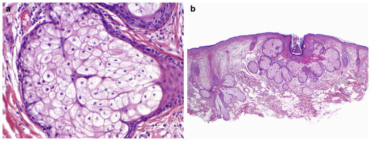
a, Sebaceous adenoma with multilobular architecture and a smooth base. b, Higher power of a sebaceous adenoma reveals an expanded peripheral basaloid component. c, The expansion of the basaloid component can be more subtle in sebaceous adenoma and is often accompanied by an increased prominence of the basaloid cells as well as an expansion of the number of cells. d, The cellularity of sebaceous adenomas ranges from more peripheral germinative basaloid cells to more central mature sebocytes with crenulated nuclei.
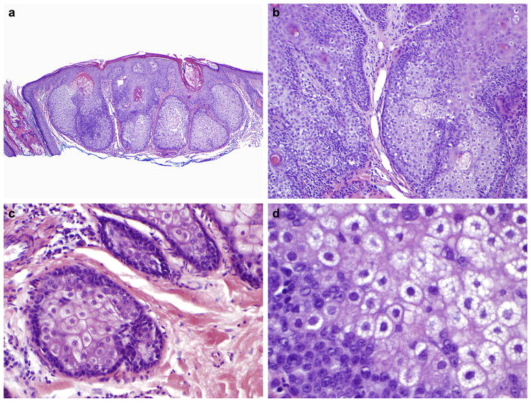
a, Sebaceomas are well-circumscribed and composed primarily of basaloid cells. b, The mature sebaceous component in sebaceoma can be rather focal.
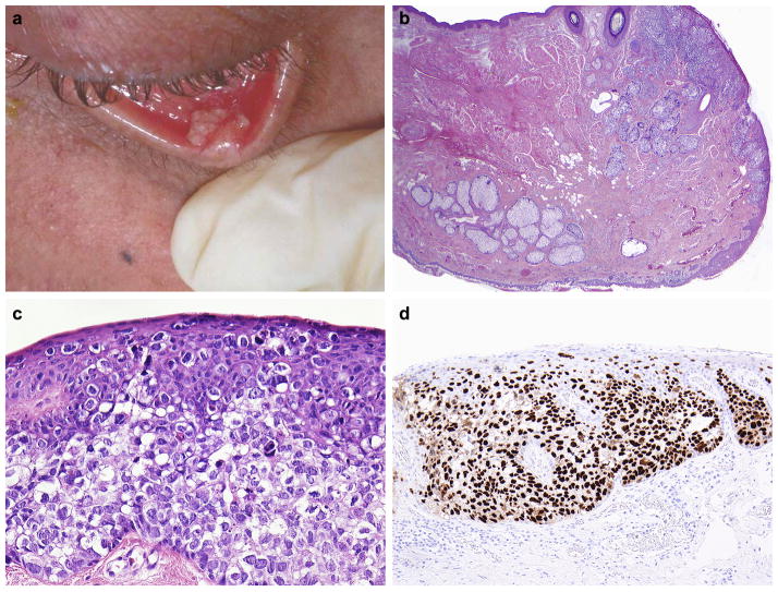
a, Periocular sebaceous carcinoma involving the lower eyelid. b, Full-thickness eyelid excision revealing sebaceous carcinoma involving the cutaneous portion of the eyelid (upper right). The mucosal surface is below. c, Periocular sebaceous carcinoma with pagetoid involvement of the overlying epithelium. d, Strong, nuclear p53 expression detected by immunohistochemistry in a periocular sebaceous carcinoma. Clinical photograph courtesy of Dr Bita Esmaeli, UT-M. D. Anderson Cancer Center, Houston, TX, USA.
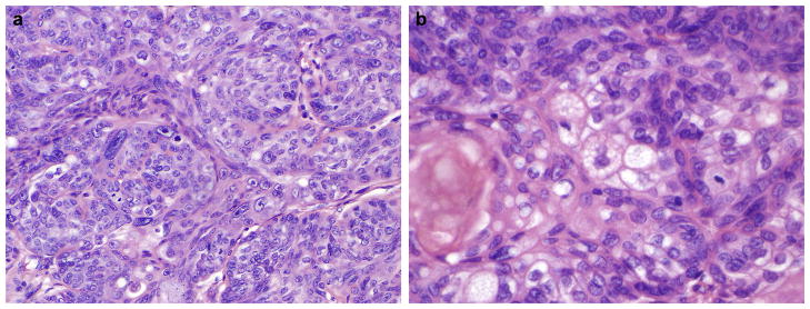
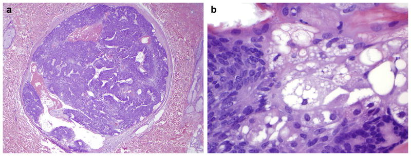
a, Extraocular sebaceous carcinoma showing scattered pleomorphic cells. b, Sebaceous differentiation can be focal.
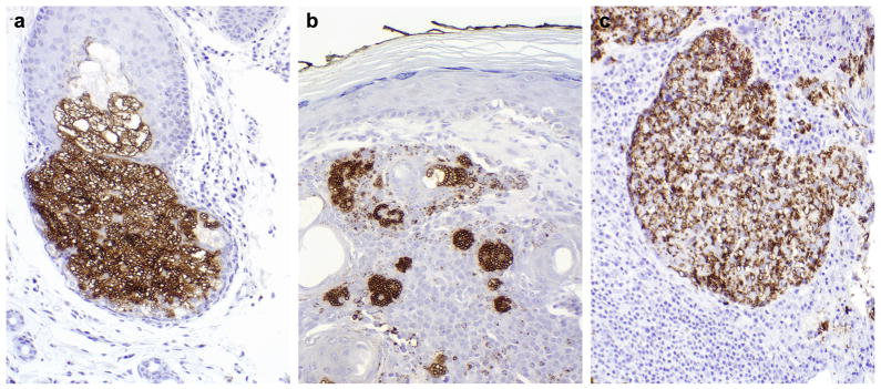
Adipophilin immunohistochemistry in a normal sebaceous gland (a), sebaceoma (b) and periocular sebaceous carcinoma (c). It is important to identify the individual outlines of the membranes that surrounded the lipid globules within the cells for specificity. The numbers of globules will vary and are often less in number in sebaceous carcinomas with limited sebaceous differentiation.
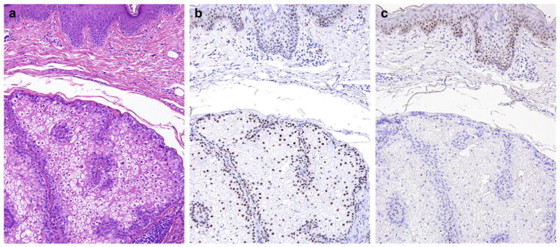
The sebaceous tumour (a) from this Muir–Torre patient shows nuclear expression of MLH1 (b) with loss of MSH2 (c) on immunohistochemistry. The overlying epidermis serves as an internal positive control.

The BAT26 microsatellite shows increased variability in a sebaceous tumour from a Muir–Torre patient (lower panel) compared with normal tissue from the same patient (upper panel). This DNA-based test uses polymerase chain reaction amplification of specific loci with chromatographic determination of amplicon sizes to detect alterations in the length of the tested microsatellite.
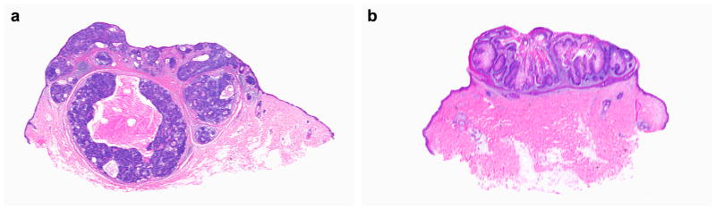
a, Sebaceous adenoma with cystic features. b, Sebaceous adenoma with keratoacanthoma-like architecture. Both of these features are thought to more specifically suggest the Muir–Torre syndrome, but more study is needed.
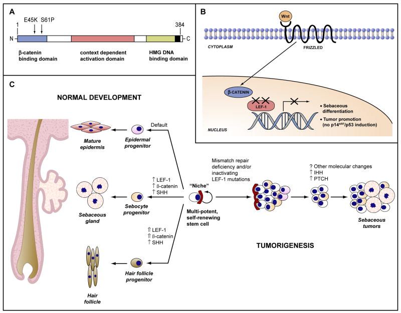
Dual inactivating mutations in the LEF-1 transcription factor have been documented in benign sebaceous neoplasms (a) leading to an attenuation of Wnt signalling in tumour cells (b). This not only promotes sebaceous differentiation, but also inhibits p53-mediated senescence and apoptosis. c, Cutaneous stem cells reside in a specialized tissue “niche” and are responsible for development and regeneration of epithelial structures including the epidermis, follicle and sebaceous gland under the influence of various signalling pathways (left). Mutations and resulting dysregulation of many of these same pathways appear to be involved in the generation of various cutaneous tumours such as that illustrated for sebaceous tumours (right). SHH, sonic hedgehog; IHH, Indian hedgehog; PTCH, patched.
Similar articles
-
Sebaceous Neoplasms.Surg Pathol Clin. 2017 Jun;10(2):367-382. doi: 10.1016/j.path.2017.01.009. Surg Pathol Clin. 2017. PMID: 28477886 Review.
-
Sebaceous lesions of the skin.Pathology. 2017 Dec;49(7):688-697. doi: 10.1016/j.pathol.2017.08.012. Epub 2017 Oct 25. Pathology. 2017. PMID: 29078997 Review.
-
[Isolated conjunctival sebaceous adenoma associated with acute endophtalmitis].Rev Esp Patol. 2022 Jan-Mar;55(1):68-72. doi: 10.1016/j.patol.2019.01.006. Epub 2019 Mar 16. Rev Esp Patol. 2022. PMID: 34980445 Spanish.
-
Extraocular sebaceous carcinoma: a report of 2 cases.Actas Dermosifiliogr. 2012 Dec;103(10):919-22. doi: 10.1016/j.adengl.2011.05.011. Epub 2012 Nov 11. Actas Dermosifiliogr. 2012. PMID: 23149052 Review.
-
Sebaceous Carcinoma Arising From Heterotopic Salivary Gland Tissue in a Patient With Muir-Torre Syndrome.Dermatol Surg. 2021 Dec 1;47(12):1659-1660. doi: 10.1097/DSS.0000000000003259. Dermatol Surg. 2021. PMID: 34608093 No abstract available.
Cited by
-
Ileocecal adenocarcinoma and ureteral transitional cell carcinoma with multiple sebaceous tumors and keratoacanthomas in a case of muir-torre syndrome.Dermatol Res Pract. 2010;2010:173160. doi: 10.1155/2010/173160. Epub 2010 Aug 15. Dermatol Res Pract. 2010. PMID: 20814549 Free PMC article.
-
[Sebaceous breast carcinoma: report of a rare histological special subtype].Pathologe. 2014 Feb;35(1):72-6. doi: 10.1007/s00292-013-1844-4. Pathologe. 2014. PMID: 24414613 Review. German.
-
Muir-Torre Syndrome Presenting as Sebaceous Adenocarcinoma and Invasive MSH6-Positive Colorectal Adenocarcinoma.Case Rep Oncol. 2016 Feb 4;9(1):95-9. doi: 10.1159/000443788. eCollection 2016 Jan-Apr. Case Rep Oncol. 2016. PMID: 26933426 Free PMC article.
-
Sebaceous carcinoma of the chest wall: A case report.Radiol Case Rep. 2021 May 25;16(7):1870-1873. doi: 10.1016/j.radcr.2021.04.060. eCollection 2021 Jul. Radiol Case Rep. 2021. PMID: 34093933 Free PMC article.
-
[Suspected malignant gastric lesion in hereditary tumor syndrome: Endoscopic resection or gastrectomy?].Chirurg. 2013 Jul;84(7):594-7. doi: 10.1007/s00104-013-2483-2. Chirurg. 2013. PMID: 23595856 German. No abstract available.
References
-
- Li MaUC. Normal skin. In: Mills SE, editor. Histology for pathologists. Philadelphia: Lippincott, Williams and Wilkins; 2007.
-
- Danby FW. Why we have sebaceous glands. J Am Acad Dermatol. 2005;52:1071–1072. - PubMed
-
- Zouboulis CC, Baron JM, Bohm M, et al. Frontiers in sebaceous gland biology and pathology. Exp Dermatol. 2008;17:542–551. - PubMed
-
- Bayer-Garner IB, Givens V, Smoller B. Immunohistochemical staining for androgen receptors: a sensitive marker of sebaceous differentiation. Am J Dermatopathol. 1999;21:426–431. - PubMed
-
- Bieniek R, Lazar AJ, Photopoulos C, Lyle S. Sebaceous tumours contain a subpopulation of cells expressing the keratin 15 stem cell marker. Br J Dermatol. 2007;156:378–380. - PubMed
Publication types
MeSH terms
Grants and funding
LinkOut - more resources
Full Text Sources
