. 2010 Mar 17;166(2):455-63.
doi: 10.1016/j.neuroscience.2009.12.059. Epub 2010 Jan 4.

Neuronostatin is co-expressed with somatostatin and mobilizes calcium in cultured rat hypothalamic neurons

Affiliations

Neuronostatin is co-expressed with somatostatin and mobilizes calcium in cultured rat hypothalamic neurons

S L Dun et al. Neuroscience. .

Abstract

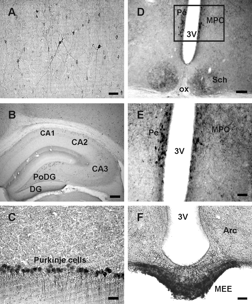
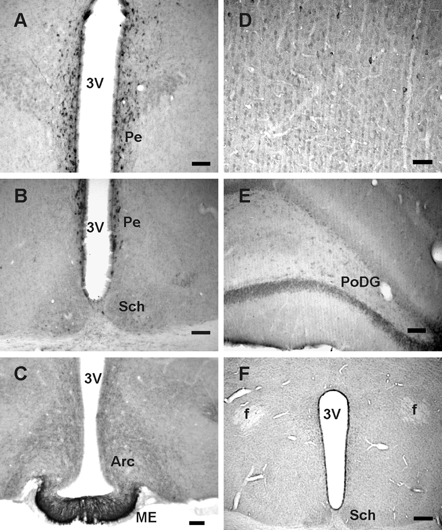
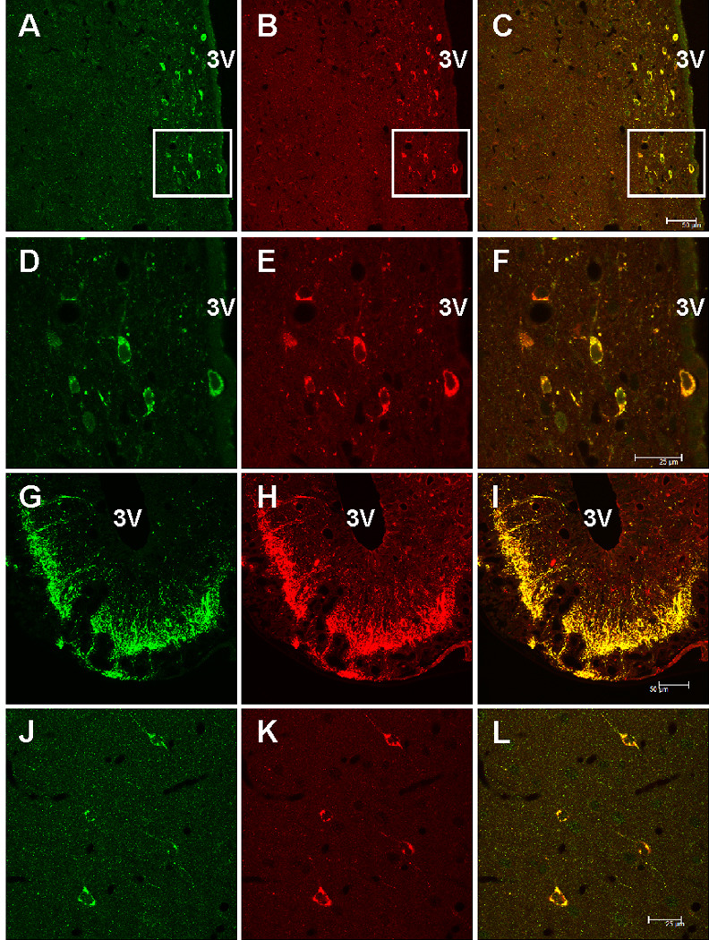
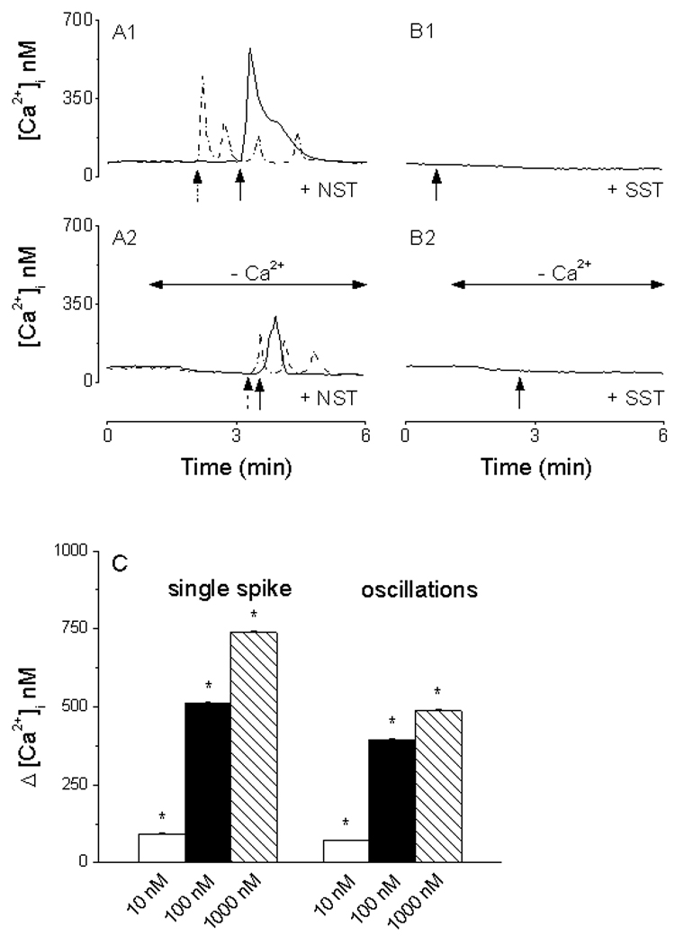
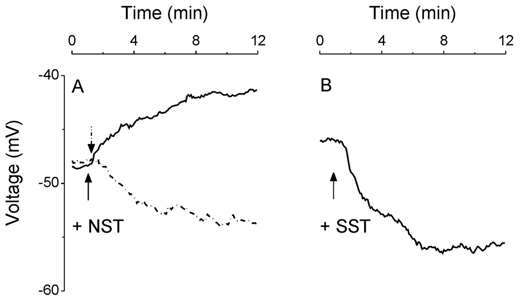
Neuronostatin (NST) is a newly identified peptide of 13-amino acids encoded by the somatostatin (SST) gene. Using a rabbit polyclonal antiserum against the human NST, neuronostatin-immunoreactive (irNST) cells comparable in number and intensity to somatostatin immunoreactive (irSST) cells were detected in the hypothalamic periventricular nucleus. Fewer and/or less intensely labeled irNST cells were noted in other regions such as the hippocampus, cortex, amygdala, and cerebellum. Double-labeling hypothalamic sections with NST- and SST-antiserum revealed an extensive overlapping of irNST and irSST cells in the periventricular nucleus. Pre-absorption of the NST-antiserum with NST (1 microg/ml) but not with SST (1 microg/ml) abrogated irNST and vice versa. The activity of NST on dissociated and cultured hypothalamic neurons was assessed by the Ca(2+) imaging method. NST (10, 100, 1000 nM) concentration-dependently elevated intracellular Ca(2+) concentrations [Ca(2+)](i) in a population of hypothalamic neurons with two distinct profiles: (1) a fast and transitory increase in [Ca(2+)](i), and (2) an oscillatory response. Whereas, SST (100 nM) reduced the basal [Ca(2+)](i) in 21 of 61 hypothalamic neurons examined; an increase was not observed in any of the cells. Optical imaging with a slow-responding voltage sensitive dye DiBAC(4)(3) showed that NST (100 nM) depolarized or hyperpolarized; whereas, SST (100 nM) hyperpolarized a population of hypothalamic neurons. The result shows that NST and SST, though derived from the same precursor protein, exert different calcium mobilizing effects on cultured rat hypothalamic neurons, resulting in diverse cellular activities.


Figures

Fig. 1
Fig. 1
Somatostatin (SST) immunoreactive cells and fibers in the rat brain. A, irSST cells and cell processes in the motor cortex. B, irSST cells in CA1, CA2 and CA3 regions and polymorphic layer of the dentate gyrus (PoDG). C, Purkinje cells of the cerebellum are irSST. D, irSST cells are concentrated in the periventricular hypothalamic nucleus (Pe) adjacent to the 3rd ventricle (3V); irSST-cells and processes are also present in the suprachiasmatic nucleus (Sch). E, enlargement of the area outlined in D where strongly labeled irSST cells are located in Pe, and fibers in medial preoptic nucleus (MPO). F, strongly labeled irSST fibers are present in median eminence, particularly the external median eminence (MEE), and in arcuate nucleus (Arc). Abbreviations: ox, optic chiasm. Calibration bar: A, C, E and F: 50 µm; B: 250 µm; D: 100 µm.
Fig. 2
Fig. 2
Neuronostatin (NST) immunoreactive cells and processes in the rat brain. A, irNST cells are concentrated in the hypothalamic periventricular nucleus (Pe). B, strongly labeled irNST cells are distributed to the periventricular nucleus, and a few lightly to moderately labeled cells and processes in the suprachiasmatic nucleus (Sch). C, numerous irNST fibers are present in the median eminence (ME) and arcuate nucleus (Arc). D, fewer or less intensely labeled irNST cells are detectable in the motor cortex. E, moderately labeled cells are seen in the polymorphic layer of the dentate gyrus (PoDG). F, irNST is not detected in a hypothalamic section processed with an NST antiserum pre-absorbed with the peptide NST (1 µg/ml) overnight. Abbreviations: 3V, 3rd ventricle; f, fornix. Calibration bar: A to C, E, 50 µm; D: 25 µm; F: 100 µm.
Fig. 3
Fig. 3
Confocal images of rat brain sections double-labeled with neuronostatin (NST, green fluorescence) and somatostatin (SST, red fluorescence) antiserum. A and B, hypothalamic periventricular cells located next to the 3rd ventricle (3V) are immunoreactive to NST and SST. C, a merged image of A and B, where cells immunoreactive to both NST and SST appear orange. D–E, higher magnification of the area outlined in A–C, where nearly all of the cells are irNST and irSST. F, a merged image of D and E, where overlapping cells appear orange color. G–I, dense fibers immunoreactive to NST and SST are seen in the median eminence, mostly in the external layer. J and K, all 4 cortical neurons in the cerebral cortex are double-labeled, but the intensity of irNST appears to be lower than that of irSS. L, a merged image of J and K. Calibration bar: A–C, and G–I, 50 µm; D–F and J–L, 25 µm.
Fig. 4
Fig. 4
Calcium responses induced by neuronostatin (NST) and somatostatin (SST) in cultured rat hypothalamic neurons. A1, NST increases cytosolic calcium [Ca2+]i with two profiles: fast and transitory (solid line) and calcium oscillations (dotted line). B1, SST slightly reduced the basal Ca2+. A2, in Ca2+-free saline, NST produced a fast and transitory (solid line) or an oscillatory response (dotted line), with amplitude lower than those produced in Ca2+-containing saline. B2, no change in [Ca2+]i was noted in response to SST in Ca2+-free saline. C, NST (10, 100 and 1000 nM) produced a concentration-dependent increase in [Ca2+]i with two profiles: single spike and oscillations. Arrows denote the administration of neuronostatin or somatostatin.
Fig. 5
Fig. 5
Changes in resting membrane potential induced by neuronostatin (NST) and somatostatin (SST) in rat hypothalamic neurons. A, examples of NST-induced depolarization (solid line) and hyperpolarization (dotted line); NST depolarized 23/257 neurons with a mean amplitude of 5.2 ± 1.6 mV and hyperpolarized 7/257 neurons with a mean amplitude of 6.1 ± 2.2 mV. B, example of SST-induced hyperpolarization; SST hyperpolarized 43/114 neurons, with a mean amplitude of 9.8 ± 2.2 mV. Arrows denote the administration of neuronostatin or somatostatin.

Similar articles

Cited by

References

    1. Augustine GJ, Santamaria F, Tanaka K. Local calcium signaling in neurons. Neuron. 2003;40:331–346. - PubMed
    1. Bakhit C, Benoit R, Bloom FE. Release of somatostatin-28(1-12) from rat hypothalamus in vitro. Nature. 1983;301:524–526. - PubMed
    1. Beaudet A, Greenspun D, Raelson J, Tannenbaum GS. Patterns of expression of SSTR1 and SSTR2 somatostatin receptor subtypes in the hypothalamus of the adult rat: relationship to neuroendocrine function. Neurosci. 1995;65:551–561. - PubMed
    1. Bennett GW, Edwardson JA, deCotte DM. Release of somatostatin from rat brain synaptosomes. J Neurochem. 1979;32:1127–1130. - PubMed
    1. Benoit R, Bohlen P, Esch F, Ling N. Neuropeptides derived from prosomatostatin that do not contain the somatostatin-14 sequence. Brain Res. 1984;311:23–29. - PubMed

Publication types