. 2010 Feb;79(2):120-30.
doi: 10.1016/j.diff.2009.11.005. Epub 2010 Jan 6.

Regeneration of acinar cells following ligation of rat submandibular gland retraces the embryonic-perinatal pathway of cytodifferentiation

Affiliations

Regeneration of acinar cells following ligation of rat submandibular gland retraces the embryonic-perinatal pathway of cytodifferentiation

Emanuele Cotroneo et al. Differentiation. 2010 Feb.

Abstract

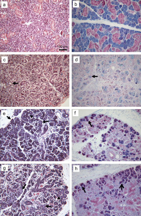
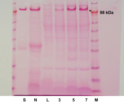
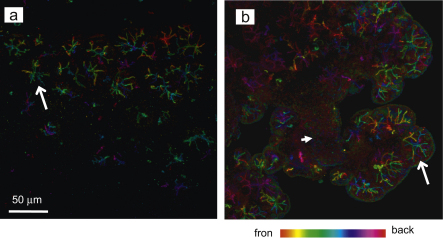
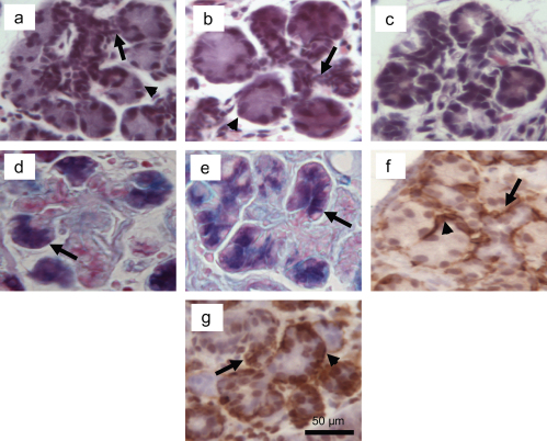
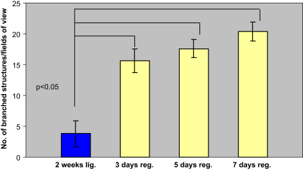
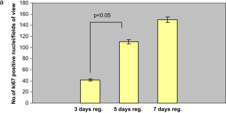
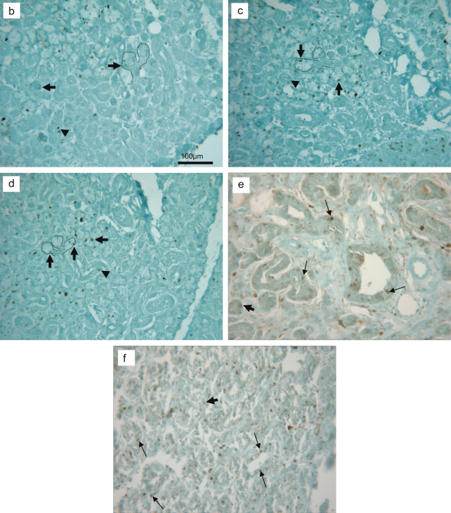
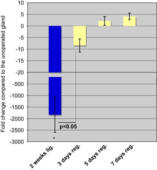
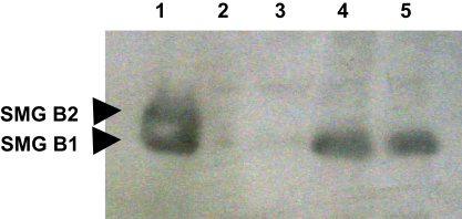
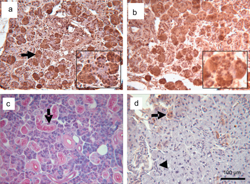
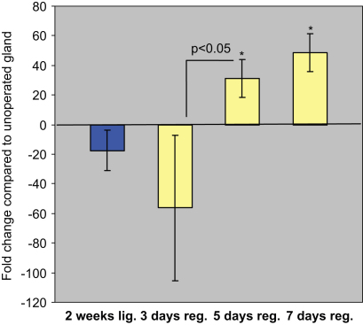
Rat submandibular gland can regenerate following ligation-induced atrophy, eventually recovering its normal morphology and function. Previous studies have suggested that the regeneration process implies both self-proliferation of existing acini and formation of new acinar cells. One hypothesis is that new acinar cells may differentiate from the ductal cells in a similar fashion to the process of cytodifferentiation occurring during submandibular glandular development. In this study atrophy was induced, under recovery anaesthesia, by applying a metal clip on the main duct of the submandibular gland without including the chorda lingual nerve. After 2 weeks the duct was deligated for 3, 5 or 7 days or 8 weeks and the glands collected. Tissue was prepared for immunohistochemistry, biochemical analysis and RNA extraction. The histology of the regenerated glands shows several normal-looking acini, which have regained their glycoprotein content (AB/PAS positive), data also confirmed by biochemical analysis (SDS-PAGE/PAS). Regenerating tissue was characterized by the presence of embryonic-like branched structures ending with AB/PAS positive acinar cells. The proteins SMG-B and PSP are normally expressed in acinar cell precursors during development but only by intercalated ductal cells in the adult stage. In the adult regenerating gland mRNA levels of both SMG-B and PSP were found to be up-regulated compared to ligated glands and SMG-B expression localized to acinar cells whilst the ductal cells were negative. This study of rat submandibular gland regeneration suggests new acinar cells have differentiated from ducts and express markers of acinar cell precursors in a similar manner to the cytodifferentiation process occurring during glandular development.


Figures

Fig. 1
Fig. 1
Histological comparison of normal gland, ligated (atrophic) gland and 5 or 7 days deligated glands. a, b Normal, unoperated gland stained with H & E and AB/PAS, respectively, showing the typical appearance of acinar and ductal cells. c, d H & E and AB/PAS-stained sections of 2 weeks ligated gland showed extensive inflammation, dilatation of the duct lumen (arrow in c), and loss of glycoproteins from the acini (arrow in d). e, g H & E staining of 5 and 7 days deligated glands respectively. Several acini and ductal cells have recovered some of their size (arrows). Branched structures were often visible (arrowheads). f, h AB/PAS staining of 5 and 7 days deligated glands respectively, showing the recovery of glycoproteins content in the acini (arrows) and in the granular ducts (open arrow).
Fig. 2
Fig. 2
PAS staining of glycoproteins resolved by SDS-PAGE gel for: Methacholine-evoked saliva from normal submandibular gland (Lane S) and submandibular gland homogenate from unoperated (normal) submandibular gland (Lane N); 2 weeks ligated (atrophic) gland (Lane L); 3 days deligated gland (Lane 3); 5 days deligated gland (Lane 5) and 7 days deligated gland (Lane 7). Molecular weight marker (M). The asterisk marks the rat low molecular mucin (∼114 KDa).
Fig. 3
Fig. 3
AQP5 expression in the (a) 5 and (b) 7 days regenerated submandibular gland. Collagenase-digested cells were incubated with an anti-AQP5 antibody. Different colours represent differences in depth of field. Aquaporin 5 is expressed in the acinar cells (arrow) whilst the ducts are negative (small arrow).
Fig. 4
Fig. 4
Morphological features of the branched structures. a, b H & E staining of the branched structures in respectively 5 and 7 days deligated glands, characterized by short ducts (arrows) ending with acini (arrowheads). c Typical embryonic (e20) branched structures. d, e AB/PAS of 5 and 7 days deligated gland respectively, showing presence of glycoprotein in the acini at the end of the branching (arrows). Smooth muscle actin immunohistochemistry of 5 (f) and 7 days (g) deligated showing presence of myoepithelial cells around the acini (arrowheads) and the acinar-duct junction (arrows); sections were counterstained with haematoxylin. Fig. g shows background staining in few nuclei.
Fig. 5
Fig. 5
Mean number (±SEM) of branched structures per field of view (×200 magnification) in the 2 weeks ligated (2 weeks lig.) and the 2 weeks ligated plus 3, 5 or 7 days deligated (3 days reg.; 5 days reg.; 7 days reg.) submandibular gland stained by H&E. The number of the branched structures increased at 3 days of regeneration compared to the 2 weeks ligated gland (p<0.05, 5 observation from 5 rats, n=25), however it did not increase further (p>0.05) during the progression of the regeneration.
Fig. 6
Fig. 6
Cell proliferation in the deligated glands. a, number of ki-67 positive nuclei per field of view (×200 magnification) in the 2 weeks ligated plus 3 or 5 or 7 days deligated (3 days reg.; 5 days reg.; 7 days reg.) submandibular gland. 5 days deligated gland showed an increase in the number of proliferating cells (p<0.05, 3 observation in 5 rats, n=15) compared to the 3 days. b, c, d ki-67 immunostaining (counterstained with Light Green dye) in the 2 weeks ligated plus 3 or 5 or 7 days deligated respectively, showing mostly proliferating acinar cells (arrows), also present at the end of the branched structures (dashed silhouettes), and occasionally ductal cells (arrowheads). e, f ki-67 immunostaining of normal adult and ligated rat submandibular gland respectively. Normal gland (e) showed proliferation of ductal cells (arrow) and occasionally acinar cells (thick arrow). The ligated gland (f) revealed proliferation of ductal cells (arrows) and some non-parenchymal cells (thick arrow). All tissue sections were counterstained with Light Green.
Fig. 6
Fig. 6
Cell proliferation in the deligated glands. a, number of ki-67 positive nuclei per field of view (×200 magnification) in the 2 weeks ligated plus 3 or 5 or 7 days deligated (3 days reg.; 5 days reg.; 7 days reg.) submandibular gland. 5 days deligated gland showed an increase in the number of proliferating cells (p<0.05, 3 observation in 5 rats, n=15) compared to the 3 days. b, c, d ki-67 immunostaining (counterstained with Light Green dye) in the 2 weeks ligated plus 3 or 5 or 7 days deligated respectively, showing mostly proliferating acinar cells (arrows), also present at the end of the branched structures (dashed silhouettes), and occasionally ductal cells (arrowheads). e, f ki-67 immunostaining of normal adult and ligated rat submandibular gland respectively. Normal gland (e) showed proliferation of ductal cells (arrow) and occasionally acinar cells (thick arrow). The ligated gland (f) revealed proliferation of ductal cells (arrows) and some non-parenchymal cells (thick arrow). All tissue sections were counterstained with Light Green.
Fig. 7
Fig. 7
SMG-B mRNA expression level in the deligated glands. Real time PCR analysis showed an increase in smg-b expression in 3 days deligated gland (3 days reg.) compared to the atrophic gland (baseline) (*p<0.05). The level of SMG-B did not increase further during the progression of the regeneration (day 5 and 7). At all time points of regeneration the level of smg-b transcript was not up-regulated compared to the normal gland.
Fig. 8
Fig. 8
Western Blotting showing the expression of both SMG-B isoforms (B1 26 kDa and B2 27.5 kDa) in the unoperated (lane 1), 2 weeks ligated (lane 2), 3 days (lane 3), 5 days (lane 4) and 7 days deligated (lane 5) submandibular gland. In the normal adult gland both isoforms are present. After atrophy SMG-B1 isoform started to re-appear at 3 days and increased in the 5 and 7 days deligated glands.
Fig. 9
Fig. 9
a, b SMG-B immunohistochemistry of 5 & 7 days and 8 weeks deligated submandibular glands respectively. In both the 5 & 7 day deligated glands SMG-B exclusively localized in the acinar cell (arrows) whilst the duct are negative. The acini at the end of the branched structures are also positive (double arrows inset). c AB/PAS staining of 8 weeks deligated glands showing recovery of the secretory granules in the acini and in the granular duct cells (arrow). d SMG-B immunohistochemistry 8 weeks deligated submandibular glands; most of the acini are now negative, except for few occasional cells (arrow). The intercalated ducts cells do not show immunoreactivity (arrowhead). All sections were counterstained with haematoxylin. The occasional nuclear staining is an artefact due to antigen retrieval pre-treatment.
Fig. 10
Fig. 10
PSP mRNA expression level in the 2 weeks ligated (2 weeks lig.), 2 weeks ligated plus 3 or 5 or 7 days deligated (3 days reg.; 5 days reg.; 7 days reg.) using the unoperated adult submandibular glands as baseline. The 5 days deligated gland (5 days reg.) revealed an increase in SMG-B expression (p<0.05) compared to the 3 days deligated gland. The level of PSP did not increase further during the progression of the regeneration (day 7). The psp transcript was up-regulated at 5 and 7 days of regeneration compared to the normal gland (30 and 50 fold change respectively; *p<0.05).

Similar articles

Cited by

References

    1. Ball W.D., Redman R.S. Two independently regulated secretory systems within the acini of the submandibular gland of the perinatal rat. Eur. J. Cell Biol. 1984;33:112–122. - PubMed
    1. Ball W.D., Hand A.R., Johnson A.O. Secretory proteins as markers for cellular phenotypes in rat salivary glands. Dev. Biol. 1988;125:265–279. - PubMed
    1. Ball W.D., Hand A.R., Moreira J.E. A neonatal secretory protein associated with secretion granule membranes in developing rat salivary glands. J. Histochem. Cytochem. 1991;39:1693–1706. - PubMed
    1. Carpenter G.H., Khosravani N., Ekstrom J., Osailan S.M., Paterson K.P., Proctor G.B. Altered plasticity of the parasympathetic innervation in the recovering rat submandibular gland following extensive atrophy. Exp. Physiol. 2009;94:213–219. - PMC - PubMed
    1. Cotroneo E., Proctor G.B., Paterson K.L., Carpenter G.H. Early markers of regeneration following ductal ligation in rat submandibular gland. Cell Tissue Res. 2008;332:227–235. - PMC - PubMed

Publication types

LinkOut - more resources