Review
. 2010 Feb;22(1):25-33.
doi: 10.1016/j.smim.2009.12.002. Epub 2010 Jan 6.

Evolution of adaptive immune recognition in jawless vertebrates

Affiliations
Review

Evolution of adaptive immune recognition in jawless vertebrates

Nil Ratan Saha et al. Semin Immunol. 2010 Feb.

Abstract

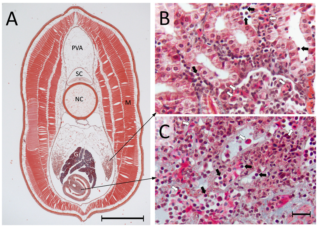
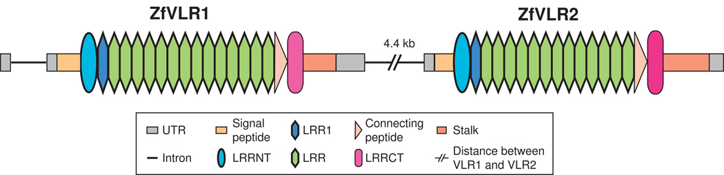
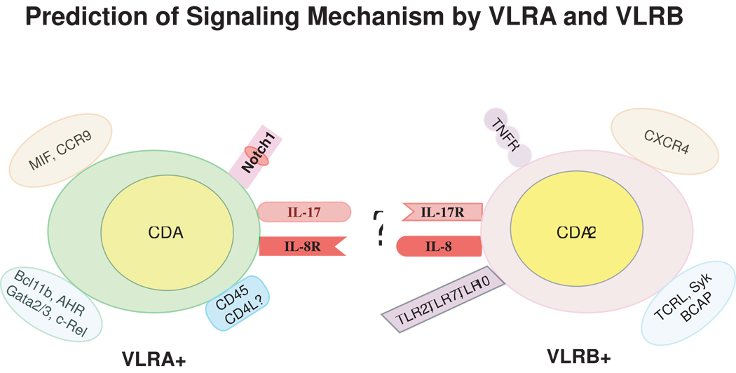
All extant vertebrates possess an adaptive immune system wherein diverse immune receptors are created and deployed in specialized blood cell lineages. Recent advances in DNA sequencing and developmental resources for basal vertebrates have facilitated numerous comparative analyses that have shed new light on the molecular and cellular bases of immune defense and the mechanisms of immune receptor diversification in the "jawless" vertebrates. With data from these key species in hand, it is becoming possible to infer some general aspects of the early evolution of vertebrate adaptive immunity. All jawed vertebrates assemble their antigen-receptor genes through combinatorial recombination of different "diversity" segments into immunoglobulin or T-cell receptor genes. However, the jawless vertebrates employ an analogous, but independently derived set of immune receptors in order to recognize and bind antigens: the variable lymphocyte receptors (VLRs). The means by which this locus generates receptor diversity and achieves antigen specificity is of considerable interest because these mechanisms represent a completely independent strategy for building a large immune repertoire. Therefore, studies of the VLR system are providing insight into the fundamental principles and evolutionary potential of adaptive immune recognition systems. Here we review and synthesize the wealth of data that have been generated towards understanding the evolution of the adaptive immune system in the jawless vertebrates.


Figures

Figure 1
Figure 1
Distribution of cells in primary hematopoietic tissues in larval lamprey. (A) A transverse section from the mid-body region of an ammocoete lamprey (~ 13 cm in length). The 10 µm processed section stained with Masson Trichrome is showing different major internal organs, which include protovertebral arch (PVA), spinal cord (SC), notochord (NC), gonad (G), kidney (K), typhlosole (T) and muscle (M). Scale bar = 1 mm. (B) A magnified view of the kidney showing the distribution of blood cells, including many lymphocytes and erythrocytes (black and white arrows, respectively). Collections of blood cells are seen in and amongst the renal tubules. (C) A magnified view of the typhlosole showing diverse blood cells (lymphocytes and erythrocytes are indicated by black and white arrows, respectively). Scale bar = 10 µm. The structures that are stained light blue are largely extracellular matrix, which is highly abundant in the typhlosole.
Figure 2
Figure 2
Genomic organization and rearrangement of a mature VLR gene of sea lamprey. The germline VLR (gVLR) configuration with 5’ and 3’-encoding LRR genetic segments (top) contains an additional 13.2 kb of non-coding intervening sequence and lacks the key LRR modules, which are essential to the structure of functional VLR genes. The inserted LRR modules lie both 5’ and 3’ of gVLR. The VLR locus undergoes stepwise assembly (middle) via recombination between short stretches of nucleotide homology found at the junctions of various LRR modules. This process may occur on either (or both) strand(s) during replication and gradually replaces the intervening sequence with all the variable LRR segments. The end product of these recombination events (bottom) is a mature VLR locus capped with an invariant 5’ end of the LRRNT module, an invariant 3’ end of the LRRCT module and a variable number of LRR cassettes (each encoding 24 amino acids) that vary in number and sequence. The illustration is not drawn to scale.
Figure 3
Figure 3
Genomic organization of two VLR-like genes in the zebrafish. The two VLR-like genes separated by 4.4 kb in the zebrafish genome assembly (Accession No. BX569794). Both genes contain all core VLR components identically juxtaposed as in cyclostome mVLRs. However, these genes contain comparatively more LRR cassettes than the average VLR molecule in cyclostomes and lack the ability to undergo genomic diversification. The exact phylogenetic relationship to cyclostome VLRs is unclear, though there is a distinct possibility that they are evolutionarily related at least on the basis of their similar genomic organizations. The functionality of these molecules is largely unknown. Color coding of structural features is presented in the inset and is identical to that in Figure 2. The illustration is not drawn to scale.
Figure 4
Figure 4
A conceptual diagram of differential gene expression by VLRA+ and VLRB+ lymphocytes. This illustration highlights the various proteins that were empirically shown [5] to be differentially expressed in flow sorted VLRA+ and VLRB+ lymphocyte populations [5]. These include possible enzymes for genomic rearrangement, signaling molecules, transcription factors, cell surface receptors, and chemokines/cytokines and their cognate receptor genes. The restriction of CDA1 and to VLRA+ and CDA2 to VLRB+ cells implies the potential for their selective involvement in the assembly of the VLRA and VLRB loci during lymphocyte development. It is speculated that the VLRA+ lymphocytes expressing IL-17 may attract IL-17R bearing VLRB+ lymphocytes, and VLRB+ lymphocytes may use IL-8 to attract and engage IL-8R bearing VLRA+ lymphocytes. Likewise, expression of TLR ligands by VLRB+ lymphocytes might trigger the activation of this cell population. Molecules are grouped on the basis of functionality and are not necessarily coexpressed (for example, TCRL, Syk and BCAP are B-cell signaling molecules, whereas Bcl11b, AHR, Gata2/3 and c-REL are T-cell transcription factors, etc).

Similar articles

Cited by

References

    1. Kasahara M, Suzuki T, Pasquier LD. On the origins of the adaptive immune system: novel insights from invertebrates and cold-blooded vertebrates. Trends Immunol. 2004 February;25(2):105–111. - PubMed
    1. Pancer Z, Amemiya CT, Ehrhardt GR, Ceitlin J, Gartland GL, Cooper MD. Somatic diversification of variable lymphocyte receptors in the agnathan sea lamprey. Nature. 2004 July 8;430(6996):174–180. - PubMed
    1. Alder MN, Rogozin IB, Iyer LM, Glazko GV, Cooper MD, Pancer Z. Diversity and function of adaptive immune receptors in a jawless vertebrate. Science. 2005 December 23;310(5756):1970–1973. - PubMed
    1. Rogozin IB, Iyer LM, Liang L, Glazko GV, Liston VG, Pavlov YI, Aravind L, Pancer Z. Evolution and diversification of lamprey antigen receptors: evidence for involvement of an AID-APOBEC family cytosine deaminase. Nat Immunol. 2007 June;8(6):647–656. - PubMed
    1. Guo P, Hirano M, Herrin BR, Li J, Yu C, Sadlonova A, Cooper MD. Dual nature of the adaptive immune system in lampreys. Nature. 2009 June 11;459(7248):796–801. - PMC - PubMed

Publication types

Substances