. 2010 Mar 5;285(10):7493-504.
doi: 10.1074/jbc.M109.083220. Epub 2010 Jan 7.

Elevated expression of Paneth cell CRS4C in ileitis-prone SAMP1/YitFc mice: regional distribution, subcellular localization, and mechanism of action

Affiliations

Elevated expression of Paneth cell CRS4C in ileitis-prone SAMP1/YitFc mice: regional distribution, subcellular localization, and mechanism of action

Michael T Shanahan et al. J Biol Chem. .

Abstract

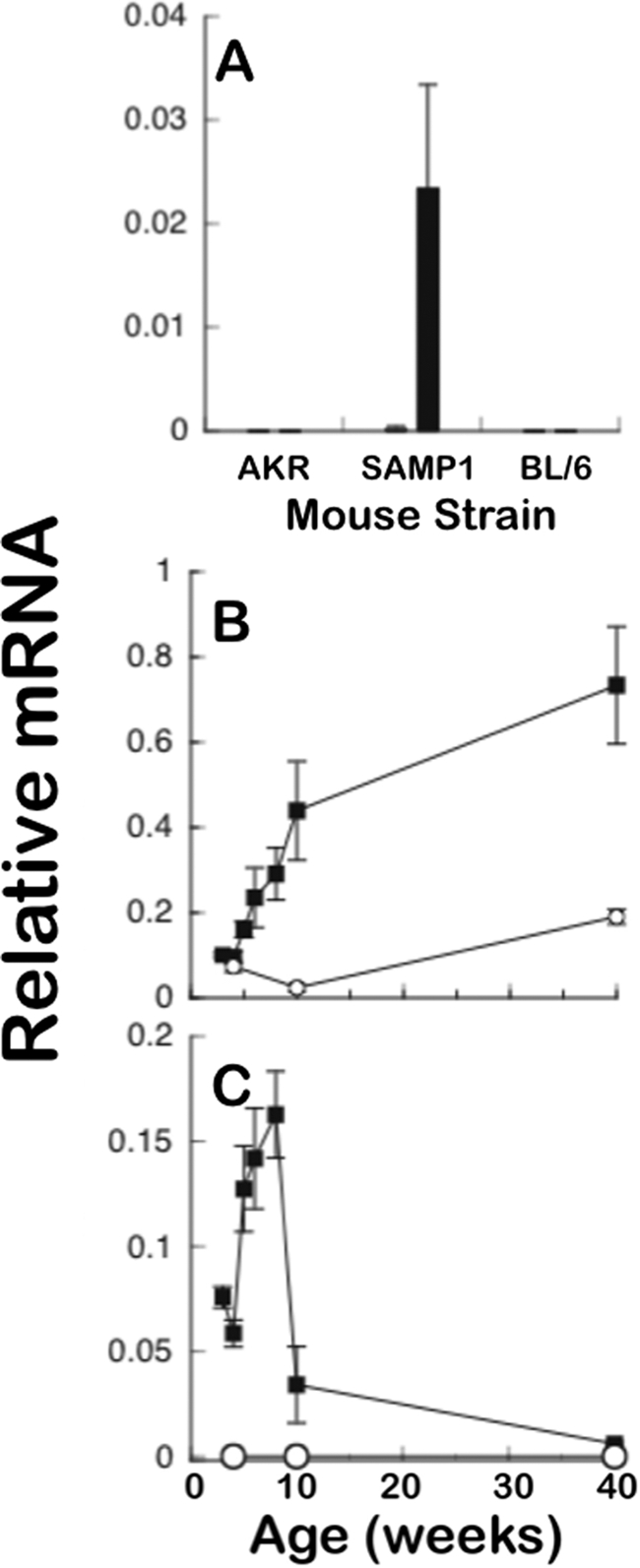
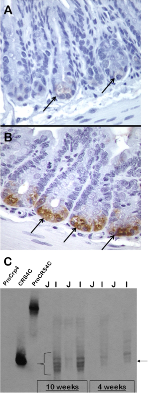
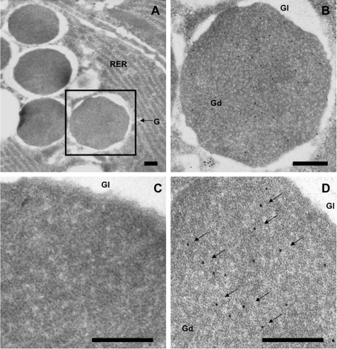
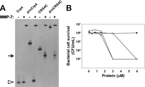
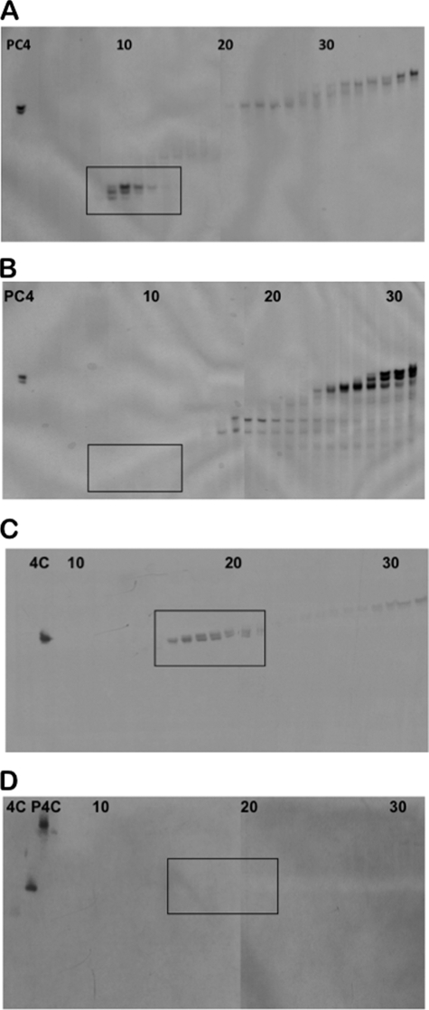
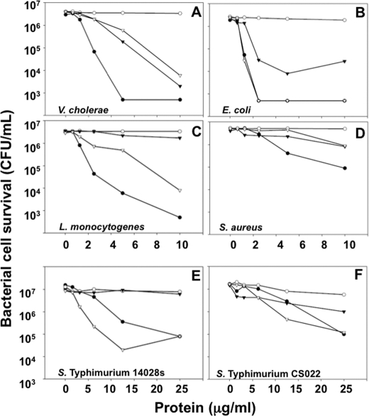
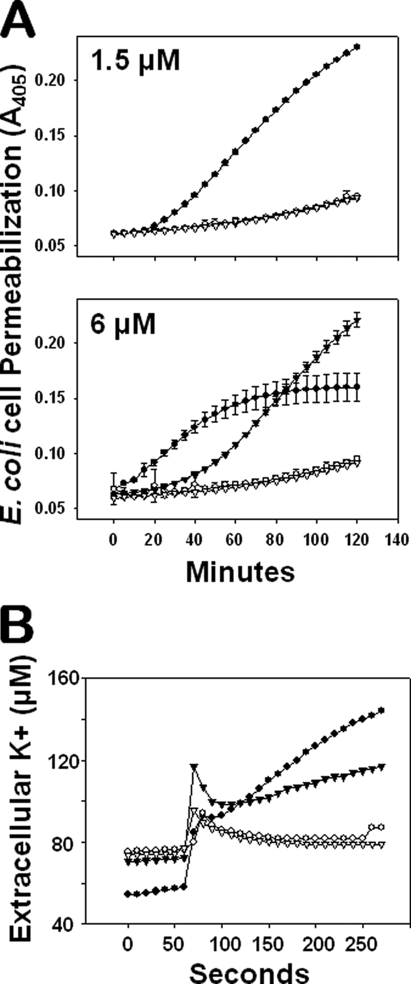
Paneth cells at the base of small intestinal crypts of Lieberkühn secrete host defense peptides and proteins, including alpha-defensins, as mediators of innate immunity. Mouse Paneth cells also express alpha-defensin-related Defcr-rs genes that code for cysteine-rich sequence 4C (CRS4C) peptides that have a unique CPX triplet repeat motif. In ileitis-prone SAMP1/YitFc mice, Paneth cell levels of CRS4C mRNAs and peptides are induced more than a 1000-fold relative to non-prone strains as early as 4 weeks of age, with the mRNA and peptide levels highest in distal ileum and below detection in duodenum. CRS4C-1 peptides are found exclusively in Paneth cells where they occur only in dense core granules and thus are secreted to function in the intestinal lumen. CRS4C bactericidal peptide activity is membrane-disruptive in that it permeabilizes Escherichia coli and induces rapid microbial cell K(+) efflux, but in a manner different from mouse alpha-defensin cryptdin-4. In in vitro studies, inactive pro-CRS4C-1 is converted to bactericidal CRS4C-1 peptide by matrix metalloproteinase-7 (MMP-7) proteolysis of the precursor proregion at the same residue positions that MMP-7 activates mouse pro-alpha-defensins. The absence of processed CRS4C in protein extracts of MMP-7-null mouse ileum demonstrates the in vivo requirement for intracellular MMP-7 in pro-CRS4C processing.


Figures

FIGURE 1.
FIGURE 1.
High ileal CRS4C mRNA levels in ileitis-prone SAMP1/YitFc mice. A, CRS4C mRNA levels were measured by quantitative real-time PCR in jejunum and ileum of 10-week-old AKR, SAMP1/YitFc, and C57BL/6 mice relative to 18 S rRNA using sequence-specific primers (see “Experimental Procedures”). Filled bars denote levels in ileum, open bars are levels in jejunum. Data are expressed as mean ± S.D. with n = 4 mice/group. p < 0.001 SAMP1/YitFc ileum versus B6 ileum; p < 0.0001 SAMP1/YitFc ileum versus AKR ileum. B, Paneth cell α-defensin mRNA levels were measured at the ages indicated as given in A. Levels in SAMP1/YitFc (filled squares) and AKR (open circles) mouse ileum were compared using pan-Crp primers Defcrp130 and Defcrm380, which amplify all known mouse Crps. Data are expressed as mean ± S.D. with n = 4–6 mice/group. C, ileal CRS4C mRNA levels were measured as in B. Levels in SAMP1/YitFc (filled squares) and AKR (open circles) mouse ileum were compared using the CRS4C-specific primer set (“Experimental Procedures”). Data are expressed as mean ± S.D. with n = 4–6 mice/group.
FIGURE 2.
FIGURE 2.
Temporal and regional CRS4C peptide levels in SAMP1/YitFc mouse small intestine. A and B, immunohistochemical staining of crypts of the non-ileitis-prone AKR (A) and SAMP1/YitFc (B) mouse strains using anti-CRS4C-1 antiserum (see “Experimental Procedures”). Arrows indicate strongly immunopositive SAMP1/YitFc Paneth cells in B and AKR Paneth cells in A that exhibit markedly lower levels of immunoreactivity. CRS4C peptides are products of Paneth cells in the intestinal crypts of both mice. C, age-related appearance and regional distribution of CRS4C peptide accumulation determined for the SAMP1/YitFc mouse by Western blotting. Samples (0.50 mg) of total organ protein-extracted jejunum (J) and ileum (I) of individual 4- or 10-week-old SAMP1/YitFc mice were separated by AU-PAGE as noted and blotted onto a nitrocellulose membrane. The blot was subjected to Western blot analysis using anti-CRS4C-1 IgG diluted 1:5 (see “Experimental Procedures”). Recombinant pro-Crp-4 (leftmost lane) was a negative control peptide, and recombinant CRS4C-1 and pro-CRS4C-1 provided positive controls as shown by their strong immunoreactivity. Immunopositive bands that co-migrate with recombinant CRS4C-1, indicated by the lower right arrow and the curly brace, are readily detected in ileum of both 4- and 10-week-old SAMP1/YitFc mice, with the intensity of the signal markedly increased by 10 weeks of age.
FIGURE 3.
FIGURE 3.
CRS4C protein localization to dense core secretory granules of mouse Paneth cells. Sections of C57/BL6 mouse small intestinal tissue were incubated with preimmune (C) and anti-CRS4C-1 antisera and anti-goat IgG secondary antibody conjugated with 10-nm gold particles (A, B, and D), as described under “Experimental Procedures.” Magnification, ×20,000 (A). Scale bars, 200 nm (all panels). Electron micrographs show that the anti-CRS4C antibody reacts specifically with the electron-dense regions of Paneth cell secretory granules (arrows).
FIGURE 4.
FIGURE 4.
In vitro enzymatic activation of pro-CRS4C-1 bactericidal activity by MMP-7. A, equimolar samples of recombinant Crp-4, pro-Crp-4, deduced mature CRS4C-1, and pro-CRS4C-1 were incubated overnight at 37 °C with (+) or without (−) 0.5-mol equivalents of MMP-7 and subjected to analytical AU-PAGE. Note that MMP-7cleaves pro-Crp-4 as well as pro-CRS4C. Pro-CRS4C peptides are processed by MMP-7. B, exponentially growing E. coli ML35 were exposed to mature CRS4C-1 (○) and pro-CRS4C-1 (●) as well as mature CRS4C-1 (▿) and pro-CRS4C-1 (▾) that were exposed to MMP-7 for 1 h at 37 °C. Surviving bacteria were quantified as colony-forming units (CFU)/ml (see “Experimental Procedures”). The displayed result is representative of three independent experiments. MMP-7 converts the inactive CRS4C precursor to a bactericidal peptide activity level equivalent to that of mature CRS4C.
FIGURE 5.
FIGURE 5.
Analysis of in vivo processing of native CRS4C peptides. Acid extracts of ileal organs from wild-type SAMP1/YitFc mice (A and C) and MMP-7 gene knock-out mice (B and D) were separated by preparative AU-PAGE and analyzed in analytical AU-polyacrylamide gels by Coomassie Blue staining (A and B) and Western blotting using anti-CRS4C-1 IgG (1:15) (C and D). Peptide controls included pro-Crp-4 (PC4), CRS4C-1 (4C), and pro-CRS4C-1 (P4C), and fraction numbers are denoted at the tops of the gels. Processed mouse enteric α-defensins (A, boxed region) and immunoreactive, processed CRS4C (C, boxed region) are found in wild-type mice but absent in MMP-7-null mice. MMP-7 is required for native CRS4C processing in vivo.
FIGURE 6.
FIGURE 6.
Bactericidal peptide activities of native and alkylated CRS4C. Exponentially growing V. cholerae (A), E. coli (B), L. monocytogenes (C), S. aureus (D), S. typhimurium 14028s (E), and S. typhimurium CS022 (F) were exposed to the peptide concentrations shown at 37 °C in 50 ml of 10 mm PIPES buffer supplemented with 1% trypticase soy broth for 1 h. Following peptide exposure, the bacteria were plated on trypticase soy broth and agar and incubated overnight at 37 °C. Surviving bacteria were counted as colony-forming units (CFU)/ml at each peptide concentration, and each panel is representative of three independent experiments. Values at or below 1 × 10³ colony-forming units/ml signify that no colonies were detected. ●, Crp-4; ○, pro-Crp-4; ▿, native CRS4C-1(54–72); ▾, alkylated CRS4C-1(54–72).
FIGURE 7.
FIGURE 7.
CRS4C-mediated bactericidal peptide activity is mediated via a membrane disruptive mechanism. A, E. coli ML35 cells were exposed to 1.5 μm (upper panel) and 6.0 μm (lower panel) pro-Crp-4 (○), Crp-4 (●), pro-CRS4C-1 (▿), and CRS4C-1 (▾) in the presence of 2.5 mm ONPG for 2 h at 37 °C. Membrane permeabilization was measured spectrophotometrically at A405 nm. Mature CRS4C-1 and Crp-4, but not their precursors, induce target cell membrane leakage. B, membrane efflux of K+ from live E. coli cells was measured in response to peptide exposure as an index of membrane disruption and cell death. Exponential-growing E. coli ML35 cells were incubated for 1 min in 10 mm PIPES (pH 7.4) supplemented with 0.1% (v/v) trypticase soy broth and subsequently exposed to 7 μm pro-Crp-4 (○), Crp-4 (●), pro-CRS4C-1 (▿), and CRS4C-1 (▾) in the same buffer for 4 min at 37 °C. Extracellular levels of potassium ions were measured using an ion-selective electrode sensitive for K+ (see “Experimental Procedures”). Mature CRS4C-1 and Crp-4, but not their precursors, induce K+ efflux of target cells.

Similar articles

Cited by

References

    1. Lehrer R. I. (2007) Curr. Opin. Hematol. 14, 16–21 - PubMed
    1. Selsted M. E., Ouellette A. J. (2005) Nat. Immunol. 6, 551–557 - PubMed
    1. Brogden K. A., Ackermann M., McCray P. B., Jr., Tack B. F. (2003) Int. J. Antimicrob. Agents 22, 465–478 - PubMed
    1. Schutte B. C., McCray P. B., Jr. (2002) Annu. Rev. Physiol. 64, 709–748 - PubMed
    1. Tomasinsig L., Zanetti M. (2005) Curr. Protein. Pept. Sci. 6, 23–34 - PubMed

Publication types

MeSH terms