. 2010 Feb;137(3):457-66.
doi: 10.1242/dev.045286. Epub 2010 Jan 7.

Multi-step control of muscle diversity by Hox proteins in the Drosophila embryo

Affiliations

Multi-step control of muscle diversity by Hox proteins in the Drosophila embryo

Jonathan Enriquez et al. Development. 2010 Feb.

Abstract

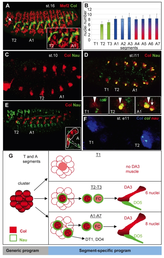
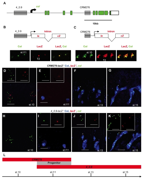
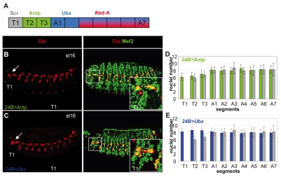
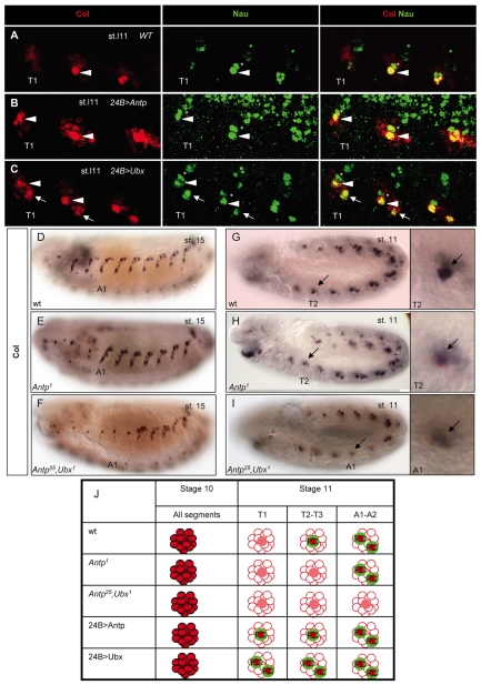
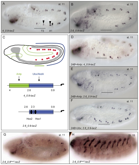
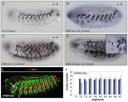
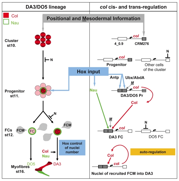
Hox transcription factors control many aspects of animal morphogenetic diversity. The segmental pattern of Drosophila larval muscles shows stereotyped variations along the anteroposterior body axis. Each muscle is seeded by a founder cell and the properties specific to each muscle reflect the expression by each founder cell of a specific combination of 'identity' transcription factors. Founder cells originate from asymmetric division of progenitor cells specified at fixed positions. Using the dorsal DA3 muscle lineage as a paradigm, we show here that Hox proteins play a decisive role in establishing the pattern of Drosophila muscles by controlling the expression of identity transcription factors, such as Nautilus and Collier (Col), at the progenitor stage. High-resolution analysis, using newly designed intron-containing reporter genes to detect primary transcripts, shows that the progenitor stage is the key step at which segment-specific information carried by Hox proteins is superimposed on intrasegmental positional information. Differential control of col transcription by the Antennapedia and Ultrabithorax/Abdominal-A paralogs is mediated by separate cis-regulatory modules (CRMs). Hox proteins also control the segment-specific number of myoblasts allocated to the DA3 muscle. We conclude that Hox proteins both regulate and contribute to the combinatorial code of transcription factors that specify muscle identity and act at several steps during the muscle-specification process to generate muscle diversity.


Figures

Fig. 1.
Fig. 1.
Segment-specific formation and properties of the DA3 muscle. (A) Mef2 (red) and Col (green) expression in a stage (st.) 16 Drosophila embryo, showing the entire muscle pattern and DA3 muscle, respectively. The arrow indicates the absence of DA3 muscle in the T1 segment. The inset shows an enlarged view of the T2, T3 and A1 segments. Note that the DA3 muscle is smaller in T than in A segments. (B) The segment-specific number of nuclei in the DA3 muscle (number of segments counted=30). The mesodermal domain of expression of Antp is in green, Ubx in blue and AbdA in red (Bate,1993) (see Fig. S3 in the supplementary material). (C-E) Comparison of expression of Col (red) and Nau (green) in stage 10 (C), late stage 11 (st.l11) (D) and stage 15 (E) embryos. Insets in D show the Col-expressing cluster in T1 (arrowhead), the DA3/DO5 progenitor in T2, and DA3/DO5 (arrowhead) and the DT1/DO4 progenitors (arrow) in A1. The inset in E is an enlarged view of the abdominal DA3 (circled) and DO5 (dotted circle) muscles that express Col and Nau, respectively. (F) Double in situ hybridization for col (green) and nau (red) primary transcripts and immunostaining for Col (blue). The T1 and T2 segments of an early stage 11 embryo are shown. Two dots per nucleus reflect the two alleles. All embryos are oriented anterior to the left and dorsal to the top. (G) Col (red) and Nau (green) expression during the process of DA3 muscle formation in different trunk segments, highlighting the segment-specific aspects. PC, progenitor; FC, founder cell.
Fig. 2.
Fig. 2.
Two separate CRMs control col transcription at different steps during DA3 muscle specification. (A) The Drosophila col genomic region. Coding exons are in green. The transcription start site is indicated by an arrow. Gray boxes represent the muscle CRMs 4_0.9 and 276. (B,C) The 4_0.9-lacZi and CRM276-lacZi transgenes, in which the lacZ coding sequence (red) is split by the Drosophila virilis βtub56D intron. The transcription start site is indicated by an arrow. Beneath are shown (B) stage 11 4_0.9-lacZi and (C) stage 10 CRM276-lacZi embryos immunostained for Col (green) and β-galactosidase (red, LacZ), or both, as indicated. (D-K) Double in situ hybridization for col (green) and lacZ (red) primary transcripts and Col immunostaining (blue) on (D-G) CRM276-lacZi and (H-K) 4_0.9-lacZi embryos. Developmental stages are indicated in each panel. The T2 and T3 segments are shown. Insets show only lacZi or col transcripts. Note that the red and green dots corresponding to lacZi and col, respectively, do not always overlap because the two genes are located at separate chromosomal positions, with two dots per gene per nucleus reflecting the two alleles. The asterisk (F,G,J,K) indicates a Col-expressing multidendritic (md) neuron. CRM276-lacZi transcription is only active in promuscular clusters and in the corresponding progenitor (D,E). 4_0.9-lacZi transcription starts in the DA3/DO5 progenitor (I) and is active in DA3 nuclei until stage 15 (J,K). (L) The temporal windows of activity of CRM276 and 4_0.9, which specifically overlap at the progenitor stage (gray).
Fig. 3.
Fig. 3.
Hox proteins are required for the formation of the DA3 muscle and allocate its number of nuclei. (A) A color-coded representation of Scr, Antp, Ubx and AbdA expression in the Drosophila trunk segments. (B,C) Col (red) and Mef2 (green) expression in stage 16 embryos expressing Antp (B) or Ubx (C) in all mesodermal cells (24B-Gal4 driver). Col staining alone is shown on the left. The arrow points to the DA3-like muscle that forms in T1. (D,E) The number of nuclei per DA3 muscle in each T1 to A7 segment. The genotypes are shown at the top, with the Hox color code as in Fig. 1. The gray bars show wild-type (wt) nuclei numbers. Number of segments counted=30.
Fig. 4.
Fig. 4.
Hox proteins control the number of muscle progenitors expressing Col and Nau. (A-C) Col (red) and Nau (green) expression in T segments of late stage 11 Drosophila embryos. Overlap (yellow) is shown on the right. (A) One progenitor expressing high levels of Col and Nau is found in wt T2 (arrowhead) and T3 and in T1 upon expression of Antp (B) or Ubx (C). (C) Ubx induces a second progenitor (arrow) to express high levels of Col and Nau in all three T segments, similar to wt A segments (see Fig. 1D). The asterisk indicates another muscle progenitor that expresses Nau, but not Col, in wt T segments and sometimes low levels of Col upon Hox overexpression. (D-I) Col expression in stage 15 (D-F) and 11 (G-I) embryos. The DA3 muscle is lacking in T2-T3 and T2-A2 in Antp (E) and Antp; Ubx (F) mutants, respectively, correlating with the absence of high-level Col-expressing progenitors (H,I). The remaining Col-positive nucleus corresponds to that of an md neuron, as shown by its expression of pickpocket (Crozatier and Vincent, 2008). Enlarged views of the Col-expressing cluster/progenitor in T2 (G,H) and A1 (I) show that Col upregulation in progenitor cells does not occur in the absence of Hox protein. Residual Col protein indicates the position of the Col-expressing cluster. (J) Col (red) and Nau (green) expression in muscle progenitors in different trunk segments in Hox mutants and Hox-overexpression conditions.
Fig. 5.
Fig. 5.
Hox regulation of col transcription at the progenitor stage is modular. (A,B,D-H) Lateral views of stage 11 Drosophila embryos expressing lacZ in the DA3/DO5 (A, arrowhead) and DT1/DO4 (A, arrow) progenitors under control of either the 4_0.9 or 2.6_0.9 CRM. (A,B) lacZ expression in T2 and T3 (horizontal bar) in wt embryos requires the 4_2.6 CRM. (D-F) 2.6_0.9-lacZ expression is induced in T2 and T3 by Ubx but not by Antp; 4_0.9-lacZ expression is induced by Antp in all segments, including in T1 (asterisk in D). (G) Mutation of the Hox2 site abolishes 2.6_0.9-lacZ expression in progenitors. (H) lacZ expression in the DA3 muscle at stage 15 is independent of the 4_2.6 CRM and Hox2. (C) Schematic of a stage 11 embryo with the Col-expressing progenitors shown as red dots and domains of mesodermal Antp and Ubx/AbdA expression as green and blue lines, respectively. Beneath are the 4_0.9-lacZ and 2.6_0.9-lacZ constructs showing the Antp- and Ubx-responsive elements in green and blue, respectively. Position of the Hox1 and Hox2 sites is indicated (see Fig. S4 in the supplementary material).
Fig. 6.
Fig. 6.
Expression of Nau and Col can bypass the Hox requirement for DA3 muscle specification. (A-D) lacZ expression in stage 16 2.6_0.9-lacZ Drosophila embryos upon pan-mesodermal expression of Nau (B), Col (C), Col plus Nau (D) or neither protein (A). Only co-expression of Col plus Nau results in the formation of a DA3-like muscle in T1 (arrow). This ectopic muscle has only two nuclei (inset in D). (E) Double immunostaining of stage 15 embryos for Mef2 (green) and Col (red) showing Col expression upon expression of Ubx in all FCs. (F) The number of nuclei per DA3 muscle in each segment of rP298>Ubx embryos is in blue. Wt values are in gray. Number of segments counted=30.
Fig. 7.
Fig. 7.
Transcriptional control of Drosophila muscle identity: the central role of Hox proteins. Expression of Col (red) and Nau (green) in the DA3 muscle lineage is shown on the left, with the corresponding cis control of col expression represented on the right. col transcription is activated in one promuscular cluster per trunk segment under control of the early-acting CRM276, which integrates A/P and D/V positional information, mesoderm-intrinsic cues and repression by N signaling. This results in Col expression in one (T segments) or two (A1-A7 segments) dorsal progenitors. Upregulation of col (red) and nau (green) transcription in the DA3/DO5 progenitor at stage 11 is controlled by Antp and Ubx/AbdA and is mediated by two separate elements of the 4_0.9 CRM. This is the key step, when segment-specific variations are imposed on the segmental muscle pattern. Following progenitor division, restriction of col transcription to the DA3 FC requires positive regulation by Col and Nau and repression by N. From this stage on, Col is required for activating its own transcription in all FCM nuclei incorporated into the DA3 myofiber. Hox activity in FCs controls the number of myoblasts allocated to the DA3 muscle, independently of the control of nau and col. The key regulatory steps are shaded and centered: early positional information is relayed by segment-specific Hox information at the progenitor stage. The active and inactive CRMs are indicated by gray and white boxes, respectively. Binding of mesodermal transcription factors (M), such as Twi and Mef2, to CRM 276 and 4_0.9 is suggested by phylogenomic footprinting (see Figs S3 and S4 in the supplementary material) and ChIP-on-chip data (Sandmann et al., 2006; Sandmann et al., 2007).

Similar articles

Cited by

References

    1. Abbott M. K. and Kaufman, T. C. (1986). The relationship between the functional complexity and the molecular organization of the Antennapedia locus of Drosophila melanogaster. Genetics 114, 919-942. - PMC - PubMed
    1. Affolter M., Slattery, M. and Mann, R. S. (2008). A lexicon for homeodomain-DNA recognition. Cell 133, 1133-1135. - PubMed
    1. Alvares L. E., Schubert, F. R., Thorpe, C., Mootoosamy, R. C., Cheng, L., Parkyn, G., Lumsden, A. and Dietrich, S. (2003). Intrinsic, Hox-dependent cues determine the fate of skeletal muscle precursors. Dev. Cell 5, 379-390. - PubMed
    1. Balagopalan L., Keller, C. A. and Abmayr, S. M. (2001). Loss-of-function mutations reveal that the Drosophila nautilus gene is not essential for embryonic myogenesis or viability. Dev. Biol. 231, 374-382. - PubMed
    1. Barolo S., Carver, L. A. and Posakony, J. W. (2000). GFP and beta-galactosidase transformation vectors for promoter/enhancer analysis in Drosophila. Biotechniques 29, 726, 728, 730, 732. - PubMed

Publication types