. 2010 Feb;65(2):147-55.
doi: 10.1093/gerona/glp203. Epub 2010 Jan 7.

Possible molecular mechanisms underlying age-related cardiomyocyte apoptosis in the F344XBN rat heart

Affiliations

Possible molecular mechanisms underlying age-related cardiomyocyte apoptosis in the F344XBN rat heart

Sunil K Kakarla et al. J Gerontol A Biol Sci Med Sci. 2010 Feb.

Abstract

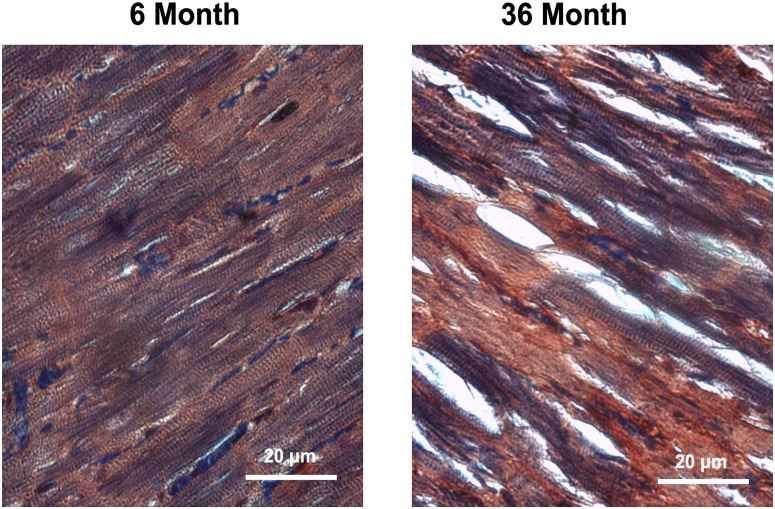
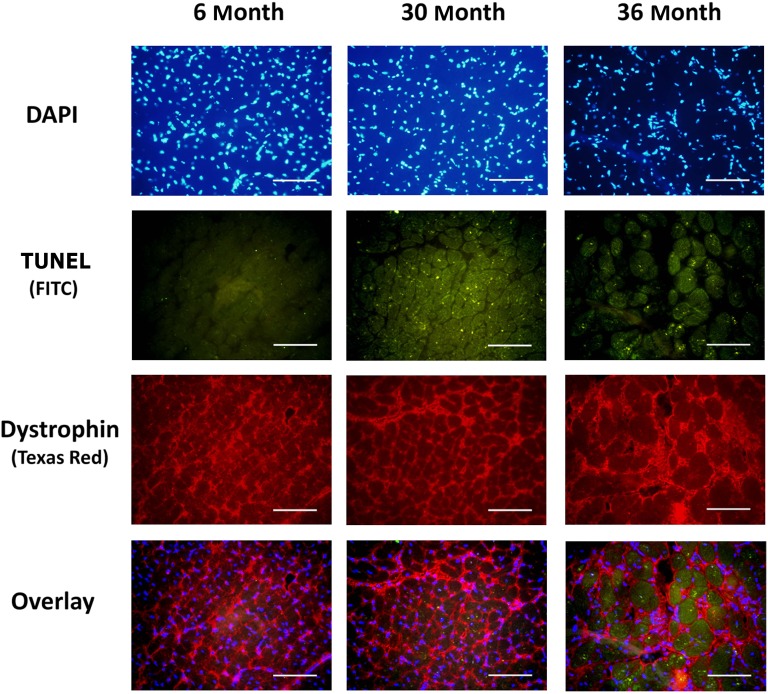
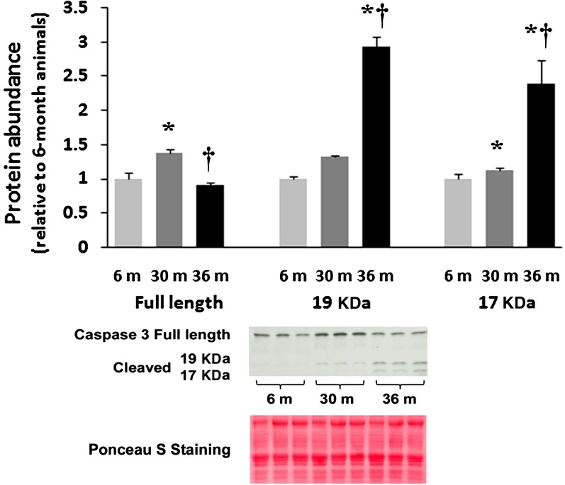
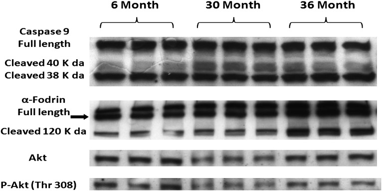
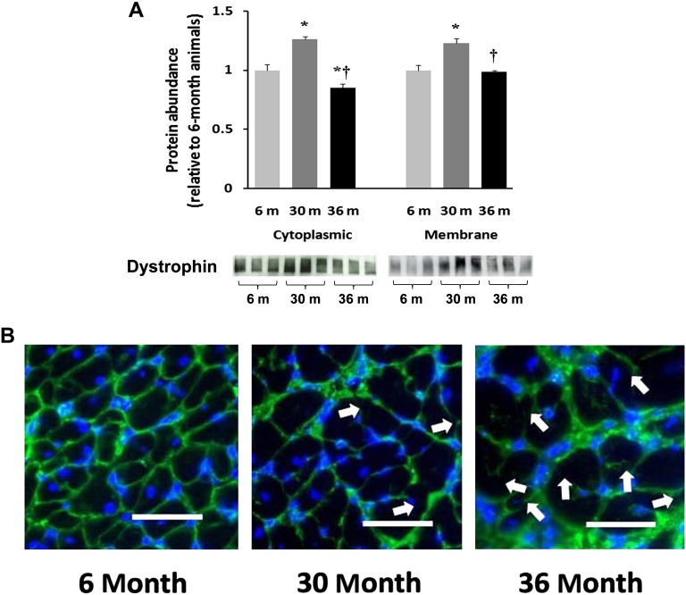
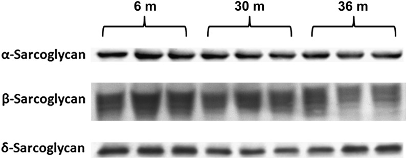
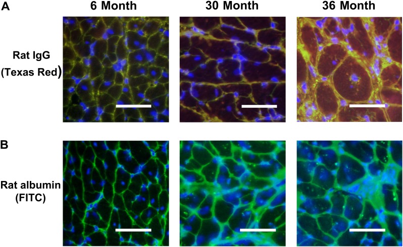
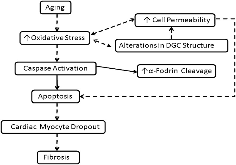
Despite advances in treatment, age-related cardiac dysfunction still remains a leading cause of cardiovascular death. Recent data have suggested that increases in cardiomyocyte apoptosis may be involved in the pathological remodeling of heart. Here, we examine the effects of aging on cardiomyocyte apoptosis in 6-, 30-, and 36-month-old Fischer344 x Brown Norway F1 hybrid rats (F344XBN). Compared with 6-month hearts, aged hearts exhibited increased TdT-mediated dUTP nick end labeling-positive nuclei, caspase-3 activation, caspase-dependent cleavage of alpha-fodrin and diminished phosphorylation of protein kinase B/Akt (Thr 308). These age-dependent increases in cardiomyocyte apoptosis were associated with alterations in the composition of the cardiac dystrophin glycoprotein complex and elevated cytoplasmic IgG and albumin immunoreactivity. Immunohistochemical analysis confirmed these data and demonstrated qualitative differences in localization of dystrophin-glycoprotein complex (DGC) molecules with aging. Taken together, these data suggest that aging-related increases in cardiac apoptotic activity model may be due, at least in part, to age-associated changes in DGC structure.


Figures

Figure 1.
Figure 1.
Aging alters the morphology of cardiac tissue in the F344XBN heart. Cross sections of hearts obtained from adult (6-month) and very aged (36-month) rats after staining with phosphotungstic acid hematoxylin to visualize cardiac morphology. Bar = 20 μm.
Figure 2.
Figure 2.
Aging increases the number of apoptotic nuclei in cardiomyocytes. TdT-mediated dUTP nick end labeling [TUNEL] staining along with immunostaining for dystrophin (Texas Red) was used to investigate myocytes apoptosis in 6-, 30-, and 36-month hearts. Bar = 50 μm.
Figure 3.
Figure 3.
Aging increases the cleavage of caspase-3. Protein isolates from cytosolic fraction of hearts excised from adult (6-month), aged (30-month), and very aged (36-month) rats were analyzed by immunoblotting for changes in caspase-3 and caspase-3 cleavage. Ponceau S staining of the nitrocellulose membrane along with densitometric analysis of protein present was done to verify equivalent protein loading between the lanes (data not shown). Asterisk indicates significant difference from adult (6-month) value (p < .05). Dagger indicates significant difference from 30-month value (p < .05). n = 8 for all groups.
Figure 4.
Figure 4.
Aging alters the regulation of mitochondrial dependent apoptotic pathway signaling proteins. Protein isolates from cytosolic fraction of hearts excised from adult (6-month), aged (30-month), and very aged (36-month) rats were analyzed by immunoblotting for changes in caspase-9, α-fodrin, Akt, and Akt phosphorylation (Thr308). Ponceau S staining of the nitrocellulose membrane along with densitometric analysis of protein present was done to verify equivalent protein loading between the lanes (data not shown). n = 8 for all groups.
Figure 5.
Figure 5.
Aging alters the regulation of dystrophin in the F344XBN heart. A). Protein isolates from cytoplasmic and membrane fractions of hearts obtained from adult (6-month), aged (30-month), and very aged (36-month) rats were analyzed by immunoblotting for changes in dystrophin protein content. Ponceau S staining of the nitrocellulose membrane along with densitometric analysis of protein present was done to verify equivalent protein loading between the lanes (data not shown). Asterisk indicates significant difference from adult (6-month) value (p < .05). Dagger indicates significant difference from 30-month value (p < .05). n = 8 for all groups. B). Dystrophin immunofluorescence in 6-, 30-, and 36-month hearts. Arrows indicate irregular and discontinuous immunoreactive signaling along the sarcolemma in the 30- and 36-month hearts. Bar = 50 μm.
Figure 6.
Figure 6.
Aging alters the regulation of dystroglycans in the F344XBN heart. Protein isolates from cytoplasmic and membrane fractions of hearts excised from adult (6-month), aged (30-month), and very aged (36-month) rats were analyzed by immunoblotting for changes in (A) α-dystroglycan and (B) β-dystroglycan protein expression. Ponceau S staining of the nitrocellulose membrane along with densitometric analysis of protein present was done to verify equivalent protein loading between the lanes (data not shown). Asterisk indicates significant difference from adult (6-month) value (p < .05). Dagger indicates significant difference from 30-month muscles (p < .05). n = 8 for all groups.
Figure 7.
Figure 7.
α-, β-, and δ-sarcoglycan protein levels are altered with aging. Protein isolates excised from adult (6-month), aged (30-month), and very aged (36-month) rats were analyzed by immunoblotting for α-, β-, and δ-sarcoglycan protein expression. n = 8 for all groups.
Figure 8.
Figure 8.
Aging is associated with a loss of cardiac myocyte membrane integrity. A). Double immunofluorescence labeling with dystrophin (FITC) and rat IgG (Texas Red) in 6-, 30-, and 36-month hearts. Increased permeability of the myocytes membrane is indicated by intracellular IgG reactivity. Note diffuse immunoreactive signaling within the cytoplasm of the 30- and 36-month hearts. B). Immunofluorescent labeling with anti-rat albumin in 6-, 30-, and 36-month hearts. Note localization of albumin in 30- and 36-month hearts. Bar = 50 μm.
Figure 9.
Figure 9.
Proposed model for how changes in the dystrophin–glycoprotein complex (DGC) may be related to age-related cardiac dysfunction. Data from the literature support the existence of increased oxidative stress and contractile dysfunction in aged cardiac muscle. Changes in the cardiac DGC could contribute to each of these processes either directly or by causing changes in cell permeability or by increasing myocyte apoptosis. Dashed arrows indicate cellular mechanisms that have been postulated to participate where as solid arrows indicate measures that were experimentally assessed in this study.

Similar articles

Cited by

References

    1. Lakatta EG. Aging and cardiovascular structure and function in healthy sedentary humans. Aging (Milano) 1998;10:162–164. - PubMed
    1. Hacker TA, McKiernan SH, Douglas PS, Wanagat J, Aiken JM. Age-related changes in cardiac structure and function in Fischer 344 x Brown Norway hybrid rats. Am J Physiol Heart Circ Physiol. 2006;290:H304–H311. - PubMed
    1. Walker EM, Jr, Nillas MS, Mangiarua EI, et al. Age-associated changes in hearts of male Fischer 344/Brown Norway F1 rats. Ann Clin Lab Sci. 2006;36:427–438. - PubMed
    1. Asano S, Rice KM, Kakarla S, et al. Aging influences multiple indices of oxidative stress in the heart of the Fischer 344/NNia x Brown Norway/BiNia rat. Redox Rep. 2007;12:167–180. - PubMed
    1. Tsutsui H, Ide T, Kinugawa S. Mitochondrial oxidative stress, DNA damage, and heart failure. Antioxid Redox Signal. 2006;8:1737–1744. - PubMed

Publication types