. 2010 Mar;21(3):419-27.
doi: 10.1681/ASN.2009060571. Epub 2010 Jan 7.

Satellite cell dysfunction and impaired IGF-1 signaling cause CKD-induced muscle atrophy

Affiliations

Satellite cell dysfunction and impaired IGF-1 signaling cause CKD-induced muscle atrophy

Liping Zhang et al. J Am Soc Nephrol. 2010 Mar.

Abstract

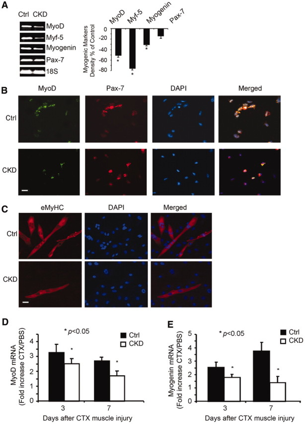
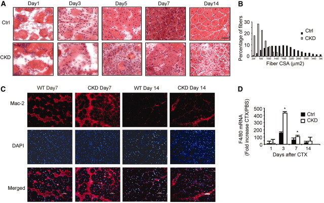
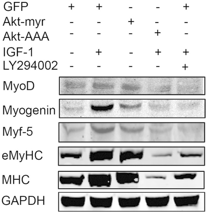
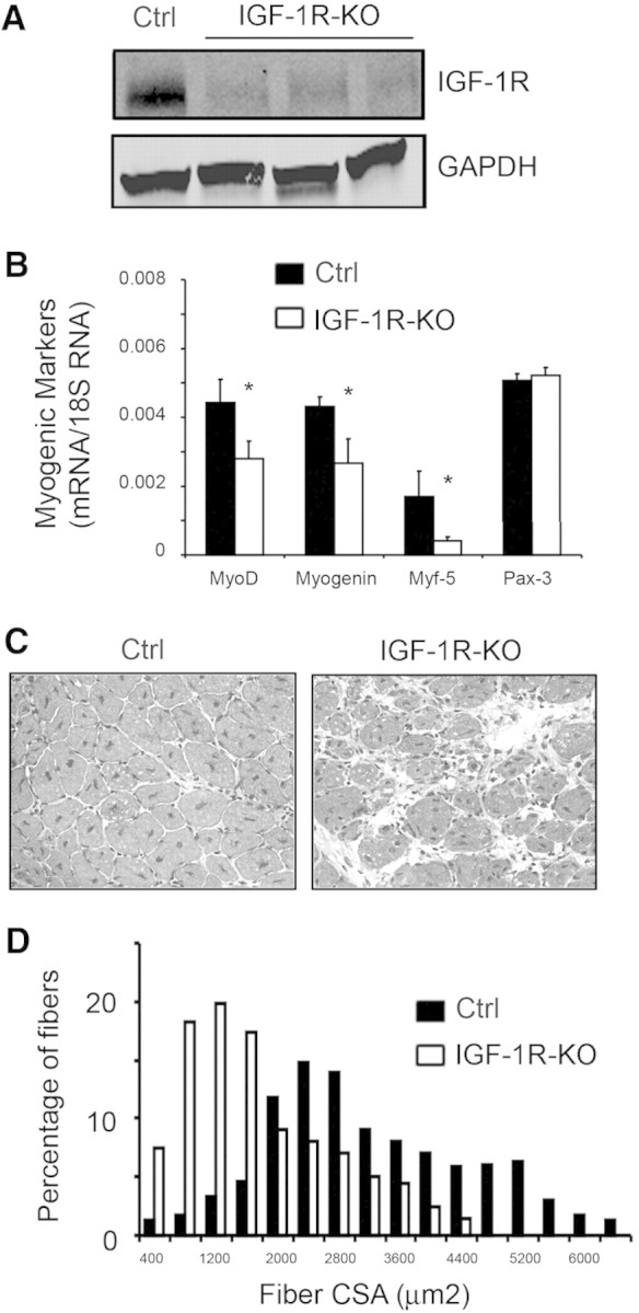
Muscle wasting in chronic kidney disease (CKD) begins with impaired insulin/IGF-1 signaling, causing abnormal protein metabolism. In certain models of muscle atrophy, reduced satellite cell function contributes to atrophy, but how CKD affects satellite cell function is unknown. Here, we found that isolated satellite cells from mice with CKD had less MyoD, the master switch of satellite cell activation, and suppressed myotube formation compared with control mice. In vivo, CKD delayed the regeneration of injured muscle and decreased MyoD and myogenin expression, suggesting that CKD impairs proliferation and differentiation of satellite cells. In isolated satellite cells from control mice, IGF-1 increased the expression of myogenic genes through an Akt-dependent pathway. CKD impaired Akt phosphorylation in satellite cells after muscle injury. To test whether impaired IGF-1 signaling could be responsible for decreased satellite cell function in CKD, we created an inducible IGF-1 receptor knockout mouse and found impaired satellite cell function and muscle regeneration. In addition, both CKD and IGF-1 receptor knockout mice developed fibrosis in regenerating muscles. Taken together, impaired IGF-1 signaling in CKD not only leads to abnormal protein metabolism in muscle but also impairs satellite cell function and promotes fibrosis in regenerating muscle. These signaling pathways may hold potential therapeutic targets to reduce CKD-related muscle wasting.


Figures

Figure 1.
Figure 1.
CKD impairs satellite cell activation. (A) mRNAs of myogenic markers (MyoD, myogenin, and Myf-5) are reduced in CKD mouse gastrocnemius muscles (left). Differences were quantified as percentage difference = [(CKD − control)/control] × 100 (right). Reverse transcriptase–PCR (RT-PCR) was repeated three times for each group of five mice (*P < 0.05, CKD versus control [Ctrl]). (B) Isolated satellite cells from muscles of CKD and control mice were cultured for 16 hours before immunostaining for MyoD (green), Pax-7 (red), and DAPI (blue). Yellow color in the merged picture indicates that Pax-7–positive cells expressed MyoD. Bar = 20 μm. (C) Satellite cells isolated as in B were cultured for 10 days before incubation in 2% horse serum to elicit differentiation. Immunostaining of eMyHC (red) identified myofibers, and DAPI (blue) detected nuclei. Bar = 20 μm. (D and E) For examination of satellite cell activation in vivo, satellite cells were isolated from CTX-injured muscles. Using RT-PCR, MyoD and myogenin mRNAs from satellite cells were measured and normalized to 18S mRNA. The ratio of satellite cell mRNAs from injured versus control muscles is shown (*P < 0.05 versus results from satellite cells of control mice; n = 5 mice per group).
Figure 2.
Figure 2.
CKD suppresses muscle regeneration in vivo induced by injuring muscle to activate satellite cells. (A) Representative hematoxylin- and eosin-stained cross-cryosections of injured TA muscles were obtained from control and CKD mice. Regenerated myofibers were identified by their central nuclei. Injured muscles from CKD mice had increased infiltration of cells and slowed regeneration. Bar = 20 μm. (B) At 1 month after injury, fibers were immunostained for laminin, and the sizes of myofibers with central nuclei were measured. The histogram of the sizes of the myofibers with central nuclei indicates that CKD impaired regeneration. (C) At days 7 and 14 after injury, muscles were immunostained for macrophages (Mac-2 antibody; red) and DAPI for nuclei (blue). At both times, macrophages were increased in muscles of CKD mice. Bar = 50 μm. (D) Sequential changes in F4/80 mRNAs after CTX injury were detected by RT-PCR and normalized for 18S mRNA. The fold increase over PBS treatment confirmed that macrophages were more plentiful in muscles of CKD mice. (*P < 0.05 versus results in injured muscles of control mice; n = 5 mice in each group).
Figure 3.
Figure 3.
IGF-1 signaling pathway regulates MyoD, myogenin, and Myf-5 expression in satellite cells. Satellite cells were isolated from muscles of C57/BL6 mice, and 106 cells were plated on matrigel-coated six-well plates. After 5 days, they were infected with an adenovirus to express myristoylated Akt or the serine Akt mutant Akt-AAA; an adenovirus expressing GFP was used as a control. In other wells, cells were treated with 100 ng of IGF-1/ml or IGF-1 plus the phosphatidylinositol 3-kinase inhibitor LY294002 (10 μM). The cells were differentiated over 48 hours before being lysed and subjected to Western blotting using the indicated antibodies.
Figure 4.
Figure 4.
Knockout of IGF-1R impairs muscle regeneration stimulated by muscle injury. (A) Western blot to detect IGF-1R in gastrocnemius muscles from IGF-1R-flox (control) or three IGF-1R-KO mice. (B) mRNAs from gastrocnemius muscles of control and IGF-1R-KO mice were analyzed by RT-PCR for measurement of mRNAs of myogenic markers after correction for 18S mRNA (*P < 0.05 in IGF-1R-KO versus control; n = 3 mice in each group). (C) At 7 days after injury, new myofibers (central nuclei) were detected by hematoxylin and eosin staining. When compared with results in control muscles, muscles of IGF-1R-KO mice exhibited more cellular infiltration and slowed regeneration. (D) At 1 month after injury, a histogram of the percentage of myofibers of different sizes of regenerating myofibers showed that myofibers in muscle of IGF-1R-KO mice were shifted to smaller sizes.
Figure 5.
Figure 5.
Fibrosis develops in muscles of CKD and IGF-1R-KO mice. (A) At various days after CTX injury, mRNAs of TGF-β1 (normalized for 18S) were higher in muscle of CKD mice. The fold increase of TGF-β1 mRNA in injured over uninjured muscles is shown (*different from values in control mice; n = 3 for each time period; P < 0.05). (B) At 1 month after injury, Sirius red was used to detect collagen accumulation in muscles of CKD and control mice. Bar = 20 μm. (C) At various days after CTX injury, mRNAs of TGF-β1 (normalized for 18S) were higher in muscle of IGF-1R-KO mice. The fold increase in TGF-β1 mRNA in injured over uninjured muscles is shown (*different from values in control mice; n = 3 for each time period; P < 0.05). (D) At 1 month after injury, Sirius red was used to detect collagen accumulation in muscles of IGF-1R-KO and control mice. Bar = 20 μm.

Similar articles

Cited by

References

    1. May RC, Kelly RA, Mitch WE: Metabolic acidosis stimulates protein degradation in rat muscle by a glucocorticoid-dependent mechanism. J Clin Invest 77: 614–621, 1986 - PMC - PubMed
    1. May RC, Kelly RA, Mitch WE: Mechanisms for defects in muscle protein metabolism in rats with chronic uremia: Influence of metabolic acidosis. J Clin Invest 79: 1099–1103, 1987 - PMC - PubMed
    1. Bailey JL, Wang X, England BK, Price SR, Ding X, Mitch WE: The acidosis of chronic renal failure activates muscle proteolysis in rats by augmenting transcription of genes encoding proteins of the ATP-dependent ubiquitin-proteasome pathway. J Clin Invest 97: 1447–1453, 1996 - PMC - PubMed
    1. Song YH, Li Y, Du J, Mitch WE, Rosenthal N, Delafontaine P: Muscle-specific expression of IGF-1 blocks angiotensin II-induced skeletal muscle wasting. J Clin Invest 115: 451–458, 2005 - PMC - PubMed
    1. Zhang L, Du J, Hu Z, Han G, Delafontaine P, Garcia G, Mitch WE: IL-6 and serum amyloid A synergy mediates angiotensin II-induced muscle wasting. J Am Soc Nephrol 20: 604–612, 2009 - PMC - PubMed

Publication types

MeSH terms